. 2012 Aug 15;32(33):11414-23.
doi: 10.1523/JNEUROSCI.6446-11.2012.

Alternative splicing of the TRPC3 ion channel calmodulin/IP3 receptor-binding domain in the hindbrain enhances cation flux

Affiliations

Alternative splicing of the TRPC3 ion channel calmodulin/IP3 receptor-binding domain in the hindbrain enhances cation flux

Youngsoo Kim et al. J Neurosci. .

Abstract

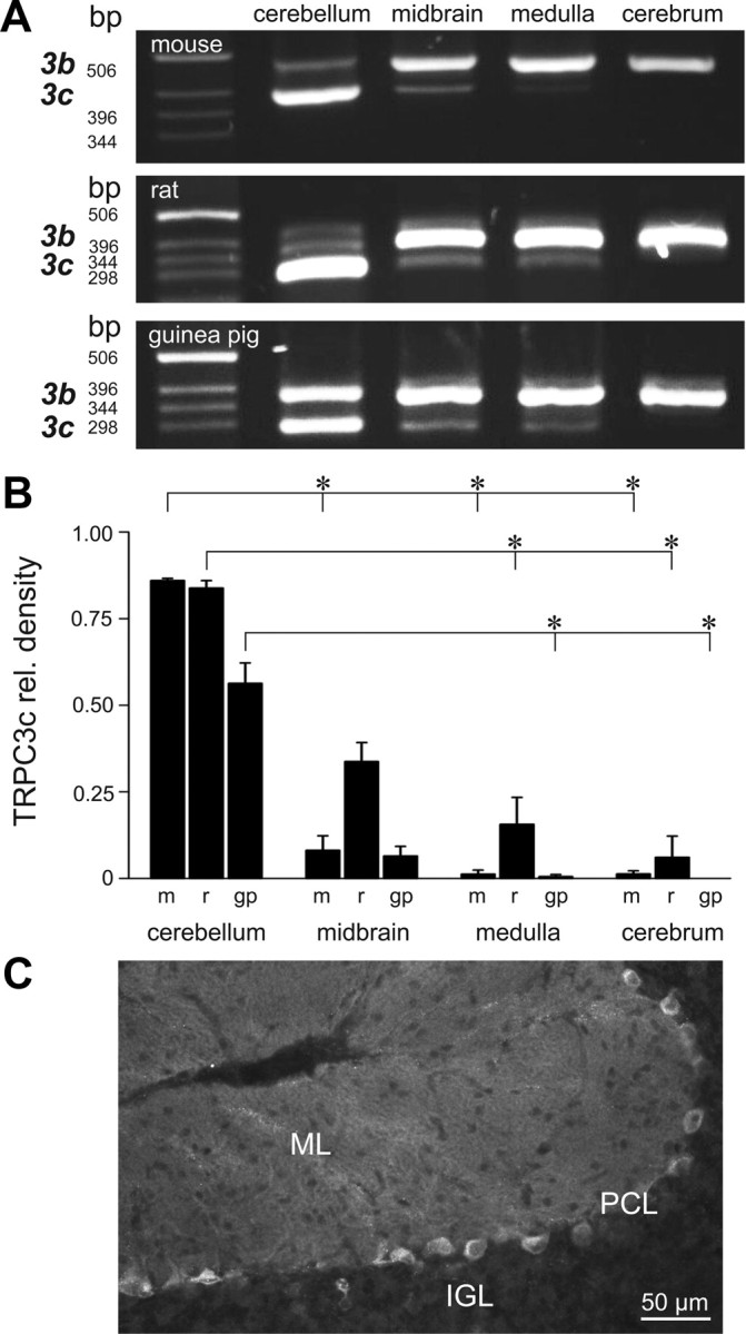
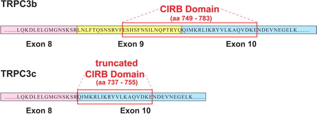
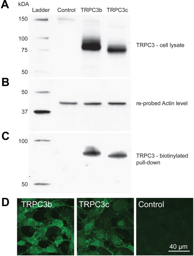
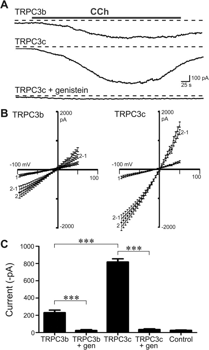
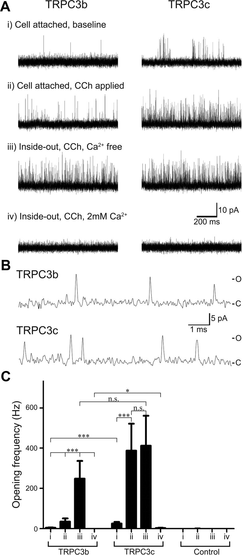
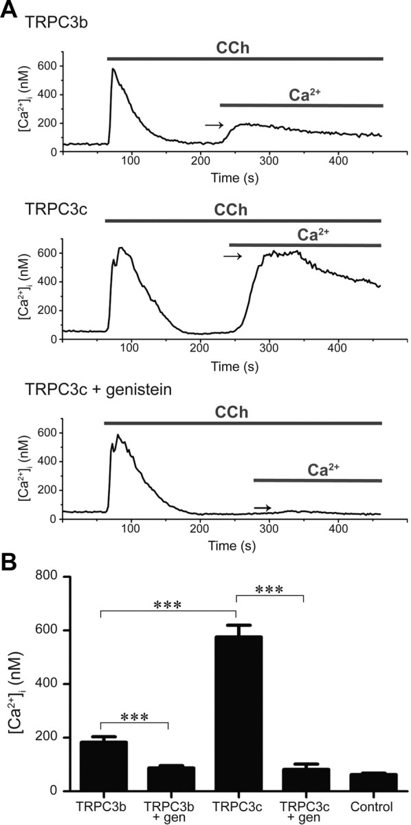
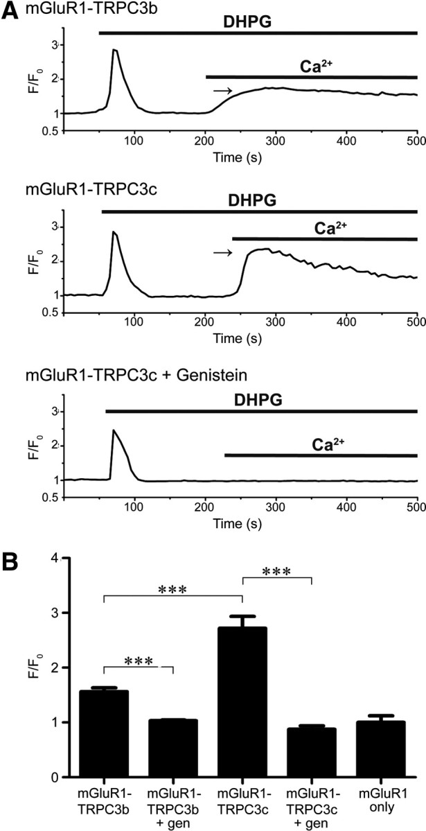
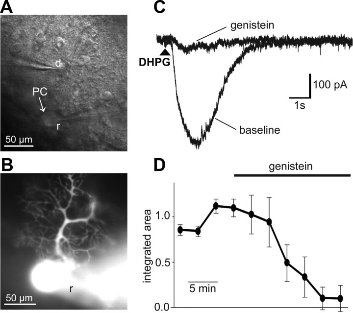
Canonical transient receptor potential (TRPC3) nonselective cation channels are effectors of G-protein-coupled receptors (GPCRs), activated via phospholipase C-diacylglycerol signaling. In cerebellar Purkinje cells, TRPC3 channels cause the metabotropic glutamate receptor (mGluR)-mediated slow EPSC (sEPSC). TRPC3 channels also provide negative feedback regulation of cytosolic Ca(2+), mediated by a C terminus "calmodulin and inositol trisphosphate receptor binding" (CIRB) domain. Here we report the alternative splicing of the TRPC3 mRNA transcript (designated TRPC3c), resulting in omission of exon 9 (approximately half of the CIRB domain) in mice, rats, and guinea pigs. TRPC3c expression is brain region specific, with prevalence in the cerebellum and brainstem. The TRPC3c channels expressed in HEK293 cells exhibit increased basal and GPCR-activated channel currents, and increased Ca(2+) fluorescence responses, compared with the previously characterized (TRPC3b) isoform when activated via either the endogenous M3 muscarinic acetylcholine receptor, or via coexpressed mGluR1. GPCR-induced TRPC3c channel opening rate (cell-attached patch) matched the maximum activation achieved with inside-out patches with zero cytosolic Ca(2+), whereas the GPCR-induced TRPC3b activation frequency was significantly less. Both TRPC3 channel isoforms were blocked with 2 mm Ca(2+), attributable to CIRB domain regulation. In addition, genistein blocked Purkinje cell (S)-2-amino-2-(3,5-dihydroxyphenyl) acetic acid (mGluR1)-activated TPRC3 current as for recombinant TRPC3c current. This novel TRPC3c ion channel therefore has enhanced efficacy as a neuronal GPCR-Ca(2+) signaling effector, and is associated with sensorimotor coordination, neuronal development, and brain injury.


Figures

Figure 1.
Figure 1.
TRPC3 isoform expression in the brain. A, Agarose gel electrophoresis showing TRPC3b (upper) and TRPC3c (lower) RT-PCR amplicons from different brain regions of mouse, rat, and guinea pig. Note the predominance of the TRPC3c isoform in the cerebellum. B, Semiquantification of the expression of TRPC3c cDNA amplicon fluorescence intensity on the agarose gel, as a proportion of the combined TRPC3c + TRPC3b signals, as shown in A (expressed as mean ± SEM). Regional differences in TRPC3c expression are apparent (*p < 0.05, Dunn's pairwise post hoc comparison of ranked data from ANOVA) with highest relative levels in cerebellum followed by midbrain. m, mouse; r, rat; g, guinea pig. C, Immunolabeling of the TRPC3 protein in the mouse cerebellum, showing the high level of staining in the Purkinje neurons including their neurite projections into the molecular layer. PCL, Purkinje cell layer; IGL, internal granule cell layer; ML, molecular layer.
Figure 2.
Figure 2.
Predicted amino acid sequences in the CIRB domain for the TRPC3b and TRPC3c splice variants (mouse; GenBank accession numbers FJ207475 and FJ207476, respectively). Alternative splicing in the TRPC3c isoform results in loss of exon 9, which codes for ∼50% of the CIRB domain (Tang et al., 2001; Zhang et al., 2001). The truncated CIRB region retained in the TRPC3c variant corresponds to the first 19/21 aa of the TRPC3 C8 fusion protein of Zhang et al. (2001), which had limited CaM binding. Further, the retained CIRB region in the TRPC3c variant also corresponds to last 19/21 aa of a fusion protein TRPC3 CΔ78 sequence necessary for trafficking of the TRPC3 protein to the plasma membrane (Wedel et al., 2003).
Figure 3.
Figure 3.
Expression of recombinant TRPC3b and TRPC3c proteins in transfected HEK293 cells detected by Western blotting and immunohistochemistry. A, Whole-cell lysate samples of transfected and untransfected HEK293 cells separated by 10% SDS-PAGE gel, blotted onto polyvinylidene difluoride membrane, and probed for TRPC3 protein with rabbit anti-TRPC3 antibody shown as ∼75 kDa protein species. Note that the TRPC3c isoform has a slightly smaller size, which is predicted based on the loss of 28 aa, encoded by exon 9 (equivalent to ∼3.1 kDa). B, Detection of actin in the same blot after anti-TRPC3 strip-off provides a control for protein loading (43 kDa). C, TRPC3 immunodetection of the membrane-bound fraction labeled with NHS-biotin and purified by adsorption onto NeutrAvidin beads, separated by a 10% SDS-PAGE gel followed by Western blotting with anti-TRPC3 antibody, ∼75 kDa. D, TRPC3b and TRPC3c expression in transfected HEK293 cells detected with anti-TRPC3 antibody by immunofluorescence confocal microscopy. The images are consistent with lower expression of TRPC3c as indicated the Western blot above (A). TRPC3-specific immunolabeling was localized to the plasma membrane and cytoplasm in the transfected cells; untransfected cells (control) were unlabeled.
Figure 4.
Figure 4.
Whole-cell voltage-clamp of HEK293 cells expressing recombinant TRPC3b or TRPC3c channels. A, Example of the larger currents produced by the TRPC3c transfected cells (currents activated by bath application of CCh (100 μm). The currents were blocked by pre-incubation with genistein (10 min; 200 μm). Example shows block of TRPC3c current; Vh = −40 mV; dashed lines indicate zero-current. B, Current/voltage relationships (I/Vs; mean ± SEM) for TRPC3b and TRPC3c (1 = control ramp before CCh; 2 = ramp during CCh response; 2–1 represents the isolated ITRPC3 I/V (trace 2-trace 1). The reversal potential (Erev) of ITRPC3 was close to 0 mV for both isoforms, indicating that the ion selectivity of the two channel isoforms was similarly nonselective. C, Mean peak whole-cell current responses for both isoforms of TRPC3 channels, genistein block for each, and control data (untransfected cells). ***p < 0.001; two-way ranked ANOVA, Holm–Sidak multiple pairwise comparisons).
Figure 5.
Figure 5.
Single-channel patch-clamp recording of HEK293 cells expressing recombinant TRPC3 channels. A, Current traces of HEK293 cells expressing TRPC3b and TRPC3c channels. Each cell group is from the same patch recording and contains four experimental modes as shown (i, ii, iii, and iv). B, Representative single-channel current transients in cell-attached mode shown at high temporal resolution, with CCh (100 μm), as for Aii. C, closed state; O, open state. C, Mean channel opening frequency of membrane patches containing TRPC3b and TRPC3c channels, as well as control patches (no recombinant TRPC3 channel). n.s. indicates that the differences were not significant (p > 0.05).
Figure 6.
Figure 6.
Ratiometric Ca2+ imaging of a field of HEK293 cells expressing recombinant TRPC3 channels using Indo-1 Ca2+ indicator dye. Cells were superfused with nominal Ca2+-free solution followed by application of CCh (100 μm), which causes release of stored Ca2+ via IP3R activation. Once released Ca2+ has been eliminated from the cell, the extracellular Ca2+ is returned to the bath, enabling TRPC3 channel-mediated Ca2+ entry (arrows). A, Greater Ca2+ entry in TRPC3c-expressing cells compared with TRPC3b-expressing cells or genistein block (200 μm; throughout the experiment). B, Mean peak [Ca2+]i arising from TRPC3b- and TRPC3c-mediated Ca2+ entry, genistein block and control (untransfected cell) data. ***p < 0.001; two-way ranked ANOVA, Holm–Sidak multiple pairwise comparison, and Mann–Whitney rank sum test.
Figure 7.
Figure 7.
Fluo-4AM Ca2+ imaging of HEK293 cells coexpressing recombinant TRPC3 channels and mGluR1. A, Rise in [Ca2+]i from Ca2+ store release is shown for a single cell with application of the mGluR1 agonist DHPG (200 μm) in Ca2+-free solution. Fluorescence signal declines as the Ca2+ is extruded from the cell, and then rises again with TRPC3-mediated Ca2+ entry upon return of Ca2+ to the bath (arrows). Greater Ca2+ entry in TRPC3c-expressing cells compared with TRPC3b-expressing cells, or genistein block (200 μm; throughout the experiment). F0 represents the Ca2+ signal just before DHPG application. B, Relative mean peak [Ca2+]i (F/Fo) arising from TRPC3b- and TRPC3c-mediated Ca2+ entry, genistein block, and control (expression of mGluR1 only, no TRPC3). ***p < 0.001; one-way ranked ANOVA, Holm–Sidak multiple pairwise comparison.
Figure 8.
Figure 8.
Whole-cell voltage-clamp recordings of DHPG-evoked inward currents in Purkinje cells. The mGluR agonist DHPG (50 μm) was applied onto the Purkinje cell's dendrites by pressure application (50 ms, 70 kPa) through a patch pipette. A, Bright-field image of the cerebellar slice shows the recording pipette (r) on the Purkinje cell (PC) soma and drug pipette (d) containing DHPG (50 μm) positioned over the dendritic field (see B). B, Fluorescence image of the Purkinje cell loaded with Alexa 594 via the patch-clamp pipette (r). C, The current (Vm = −70 mV) evoked by DHPG before and after bath application of the TRPC3 channel blocker genistein (100 μm). Arrowhead indicates the timing of DHPG application. D, Time-course plot showing the normalized integrated area of repeated (3 min intervals) DHPG-evoked currents before and during application of genistein (indicated by bar). Mean ± SEM (n = 3) responses.

Similar articles

Cited by

References

    1. Abramowitz J, Birnbaumer L. Physiology and pathophysiology of canonical transient receptor potential channels. FASEB J. 2009;23:297–328. - PMC - PubMed
    1. Becker EB, Oliver PL, Glitsch MD, Banks GT, Achilli F, Hardy A, Nolan PM, Fisher EM, Davies KE. A point mutation in TRPC3 causes abnormal Purkinje cell development and cerebellar ataxia in moonwalker mice. Proc Natl Acad Sci U S A. 2009;106:6706–6711. - PMC - PubMed
    1. Birnbaumer L. The TRPC class of ion channels: a critical review of their roles in slow, sustained increases in intracellular Ca2+ concentrations. Annu Rev Pharmacol Toxicol. 2009;49:395–426. - PubMed
    1. Bosman LW, Hartmann J, Barski JJ, Lepier A, Noll-Hussong M, Reichardt LF, Konnerth A. Requirement of TrkB for synapse elimination in developing cerebellar Purkinje cells. Brain Cell Biol. 2006;35:87–101. - PMC - PubMed
    1. Chung YH, Sun Ahn H, Kim D, Hoon Shin D, Su Kim S, Yong Kim K, Bok Lee W, Ik Cha C. Immunohistochemical study on the distribution of TRPC channels in the rat hippocampus. Brain Res. 2006;1085:132–137. - PubMed

Publication types

MeSH terms

LinkOut - more resources