. 2012 Aug 15;32(33):11478-85.
doi: 10.1523/JNEUROSCI.1423-12.2012.

A retinal ganglion cell that can signal irradiance continuously for 10 hours

Affiliations

A retinal ganglion cell that can signal irradiance continuously for 10 hours

Kwoon Y Wong. J Neurosci. .

Abstract

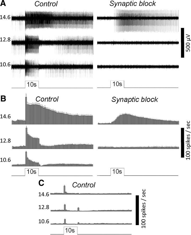
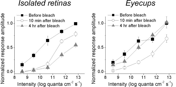
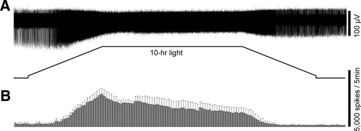
A recently discovered type of mammalian retinal ganglion cell encodes environmental light intensity and mediates non-image-forming visual behaviors, such as the pupillary reflex and circadian photoentrainment. These intrinsically photosensitive retinal ganglion cells (ipRGCs) generate endogenous, melanopsin-based photoresponses as well as extrinsic, rod/cone-driven responses. Because the ipRGCs' light responses and the behaviors they control are both remarkably tonic, these cells have been hypothesized to be capable of irradiance detection lasting throughout the day. I tested this hypothesis by obtaining multielectrode-array recordings from ipRGCs in a novel rat eyecup preparation that enhances the regeneration of rod/cone photopigments. I found that 10 h constant light could continuously evoke action potentials in these ganglion cells under conditions that stimulated (1) only melanopsin, (2) mainly the rod input, and (3) both intrinsic and extrinsic responses. In response to a 10 h stimulus with gradual intensity changes to simulate sunrise and sunset, ipRGC firing rates slowly increased during the "sunrise" phase and slowly decreased during the "sunset" phase. Furthermore, I recorded from putative ipRGCs of melanopsin-knock-out mice and found that these cells retained the ability to respond in a sustained fashion to 20 min light steps, indicating that melanopsin is not required for such tonic responses. In conclusion, ipRGCs can signal light continuously for at least 10 h and can probably track gradual irradiance changes over the course of the day. These results further suggest that the photoreceptors and ON bipolar cells presynaptic to ipRGCs may be able to respond to light continuously for 10 h.


Figures

Figure 1.
Figure 1.
MEA recordings of the novel rat eyecup preparation can detect the light responses of ipRGCs. A, The responses of an ipRGC to 10 s 480 nm light steps of three different intensities (expressed in log quanta cm−2 s−1), first in normal Ames' medium (Control) and then in the presence of l-(+)-2-amino-4-phosphonobutyric acid, d-(-)-2-amino-5-phosphonopentanoic acid, and 6,7-dinitroquinoxaline-2,3-dione to block rod/cone signaling (Synaptic block). This ganglion cell was identified as an ipRGC based on its ability to generate sluggish, high-threshold photoresponses during synaptic block, indicative of melanopsin phototransduction. B, Spike histograms illustrating the averaged light responses of all the ipRGCs detected in this experiment (n = 18). The bin size of these histograms is 1 s, and the error bars represent SEM. C, The averaged light responses of all the conventional ON and ON–OFF ganglion cells analyzed in this experiment (n = 29).
Figure 2.
Figure 2.
The rat eyecup preparation improves the postbleach recovery of rod/cone-mediated ipRGC light responses. In this experiment, an intensity series (8.6–12.8 log quanta cm−2 s−1) of 10 s light steps was presented three times to every ipRGC, first during the dark-adapted state, then ∼10 min after the presentation of a 1 min 13.8 log quanta cm−2 s−1 light to bleach rod/cone photopigments, and finally ∼4 h after this bleaching light. These plots are averages of all the ipRGCs tested, with each cell's highest-amplitude light response normalized to 1. For the ipRGCs in isolated retinas (n = 9; left), their responses recorded 10 min after the bleach were somewhat larger those obtained ∼4 h later, indicating not only a lack of rod/cone photopigment regeneration but also a gradual rundown between these time points. By contrast, the light responses of ipRGCs in eyecups (n = 9; right) increased during these two time points, suggesting the RPE enabled some degree of photopigment regeneration during dark adaptation.
Figure 3.
Figure 3.
The ipRGCs can spike continuously in response to 10 h step increases in light intensity. A, The response of an ipRGC to a 10 h 10.6 log quanta cm−2 s−1 light step, recorded in normal Ames' medium to allow rod/cone signaling. B, Averaged spike histograms of all the ipRGCs recorded in normal Ames' medium, with a bin size of 5 min. The number of cells that contributed to these histograms was six for 12.8 log quanta cm−2 s−1, three for 10.6 log quanta cm−2 s−1, and three for 7.6 log quanta cm−2 s−1. C, Total spike counts in the 10 h responses recorded in normal Ames' medium were estimated (see Materials and Methods) and plotted versus light intensity. In the linear regression fit, r equals 0.999 and the slope is 49,039 spikes per log unit. D, Averaged spike histograms of all the ipRGCs tested in the presence of synaptic blockers to isolate melanopsin photoresponses, with a bin size of 5 min. The number of cells used in these histograms was four for 12.8 log quanta cm−2 s−1 and four for 10.6 log quanta cm−2 s−1. Notice that the response amplitude scale bars are different for the two intensities.
Figure 4.
Figure 4.
The ipRGCs can approximately track gradual changes in light intensity. A, The response of an ipRGC to a 10 h light stimulus designed to simulate gradual light intensity changes over the course of the day. In this stimulus, the intensity was slowly ramped up from 8.1–12.8 log quanta cm−2 s−1 over 2.5 h, held constant at 12.8 log quanta cm−2 s−1 for 5 h, and slowly ramped back down to 8.1 log quanta cm−2 s−1 over 2.5 h. This response was recorded in normal Ames' medium to allow rod/cone input. B, The averaged spike histogram of all ipRGCs subjected to this ramp stimulus (n = 3), with a bin size of 5 min. All cells were superfused with normal Ames' medium.
Figure 5.
Figure 5.
Melanopsin is not required for prolonged ipRGC responses to bright light. A, Whole-cell recordings showed that in melanopsin-knock-out retinas, ganglion cells with ipRGC-like morphologies could respond continuously to bright light for 1 min. Left, The response of a sustained ON ganglion cell to a 1 min 12.5 log quanta cm−2 s−1 light step (top) and its M3-like morphology (bottom). The solid black lines depict dendrites near the retinal surface, whereas the dashed gray lines depict deeper dendrites. Right, The response of another sustained ON ganglion cell to the same 1 min stimulus (top) and its M4-like morphology (bottom). Both cells' axons are indicated by arrows. B, C, For each piece of melanopsin-knock-out mouse retina recorded on the MEA, the vast majority of channels exhibited only transient photoresponses and only several channels contained sustained responses. Thus, the transient units were probably conventional RGCs while the sustained ones were probably ipRGCs. B, Top, The transient response of a presumed conventional RGC to a 1 min 15.6 log quanta cm−2 s−1 light step, recorded in normal Ames' medium. Bottom, A histogram averaging the spiking responses of 30 transient ON or ON–OFF RGCs to this 1 min light step, with a bin size of 1 s. C, Top, The response of a putative melanopsin-knock-out ipRGC to a 1 min 15.6 log quanta cm−2 s−1 light step in normal Ames' medium. Middle, A histogram averaging the spiking responses of all the presumed melanopsin-knock-out ipRGCs to this 1 min light step, with a bin size of 1 s (n = 13). Bottom, During superfusion with synaptic blockers, the light response of the cell shown in C, top, was completely abolished, confirming the lack of intrinsic photosensitivity. D, Synaptic input to putative melanopsin-knock-out ipRGCs could evoke action potentials throughout a 20 min 12.8 log quanta cm−2 s−1 light step. Top, The response of one of these cells, recorded in normal Ames' medium. Bottom, A spike histogram averaging all the putative melanopsin-knock-out ipRGCs' responses to the 20 min 12.8 log quanta cm−2 s−1 light, with a bin size of 10 s (n = 13).

Similar articles

Cited by

References

    1. Barlow HB, Levick WR. Changes in the maintained discharge with adaptation level in the cat retina. J Physiol. 1969;202:699–718. - PMC - PubMed
    1. Berson DM, Dunn FA, Takao M. Phototransduction by retinal ganglion cells that set the circadian clock. Science. 2002;295:1070–1073. - PubMed
    1. Dacey DM, Liao HW, Peterson BB, Robinson FR, Smith VC, Pokorny J, Yau KW, Gamlin PD. Melanopsin-expressing ganglion cells in primate retina signal colour and irradiance and project to the LGN. Nature. 2005;433:749–754. - PubMed
    1. DeVries SH, Schwartz EA. Kainate receptors mediate synaptic transmission between cones and ‘Off’ bipolar cells in a mammalian retina. Nature. 1999;397:157–160. - PubMed
    1. Do MT, Kang SH, Xue T, Zhong H, Liao HW, Bergles DE, Yau KW. Photon capture and signalling by melanopsin retinal ganglion cells. Nature. 2009;457:281–287. - PMC - PubMed

Publication types

MeSH terms

LinkOut - more resources