. 2012 Oct 15;303(8):E994-1005.
doi: 10.1152/ajpendo.00291.2012. Epub 2012 Aug 14.

Glucagon-like peptide 2 induces vasoactive intestinal polypeptide expression in enteric neurons via phophatidylinositol 3-kinase-γ signaling

Affiliations

Glucagon-like peptide 2 induces vasoactive intestinal polypeptide expression in enteric neurons via phophatidylinositol 3-kinase-γ signaling

Elaine de Heuvel et al. Am J Physiol Endocrinol Metab. .

Abstract

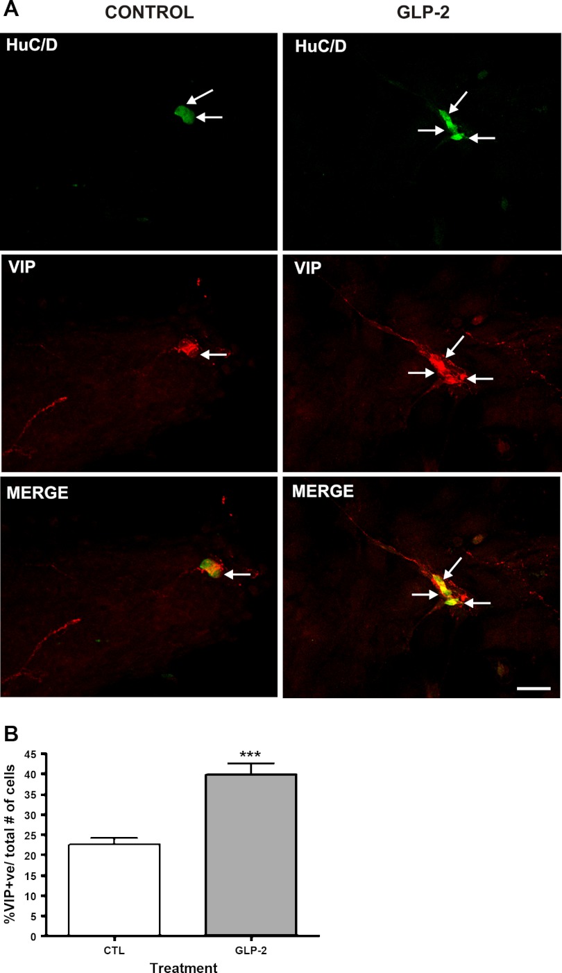
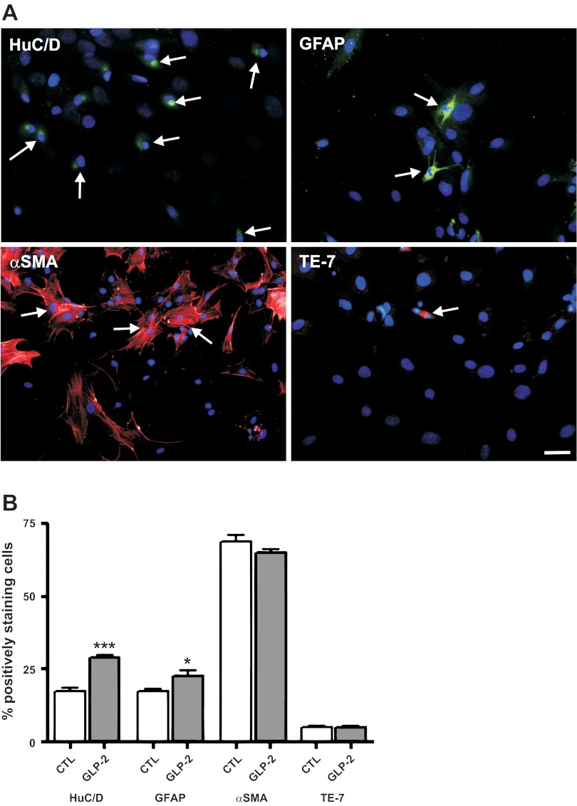
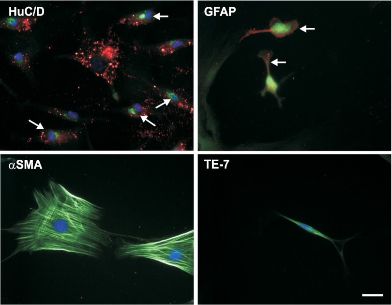
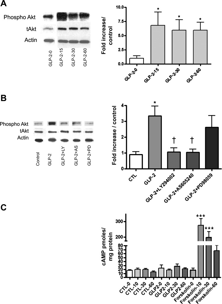
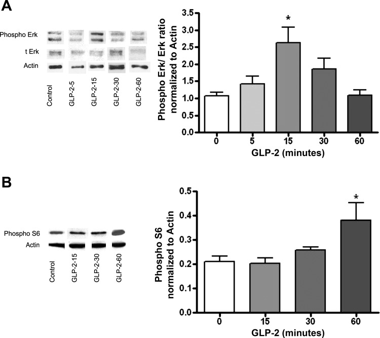
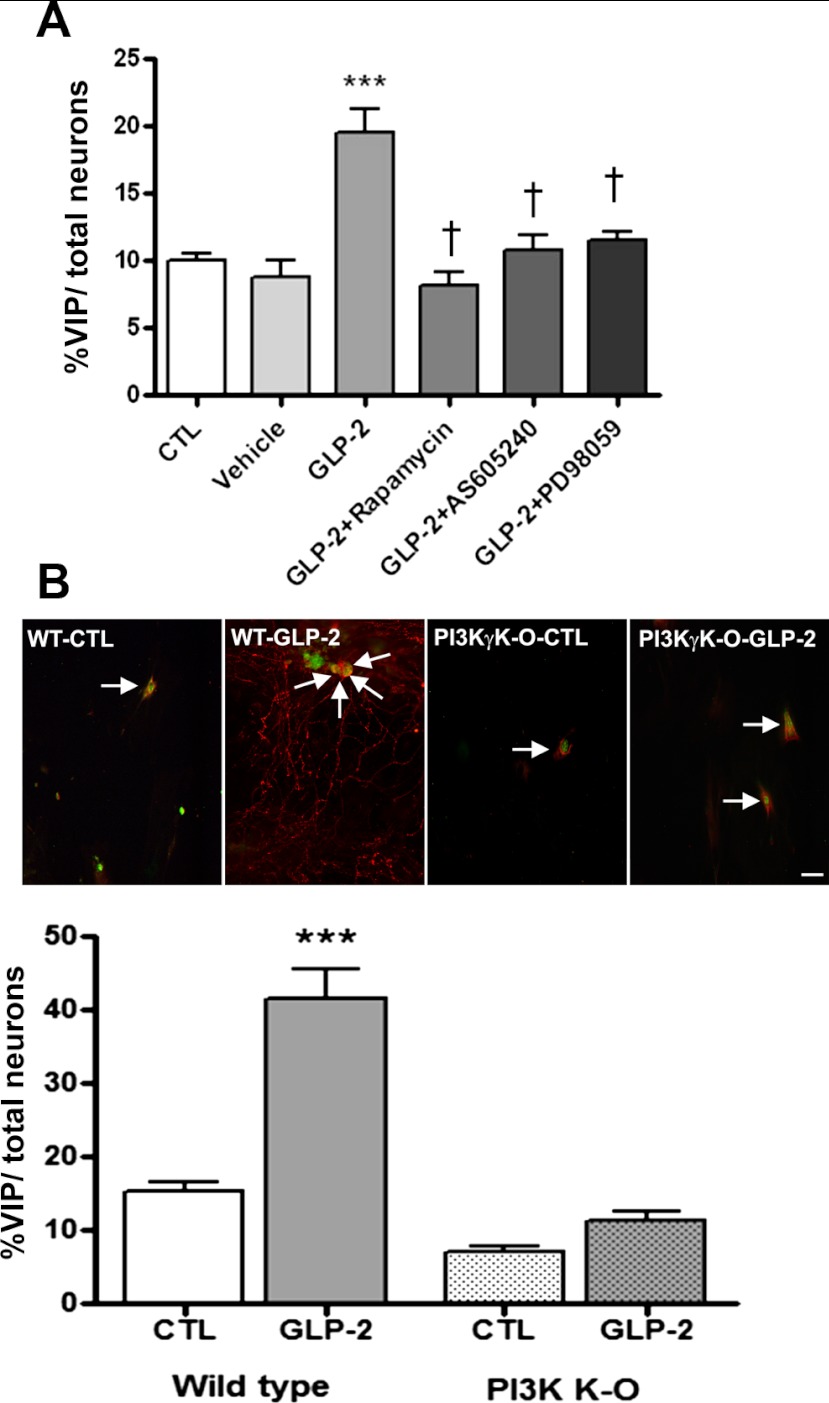
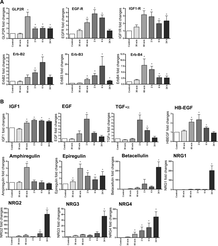
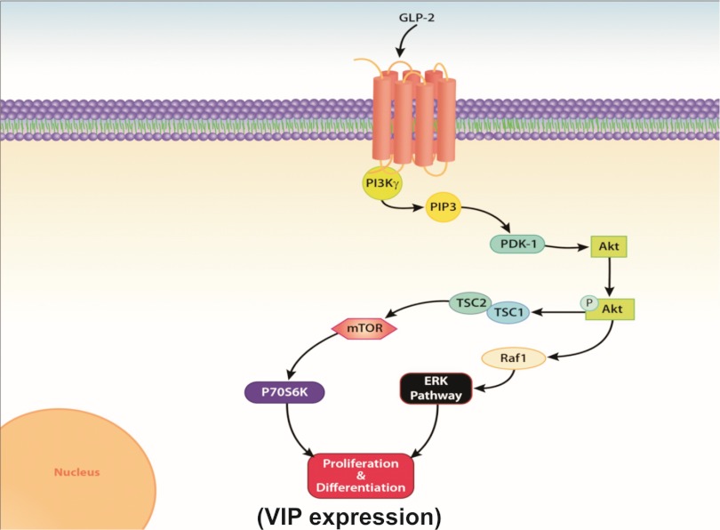
Glucagon-like peptide 2 (GLP-2) is an enteroendocrine hormone trophic for intestinal mucosa; it has been shown to increase enteric neuronal expression of vasoactive intestinal polypeptide (VIP) in vivo. We hypothesized that GLP-2 would regulate VIP expression in enteric neurons via a phosphatidylinositol-3 kinase-γ (PI3Kγ) pathway. The mechanism of action of GLP-2 was investigated using primary cultures derived from the submucosal plexus (SMP) of the rat and mouse colon. GLP-2 (10(-8) M) stimulation for 24 h increased the proportion of enteric neurons expressing VIP (GLP-2: 40 ± 6% vs. control: 22 ± 5%). GLP-2 receptor expression was identified by immunohistochemistry on neurons (HuC/D+) and glial cells (GFAP+) but not on smooth muscle or fibroblasts in culture. Over 1-4 h, GLP-2 stimulation of SMP increased phosphorylated Akt/Akt ratios 6.1-fold, phosphorylated ERK/ERK 2.5-fold, and p70S6K 2.2-fold but did not affect intracellular cAMP. PI3Kγ gene deletion or pharmacological blockade of PI3Kγ, mammalian target of rapamycin (mTOR), and MEK/ERK pathways blocked the increase in VIP expression by GLP-2. GLP-2 increased the expression of growth factors and their receptors in SMP cells in culture [IGF-1r (3.2-fold increase), EGFr (5-fold), and ErbB-2-4r (6- to 7-fold)] and ligands [IGF-I (1.5-fold), amphiregulin (2.5-fold), epiregulin (3.2-fold), EGF (7.5-fold), heparin-bound EGF (2.0-fold), β-cellulin (50-fold increase), and neuregulins 2-4 (300-fold increase) (by qRT-PCR)]. We conclude that GLP-2 acts on enteric neurons and glial cells in culture via a PI3Kγ/Akt pathway, stimulating neuronal differentiation via mTOR and ERK pathways, and expression of receptors and ligands for the IGF-I and ErbB pathways.


Figures

Fig. 1.
Fig. 1.
Vasoactive intestinal polypeptide (VIP) expression in cells derived from the submucosal plexus following glucagon-like peptide-2 (GLP-2) stimulation. The effects of GLP-2 on cells isolated from submucosal ganglia prepared from rat colon were studied in vitro. The submucosal neurons were isolated by microdissection, separated, and cultured for 8 days, being treated with cytosine arabinoside over the last 3 days. Medium was replaced with 0.1% FCS with or without added GLP-2 (10−8 M), and cells were studied by immunohistochemistry after 24 h. Labeled cells were imaged using a confocal microscope (Olympus FV1000) and Olympus Fluoview version 3.0 software. A, top: staining for HuC/D (in green). A, middle: staining for VIP (red). A, bottom: merged images. Arrows indicate nuclei. Scale bar, 50 μm. B: effects of GLP-2 on the proportion of cells expressing VIP. Nos. of cells costaining for VIP, as a proportion of all nuclei detected by 4,6-diamidino-2-phenylindole (DAPI) staining, in 10 high-powered fields from 8 coverslips/determination. Data are means ± SE; n = 8. –––P < 0.01 vs. controls (CTL).
Fig. 2.
Fig. 2.
Identity of cells in primary cultures derived from the submucosal plexus and the effects of GLP-2 stimulation. Primary cultures of cells from the submucosal neuronal plexus were prepared from rat colon, as outlined in Fig. 1. A: cell identification by immunohistochemistry. Coverslips were stained with antibody against the neuronal nuclear marker HuC/D (green), the glial marker glial fibrillary acidic protein (GFAP; green), α-smooth muscle actin (α-SMA; red), or the fibroblast marker TE-7 (red) and costained with the nuclear stain DAPI (blue). Double-labeled cells are marked by arrows. Scale bar, 100 μm. B: effects of GLP-2 on cells derived from the submucosal plexus in culture. Cell types detected by immunohistochemical labeling after 24 h of stimulation with 0.1% FCS (controls) or 0.1% FCS + GLP-2 (10−8 M). Values are the proportion of cells labeled by the antibody as %total cells (detected by DAPI staining) from 10 high-powered fields from 8 coverslips/determination. Data are means ± SE; n = 8. –P < 0.05 vs. controls; –––P < 0.001 vs. controls.
Fig. 3.
Fig. 3.
Expression of the GLP-2 receptor on primary cell cultures derived from the submucosal plexus. Primary cell cultures derived from the submucosal enteric plexus were prepared as outlined in Fig. 1. Coverslips were stained with antibody against the neuronal nuclear marker HuC/D (green), the glial marker GFAP (green), α-SMA (green), or the fibroblast marker TE-7 (green) and colabeled with antibody against the GLP-2 receptor (red) and the nuclear stain DAPI (blue). Double-labeled cells are marked by arrows. Scale bar, 25 μm.
Fig. 4.
Fig. 4.
GLP-2 effects on Akt phosphorylation and cAMP accumulation. Primary cultures of cells from the submucosal enteric neuronal plexus were prepared from rat colon, as outlined in Fig. 1, and treated with control medium or GLP-2 (10−8 M) for the times indicated. A: time course of phospho-Akt expression; Western blots of representative gels taken from lysates of cells exposed to GLP-2 for the times indicated and probed sequentially with antibodies against phospho-Akt, total Akt (t-Akt), and actin, with stripping and restaining of the original gel. Blots from a single gel are rearranged for clarity. Graph represents phospho-Akt/t-Akt normalized to controls, with actin used a loading control. B: effects of phosphatidylinositol 3-kinase (PI3K) inhibition. Cultured cells were pretreated for 1 h with the nonselective PI3K inhibitor LY-294002 (50 μM), the selective PI3Kγ inhibitor AS-605240 (50 μM), or the ERK1/2 inhibitor PD-98059 (50 μM) and then stimulated with GLP-2 (10−8 M) for 15 min. Representative Western blots of phospho-Akt, t-Akt, and actin staining are presented, and blots from a single gel are arranged for clarity. Results of phospho-Akt/t-Akt normalized to controls are presented. Methods are as outlined in A; n = 6. C: cultured cells were treated with GLP-2 or control solvent vehicle for the times indicated. Positive controls were treated with forskolin (20 μM), cells lysed at the times indicated, and cAMP levels determined. Data are means ± SE; n = 8/condition. –P < 0.05 vs. controls; –––P < 0.0001 vs. controls; †P < 0.05 vs. GLP-2 treated.
Fig. 5.
Fig. 5.
GLP-2 effects on ERK and mammalian target of rapamycin (mTOR) pathways. Primary cultures of cells from the submucosal enteric neuronal plexus were prepared from rat colon, as outlined in Fig. 1, and treated with control medium or GLP-2 (10−8 M) for the times indicated. A: time course of ERK/phospho-ERK expression. Western blots of representative gels taken from lysates of cells exposed to GLP-2 for the times indicated, probed with antibodies against phospho-ERK, t-ERK, and actin; blots are from a single gel and arranged for clarity. Graph represents phospho-ERK/t-ERK normalized to controls, with actin used a loading control. B: time course of mTOR activity. Using methods similar as in A, gels were probed with antibodies against p70S6K and actin. Graph represents densiometer readings of p70S6K/actin, with actin used as the loading control. Data are means ± SE. –P < 0.05 vs. controls.
Fig. 6.
Fig. 6.
Effects of pharmacological antagonists or gene knockout of intracellular signaling pathways on GLP-2-induced VIP expression. A: effect of pharmacological blockers of intracellular signaling. Primary cultures of cells from the submucosal enteric neuronal plexus derived from rat colon were grown on coverslips and pretreated for 1 h with the selective PI3Kγ inhibitor AS-605240 (50 μM), the ERK1/2 inhibitor PD-98059 (50 μM) or the mTOR inhibitor rapamycin (50 μM) or solvent vehicle, followed by additional GLP-2 (10−8 M) or CTL (GLP-2) for 24 h. The proportion of cells costaining for VIP and HuC/D with each condition is shown. B: effect of PI3Kγ knockout (KO). Primary cultures of cells from submucosal ganglia from mouse ileum from wild-type and PI3Kγ−/− animals were isolated and grown on coverslips, as outlined in Fig. 1, culturing cells for 14 days prior to study. Wells were treated with control medium or medium + GLP-2 (10−8 M) for 7 days. The images and the corresponding counts of cells costaining for VIP and HuC/D with each condition is shown. Scale bar, 50 μm. Data are means ± SE; n = 6–8/group. –––P < 0.001 vs. controls; †P < 0.001 vs. GLP-2 treated.
Fig. 7.
Fig. 7.
GLP-2 treatment of isolated enteric neurons increases expression of growth factor and receptors. Submucosal enteric neurons isolated from rat colon were cultured and treated with GLP-2 (10−8 M) for the times indicated, with extraction of total RNA and analysis for the receptor (A) or ligand systems (B) by qRT-PCR. Data are expressed as the fold increase from baseline relative to 18S rRNA. Data are means ± SE; n = 6 at each time point. –P < 0.05 vs. basal expression. GLP-2R, GLP-2 receptor; EGF-R, epidermal growth factor receptor.
Fig. 8.
Fig. 8.
Schematic of GLP-2 activation pathways in enteric neurons. GLP-2 binds to the G protein-coupled receptor, which activates the PI3Kγ moiety, leading to phosphorylation of Akt and downstream stimulation of the mTOR and ERK pathways, which leads to the observed increase in neuronal VIP expression. PIP3, phosphatidylinositol-3,4,5-triphosphate; PDK-1, phosphoinositide-dependent kinase-1; TSC, tuberous sclerosis complex.

Similar articles

Cited by

References

    1. Amin H, Holst JJ, Hartmann B, Wallace L, Sigalet D. Functional ontogeny of the proglucagon derived peptide axis in human neonates. Pediatrics 121: e180–e186, 2008 - PubMed
    1. Anini Y, Izzo A, Oudit GY, Backx PH, Brubaker PL. Role of phosphatidylinositol-3 kinase-γ in the actions of glucagon-like peptide-2 on the murine small intestine. Am J Physiol Endocrinol Metab 292: E1599–E1606, 2007 - PubMed
    1. Birchmeier C. ErbB receptors and the development of the nervous system. Exp Cell Res 315: 611–618, 2009 - PubMed
    1. Britsch S, Li L, Kirchhoff S, Theuring F, Brinkmann V, Birchmeier C, Riethmacher D. The ErbB2 and ErbB3 receptors and their ligand, neuregulin-1, are essential for development of the sympathetic nervous system. Genes Dev 12: 1825–1836, 1998 - PMC - PubMed
    1. Chevalier J, Derkinderen P, Gomes P, Thinard R, Naveilhan P, Vanden Berghe P, Neunlist M. Activity-dependent regulation of tyrosine hydroxylase expression in the enteric nervous system. J Physiol 586: 1963–1975, 2008 - PMC - PubMed

Publication types

MeSH terms

LinkOut - more resources