. 2013 Apr;250(2):601-11.
doi: 10.1007/s00709-012-0445-z. Epub 2012 Aug 16.

The effect of pre-incubation of Allium cepa L. roots in the ATH-rich extract on Pb uptake and localization

Affiliations

The effect of pre-incubation of Allium cepa L. roots in the ATH-rich extract on Pb uptake and localization

Sława Glińska et al. Protoplasma. 2013 Apr.

Abstract

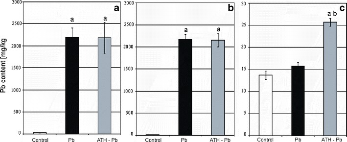
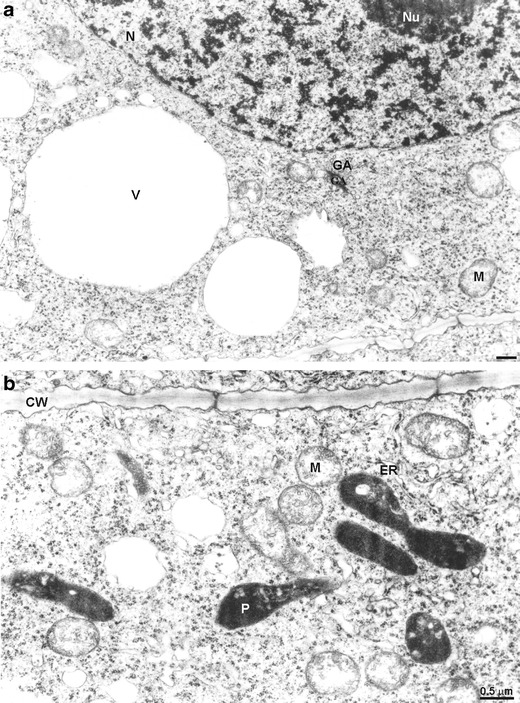
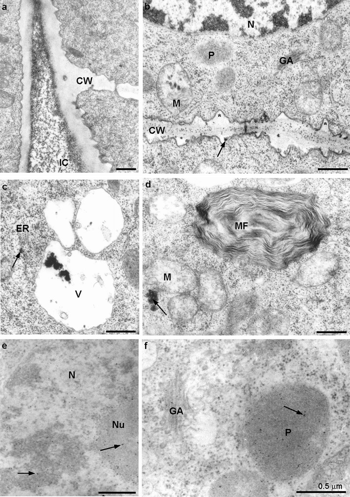
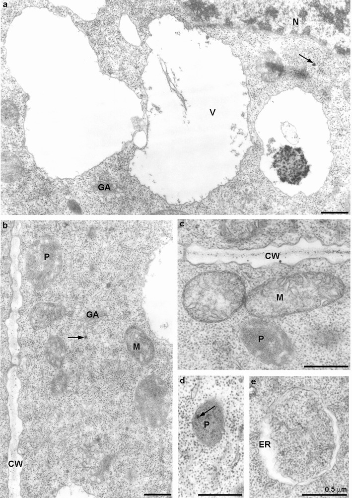
The positive influence of anthocyanin (ATH) on toxic metal-treated plant material is well documented; however, it is still not explained if it is caused by changes in element absorption and distribution. Therefore, detailed analysis of the effect of the ATH-rich extract from red cabbage leaves on Pb uptake and localization at morphological, anatomical and ultrastructural level was the goal of this study. Two-day-old adventitious roots of Allium cepa L. (cv. Polanowska) were treated for 2 h with the aqueous solution of Pb(NO3)2 at the concentration of 100 μM with or without preliminary incubation in the anthocyanin-rich extract from Brassica oleracea L. var. capitata rubra leaves (250 μM, 3 h). The red cabbage extract did not change the total Pb uptake but it enhanced the translocation of accumulated metal from roots to shoots. Within the pretreated roots, more Pb was deposited in their basal part and definitely smaller amount of the metal was bound in the apoplast of the outer layers of cortex cells. The ultrastructural analysis (transmission electron microscopy and X-ray microanalysis) revealed that the ATH-rich extract lowered the number of Pb deposits in intracellular spaces, cell wall and cytoplasm of root meristematic cells as well as in such organelles important to cell metabolism as mitochondria, plastids and nucleus. The Pb deposits were preferably localised in those vacuoles where ATH also occurred. This sequestration of Pb in vacuoles is probably responsible for reduction of metal cytotoxicity and consequently could lead to better plant growth.


Figures

Fig. 1
Fig. 1
The effect of pre-incubation in the ATH-rich extract from red cabbage leaves on the concentration of Pb in a whole plant (a), roots (b) and shoot (c) of A. cepa growing in the presence of Pb(NO3)2. Values are means ± SE (n = 3). Letters denote statistically significant differences at the 0.05 level with Student’s t test between: the control and Pb-treated material (a), Pb-treated material with or without pre-incubation in the ATH-rich extract (b)
Fig. 2
Fig. 2
A. cepa roots stained with sodium rhodizonate. Brown colour indicates the presence of lead. a Control; b 100 μM Pb(NO3)2, 2 h; c 250 μM ATH-rich extract 3 h → 100 μM Pb(NO3)2, 2 h
Fig. 3
Fig. 3
A. cepa roots stained with sodium rhodizonate—cross sections (0.5 cm from tip). a, b Control; c, d 100 μM Pb(NO3)2, 2 h; e, f 250 μM ATH-rich extract 3 h → 100 μM Pb(NO3)2, 2 h. a, c, e Differentiation zone of roots. b, d, f Cortex cells; C cortex, R rhizodermis, S stele
Fig. 4
Fig. 4
Meristematic cells of the control A. cepa root; a, b note lack of electron-dense deposits; CW cell wall, ER endoplasmic reticulum, GA Golgi apparatus, M mitochondrium, N nucleus, Nu nucleolus, P plastid, V vacuole
Fig. 5
Fig. 5
Meristematic cells of A. cepa roots treated with 100 μM Pb(NO3)2 for 2 h. a Numerous electron-dense deposits in the intercellular space; b numerous deposits in the cell wall (arrow) irregularly thickened (asterisk) and in mitochondria; c a small deposit in the lumen of ER (arrow), large ones in vacuole; d an electron-dense deposit in cytoplasm (arrow), myelinous figure visible; e small electron-dense deposits in nucleus and nucleolus (arrows); f small deposits in Golgi apparatus and in plastid (arrow); CW cell wall, ER endoplasmic reticulum, GA Golgi apparatus, IC intercellular space, M mitochondrium, MF myelinous figure, N nucleus, Nu nucleolus, P plastid, V vacuole
Fig. 6
Fig. 6
Meristematic cells of A. cepa roots pre-incubated with 250 μM ATH-rich extract for 3 h and subsequently treated with 100 μM Pb(NO3)2 for 2 h. a Large electron-dense deposits in the vacuole and a small one in cytoplasm (arrow); b very small deposits in the cell wall and vesicle of Golgi apparatus (arrow); c small electron-dense deposits in the cell wall as well as in mitochondria, and plastid deprived of deposits; d plastid with small electron-dense deposits (arrow); e swollen lumen of ER without deposits; CW cell wall, ER endoplasmic reticulum, GA Golgi apparatus, M mitochondrium, N nucleus, P plastid, V vacuole
Fig. 7
Fig. 7
Electron micrographs illustrating distribution of electron-dense deposits in the root meristematic cells of A. cepa on unstained sections. The circles indicate the regions subjected to the X-ray microanalysis and spectra collected over those regions are presented on the right. The spectra (80 keV) show the L family of X-ray for Pb. The family consist of the L α (10,540 keV) and L β1 (12,611 keV) peaks, which are indicated with vertical lines. a Numerous electron-dense lead deposits in a cell wall of meristematic cell of A. cepa root treated with 100 μM Pb(NO3)2 for 2 h, X-ray spectrum collected over the circled region of the cell wall shows lead characteristic peaks proving the presence of lead in the electron-opaque deposits; b large granular deposits in a vacuole of a meristematic cell of A. cepa root treated with 100 μM Pb(NO3)2 for 2 h and X-ray spectrum collected over them proving the presence of lead; c compact large deposits in a vacuole of a meristematic cell of A. cepa root pre-incubated with 250 μM ATH-rich extract for 3 h and subsequently treated with 100 μM Pb(NO3)2 for 2 h and X-ray spectrum collected over those precipitates proving the presence of lead; CW cell wall, N nucleus, V vacuole

Similar articles

Cited by

References

    1. Alkorta I, Hernández Allica J, Becerri JM, Amezaga I, Albizu I, Garbisu C. Recent findings on the phytoremediation of soils contaminated with environmentally toxic heavy metals and metalloids such as zinc, cadmium, lead, and arsen. Rev Environ Sci Bio/Tech. 2004;3:71–90. doi: 10.1023/B:RESB.0000040059.70899.3d. - DOI
    1. Antosiewicz D, Wierzbicka M. Localization of lead in Allium cepa L. cells by electron microscopy. J Microsc. 1999;195:139–146. doi: 10.1046/j.1365-2818.1999.00492.x. - DOI - PubMed
    1. Baranowska-Morek A, Wierzbicka M. Localization of lead in root tip of Dianthus carthusianorum. Acta Biol Cracov Bot. 2004;46:45–56.
    1. Brännvall ML, Bindler R, Renberg I, Emteryd O, Bartnicki J, Billström K. The Medieval metal industry was the cradle of modern large-scale atmospheric lead pollution in northern Europe. Environ Sci Technol. 1999;33:4391–4395. doi: 10.1021/es990279n. - DOI
    1. Buchauer MJ. Contamination of soil and vegetation near a zinc smelter by zinc, cadmium, copper and lead. Environ Sci Technol. 1973;7:131–135. doi: 10.1021/es60074a004. - DOI

Publication types

LinkOut - more resources