. 2012 Dec;2(12):1150-65.
doi: 10.1158/2159-8290.CD-12-0216. Epub 2012 Aug 15.

Identification of luminal breast cancers that establish a tumor-supportive macroenvironment defined by proangiogenic platelets and bone marrow-derived cells

Affiliations

Identification of luminal breast cancers that establish a tumor-supportive macroenvironment defined by proangiogenic platelets and bone marrow-derived cells

Hanna S Kuznetsov et al. Cancer Discov. 2012 Dec.

Abstract

Breast cancer recurrence rates vary following treatment, suggesting that tumor cells disseminate early from primary sites but remain indolent indefinitely before progressing to symptomatic disease. The reasons why some indolent disseminated tumors erupt into overt disease are unknown. We discovered a novel process by which certain luminal breast cancer (LBC) cells and patient tumor specimens (LBC "instigators") establish a systemic macroenvironment that supports outgrowth of otherwise-indolent disseminated tumors ("responders"). Instigating LBCs secrete cytokines that are absorbed by platelets, which are recruited to responding tumor sites where they aid vessel formation. Instigator-activated bone marrow cells enrich responding tumor cell expression of CD24, an adhesion molecule for platelets, and provide a source of VEGF receptor 2(+) tumor vessel cells. This cascade results in growth of responder adenocarcinomas and is abolished when platelet activation is inhibited by aspirin. These findings highlight the macroenvironment as an important component of disease progression that can be exploited therapeutically.

Significance: Currently, processes that mediate progression of otherwise indolent tumors are not well understood, making it difficult to accurately predict which cancer patients are likely to relapse. Our findings highlight the macroenvironment as an important component of disease progression that can be exploited to more accurately identify patients who would benefit from adjuvant therapy.


Figures

Figure 1
Figure 1
Breast cancer subtype-specific systemic environments affect bone marrow derived cells and phenotype of otherwise indolent tumors at distant sites. A, Systemic instigation human tumor xenograft model. Aggressively growing “Instigating” human tumors or controls are injected into one site of host Nude mice; otherwise indolent “Responding” human tumor cells injected into distant anatomical locations. B, Mass of responding tumors that formed in the systemic environments established by control Matrigel, triple-negative breast cancer (TNBC), or luminal breast cancer (LBC). Incidence of responding tumor formation is indicated above each bar; n=4 mice per group. C, Hematoxylin and eosin (H&E) stains of responding tumors after growth in indicated systemic environments; scale bar = 200 μm; inset = 100 μm. D, Staining for the proliferation marker, Ki67 (brown) in indicated responding tumors; nuclei counterstained with hematoxylin. Areas of necrosis (“N”) and edema (“E”) are indicated. Scale bar = 100 μm. E, Average number of vessels per area in indicated tumors and tissues. Vessels were counted under 40x magnification in 3 different areas from each of 3 different tumors per group (n=9 images per group). The two tissue plugs recovered opposite Matrigel that did not contain focal tumors were also counted. F, Serial sections of LBC-instigated responding tumors stained for CD31-positive endothelial cells (red, left), αSMA-positive pericytes (red, center), and VEGFR2-positive endothelial progenitor cells (red, right). Nuclei counterstained with DAPI (blue). Arrowheads indicate blood vessels. Top row scale bar = 100μm; bottom row represents high magnification of images in top row. G, Whole mount fluorescent images (4x) to visualize GFP+ BMCs recruited to responding tumors after 4 weeks of exposure to indicated systemic environments. Numbers indicate average percentage of total tissue cells that were comprised of GFP+ BMCs; n=4 per group. H, Results from flow cytometric analysis of GFP+ bone marrow-derived cells recruited into responding tumors in (G); n=4 per group. I, Flow cytometric analysis of indicated cells in the marrow of mice bearing Matrigel control or instigating LBC. Graph represents average fold change in numbers of indicated cell types in bone marrow of mice bearing LBC relative to those bearing Matrigel control; n = 4 mice per group. Also see Figs. S1, S2, S3 and S5.
Figure 2
Figure 2
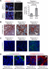
Platelets are recruited to responding tumors during LBC systemic instigation. A, Left: Immunofluorescent images of responding tumors in indicated macroenvironments stained for p-selectin to visualize platelets (red); nuclei counterstained with DAPI (blue). Scale bar = 100μm. Right: CellProfiler software outlines of p-selectin-positive areas used for quantification. Graph represents average p-selectin-positive area per image; TNBC n=15 images; LBC n=19 images. B, Masson's Trichrome to visualize collagen (blue), red blood cells (bright red), and cell nuclei (dark brown). Blood vessels (“BV”), and necrotic areas (“N”) are indicated. Scale bar = 100μm. C, CD24 staining (green) of responding tumors under indicated conditions; nuclei counterstained with DAPI (blue); scale bar = 100μm. D, Merged immunofluorescent images of responding tumors or cell plugs that had formed under indicated conditions. Left: CD24 (green), p-selectin (purple). Right: collagen IV (red), p-selectin (purple). BV denotes blood vessel. In all cases, cell nuclei counterstained with DAPI (blue); scale bars = 20μm.
Figure 3
Figure 3
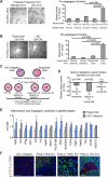
Platelets in the LBC environment are enhanced for pro-angiogenic function and take up pro-angiogenic factors secreted by LBC tumor cells. A, Left: representative images of capillary tubes formed by human umbilical vein endothelial cells (HUVECs) after 6 hr exposure to platelet releasates prepared from indicated mice; 4x magnification. Right: Quantification of HUVEC branch points over a 4-7 hour time course (see Methods) induced by platelet releasates from indicated tumor-bearing mice. Mouse and platelet status indicated below graph; n=3 samples per group, tested in duplicate. B, Left: representative 4x images of capillary tubes formed by HUVECs after 7 hr exposure to 48-hr conditioned medium (CM) from indicated cell lines. Right: Quantification of HUVEC branch points over a 4-7 hour time course (see Methods) induced by CM from indicated cell lines. Releasates from resting mouse or human platelets were used as controls. All samples analyzed in duplicate. C, Assay to test ability of platelets to absorb pro-angiogenic factors from CM of indicated cell lines. 48-hour CM was collected from LBC instigator cells or responder cells and exposed to naïve platelets from cancer-free humans or mice for 10 min at 37°C. Various media (1A-2B) were tested for ability to induce angiogenesis in the HUVEC assay. D, Relative number of capillary tube branch points induced by CM from (C). HUVECs were subjected in vitro to indicated CM and the number of branch points quantified during a 4-7 hour time course. Data represent relative number of branch points: 1B/1A for resting naïve mouse and human platelets and 2A/2B for resting naïve mouse platelets. All samples were tested in duplicate. E, Relative levels of indicated cytokines in platelet lysates from mice bearing Matrigel or LBC tumors; n=3 mice per group. Significant values: GRO (p=0.012), IFNγ (p=0.050), IL6 (p=0.044), PDGF-BB (p=0.033), and PlGF (p=0.044). F, Instigating or Responding tumors under indicated conditions stained for phosho-STAT3 (red), SV40 LgTAg-positive responder cells (green), and cell nuclei (DAPI, blue); scale bar = 100μm. Also see Fig. S4.
Figure 4
Figure 4
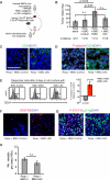
BMCs from mice bearing LBC tumors enrich responding tumor cells for CD24 surface expression but lack instigating ability. A, In vivo test of BMC tumor promoting function. BMCs harvested from mice bearing indicated systemic environments were immediately mixed with responder cells and injected subcutaneously into secondary recipients. B, Tumor mass 12 wk following injection of responder cells admixed with indicated BMCs. Numbers of mice and incidence of tumor formation indicated below graph for collective data from 2 separate experiments. C and D, Responding tumors resulting from admixture with indicated BMCs stained for CD24 (green) (C), or CD24 (green) and pselectin (red) (D). Cell nuclei stained with DAPI (blue); scale bars = 100μm. E, Representative flow cytometry histograms of CD24 expression on GFP+ responder cells after 4 d in vitro co-culture with BMCs harvested from indicated mice. Gate represents CD24+ populations. Graph represents percent change in responding tumor cell CD24 surface expression under indicated conditions relative to co-culture with BMCs from cancer-free mice; n=10 BMC samples per group. F and G, VEGFR2-positive cells (red, F) and phospho-STAT3 (red) and LgT-positive tumor cells (green) (G) in responding tumors admixed with indicated BMCs; nuclei counterstained with DAPI (blue). H, Vessel density in indicated responding tumors; differences were not significant (n.s.). Also see Fig. S4.
Figure 5
Figure 5
Instigating, non-instigating, and responding human tumor specimens. A, Human luminal breast tumor (hBRCA-LBC) xenotransplantation model. Each of 4 surgical specimens (hBRCA-LBC 1 through 4) was implanted into 3 mice per cohort. B, Growth of responding tumors in environment established by hBRCALBC1 tumor specimens; n=3 mice. Inset: Ki67 (brown) of responding tumor formed in the hBRCA-LBC1 environment; nuclei counterstained with hematoxylin (blue). C, Responding tumors exposed to the non-instigating hBRCA-LBC2 or instigating hBRCA-LBC1 environments (top two rows); responding human LBC specimen (hBRCA-LBC 5) implanted into either instigating or control environments (bottom two rows). Tumors stained for phospho-STAT3 (red, column 1), VEGFR2 (red, column 2), CD24 (red, column 3), collagens (blue, column 4), and p-selectin (green, column 5); nuclei counterstained with DAPI (blue, columns 1, 2, 5), hematoxylin (blue, column 3), or Masson's Trichrome (red, column 4). Yellow arrows indicate blood vessels with exposed collagen; white arrows indicate blood vessels with intact endothelium. Scale bar = 100 μm for all images, except for p-selectin, where scale bar = 25 μm. D, Microvessel density of responding tumors in indicated hBRCA-LBC tumor environments. Tumors were examined under 40x magnification and 3 representative areas per tumor were analyzed; n=3 tumors (9 images) for Matrigel, n=3 tumors (9 images) for hBRCA-LBC1, n=1 tumor (3 images) for hBRCA-LBC2, no tumors were recovered opposite hBRCA-LBC3 or hBRCA-LBC4. E, Xenotransplantation model for responding human primay LBC (hBRCA-LBC 5); n = 5 mice per group. F, Growth kinetics of hBRCA-LBC5 implanted into either control Matrigel (black line) or LBC (red line) systemic environments. G, hBRCA-LBC 5 vessel density in tumors recovered from indicated environments. Tumors were examined under 40x magnification and 5 representative areas per tumor were analyzed. H, Human clear cell renal cell carcinoma (cRCC) xenotransplantation model. Each surgical specimen was implanted into 4 mice per cohort. I, Growth kinetics of responding cRCC specimens in the Matrigel control or LBC systemic environments as measured by tumor volume at indicated time points. J, Histopathologic features of human cRCC tissues recovered from mice bearing the LBC systemic environment (left) or in mice bearing Matrigel plugs (right). Top panels: hematoxylin and eosin (H&E) staining; Bottom panels: immunohistochemical labeling of CD34-positive endothelial cells; (X200 magnification). Inset: tumors cells within grafts grown in LBC tumor bearing mice express the human cRCC marker CAIX (X600 magnification). Also see Figs. S5, S6, S7.
Figure 6
Figure 6
Aspirin treatment inhibits LBC-mediated systemic instigation. A, Experimental scheme to test effects of aspirin on LBC-mediated systemic instigation. All mice were injected with responders and LBC instigating tumors and treated with either 100 mg/kg aspirin or vehicle control; n=10 (5 mice per cohort for 2 independent experiments). B, Average mass of responding tumors recovered from indicated mice; incidence of tumor formation is indicated below graph. C, Responding tumors from indicated mice stained for VEGFR2 (red; top panels); pSTAT3 (red; bottom panels); nuclei counterstained with DAPI (blue). D, Numbers of VEGFR2+ cells in the bone marrow of experimental mice relative to those of cancer-free mice. Differences not statistically significant (n.s.). E, Flow cytometric analysis of CD24 expression on GFP+ responder cells co-cultured with indicated BMCs. Values represent percent increase in tumor cell CD24 levels using BMCs from indicated experimental mice relative to those using BMCs from cancer-free mice; n=3 per group; p values represent differences between indicated cohorts and control; values between the different conditions (aspirin v. vehicle) were not significant (n.s.). See also Fig. S8.
Figure 7
Figure 7
Model of LBC-mediated systemic instigation. Instigating luminal breast cancers (LBC) establish a tumor-supportive host systemic macroenvironment by modulating circulating platelets and bone marrow cells. Circulating platelets are loaded with a repertoire of cytokines, derived at least in part from the LBC tumor, that render them pro-angiogenic. Platelets are recruited to sites where otherwise indolent tumors reside, ostensibly in response to exposed collagen IV as well as CD24 glycoprotein expression, where there is evidence that platelet-derived factors are released into the responding tumor microenvironent (e.g., activation of STAT3). Bone marrow cells, specifically VEGFR2+ cells, are present in elevated numbers in the marrow and are subsuquently mobilized to responding tumor sites where they contribute to the tumor vasculature. At the tumor site, BMCs enrich tumor cell surface expression of CD24, which can serve as a ligand for p-selectin expressed on platelets. This cascade of events results in the growth of highly vascularized responding tumors, which would have otherwise remained indolent. At present, the exact chronological sequence of these events is unclear. Aspirin treatment prevents responding tumor formation and disrupts recruitment of VEGFR2+ cells and activation of STAT3 in the responding tumors, without altering the numbers or function of VEGFR2+ cells in the marrow.

Comment in

  • Hello out there...is anybody listening?
    DeFilippis RA, Tlsty TD. DeFilippis RA, et al. Cancer Discov. 2012 Dec;2(12):1084-6. doi: 10.1158/2159-8290.CD-12-0434. Cancer Discov. 2012. PMID: 23230186 Free PMC article.

References

    1. Perou CM, Sorlie T, Eisen MB, van de Rijn M, Jeffrey SS, Rees CA, et al. Molecular portraits of human breast tumours. Nature. 2000;406:747–52. - PubMed
    1. Foulkes WD, Smith IE, Reis-Filho JS. Triple-negative breast cancer. N Engl J Med. 2010;363:1938–48. - PubMed
    1. Lowery AJ, Kell MR, Glynn RW, Kerin MJ, Sweeney KJ. Locoregional recurrence after breast cancer surgery: a systematic review by receptor phenotype. Breast Cancer Res Treat. 2011;133:831–41. - PubMed
    1. Sorlie T, Wang Y, Xiao C, Johnsen H, Naume B, Samaha RR, et al. Distinct molecular mechanisms underlying clinically relevant subtypes of breast cancer: gene expression analyses across three different platforms. BMC Genomics. 2006;7:127. - PMC - PubMed
    1. Pantel K, Brakenhoff RH, Brandt B. Detection, clinical relevance and specific biological properties of disseminating tumour cells. Nat Rev Cancer. 2008;8:329–40. - PubMed

Publication types

MeSH terms

Substances