. 2012 Nov 1;303(9):F1341-52.
doi: 10.1152/ajprenal.00349.2012. Epub 2012 Aug 15.

Loss of TIMP3 selectively exacerbates diabetic nephropathy

Affiliations

Loss of TIMP3 selectively exacerbates diabetic nephropathy

Ratnadeep Basu et al. Am J Physiol Renal Physiol. .

Abstract

Diabetic nephropathy is the most common cause of end-stage renal disease. Polymorphism in the tissue inhibitor of metalloproteinase-3 (TIMP3) gene, and the ECM-bound inhibitor of matrix metalloproteinases (MMPs), has been linked to diabetic nephropathy in humans. To elucidate the mechanism, we generated double mutant mice in which the TIMP3 gene was deleted in the genetic diabetic Akita mouse background. The aggravation of diabetic injury occurred in the absence of worsening of hypertension or hyperglycemia. In fact, myocardial TIMP3 levels were not affected in Akita hearts, and cardiac diastolic and systolic function remained unchanged in the double mutant mice. However, TIMP3 levels increased in Akita kidneys and deletion of TIMP3 exacerbated the diabetic renal injury in the Akita mouse, characterized by increased albuminuria, mesangial matrix expansion, and kidney hypertrophy. The progression of diabetic renal injury was accompanied by the upregulation of fibrotic and inflammatory markers, increased production of reactive oxygen species and NADPH oxidase activity, and elevated activity of TNF-α-converting enzyme (TACE) in the TIMP3(-/-)/Akita kidneys. Moreover, while the elevated phospho-Akt (S473 and T308) and phospho-ERK1/2 in the Akita mice was not detected in the TIMP3(-/-)/Akita kidneys, PKCβ1 (but not PKCα) was markedly elevated in the double mutant kidneys. Our data provide definitive evidence for a critical and selective role of TIMP3 in diabetic renal injury consistent with gene expression findings from human diabetic kidneys.


Figures

Fig. 1.
Fig. 1.
Selective upregulation of tissue inhibitor of metalloproteinase-3 (TIMP3) levels in the kidneys of diabetic Akita mice. Shown are representative Western blots and averaged protein quantification for TIMP3 protein in hearts (A) and kidneys (B) of wild-type (WT) and Akita mice at 3 (i) and 6 mo (ii) of age demonstrating increased TIMP3 levels in the kidneys but not the hearts of Akita mice. α-Tubulin and β-actin were used as loading controls for the heart and kidney, respectively. AU, arbitrary units. Values are means ± SE; n = 5. *P < 0.05 vs. WT.
Fig. 2.
Fig. 2.
Loss of TIMP3 does not exacerbate diabetic cardiomyopathy in 6-mo-old Akita kidneys. Echocardiographic assessment is shown of heart function with representative M-mode (A), transmitral Doppler flow profile (B), and tissue Doppler imaging (C) showing normal systolic function with diastolic dysfunction in Akita and Akita/TIMP3−/− mice. LVEDD, left ventricular end-diastolic diameter; LVESD, left ventricular end-systolic diameter; E-wave, peak early transmitral inflow velocity; A-wave, transmitral inflow velocity due to atrial contraction; E′, early diastolic tissue Doppler velocity.
Fig. 3.
Fig. 3.
Loss of TIMP3 exacerbates diabetic renal injury in 6-mo-old Akita mice. Representative images are shown of whole kidneys (A) and averaged kidney weight-to-tibial length (TL) ratio (B) of the various genotypes showing greater enlargement of the kidneys in Akita/TIMP3−/− mice. Also shown are representative periodic acid-Schiff staining (C), glomerular mesangial matrix score (D), and urinary albumin excretion (E) showing exacerbation of glomerular mesangial injury and worsening of albuminuria in the absence of a differential effect on systolic blood pressure (F) and fasting blood glucose (G) in the Akita/TIMP3−/− mice. Values are means ± SE; n = 10. *P < 0.05 compared with WT or TIMP3−/−. #P < 0.05 compared with all other groups.
Fig. 4.
Fig. 4.
Wilms tumor (WT)-1 and nephrin levels are not altered by the loss of TIMP3 in 6-mo-old Akita kidneys. Shown are mRNA expression levels (A; n = 8/group) and representative Western blot and averaged quantification (B; n = 4/group) of nephrin protein levels in the indicated genotypes. B: representative images of WT-1 staining (i), analysis of WT-1-positive cells per glomeruli (ii), and WT-1 mRNA expression levels (iii) in the indicated genotypes. RE, relative expression. Values are means ± SE; n = 8/group. *P < 0.05 compared with WT. #P < 0.05 compared with all other groups.
Fig. 5.
Fig. 5.
TIMP3 deficiency reverses renal-protective signaling pathways and increases PKC expression in 6-mo-old Akita kidneys. Shown are representative blot and quantification of Western blot analysis of phosphorylation of Akt at serine-473 (A) and threonine-308 (B) and ERK1/2 (C) signaling pathways showing increased phosphorylation in Akita kidneys which was lost in the Akita/TIMP3 double mutant kidneys. Also shown are representative blot and quantification of Western blot analysis of PKCα (D) and PKCβ1 (E) levels showing no change in PKCα levels with an increased PKCβ1 levels in Akita/TIMP3 double mutant kidneys. Values are means ± SE; n = 6. *P < 0.05 vs. WT. #P < 0.05 vs. all other groups.
Fig. 6.
Fig. 6.
Loss of TIMP3 increases the expression of profibrotic, tubulointerstitial fibrosis and inflammatory markers in 6-mo-old Akita kidneys. Expression analysis shows greater mRNA levels of profibrotic markers, pro-collagen type I-α1 (A), connective tissue growth factor (CTGF; B), and transforming growth factor (TGF)-β1 (C), while histological analysis using picrosirius red (D) and Gomori trichrome (E) staining illustrate greater tubulointerstitial fibrosis in the Akita/TIMP3−/− kidneys. Expression analysis shows greater mRNA levels of monocyte chemotactic protein-1 (MCP-1; F) without altering IL-1β (G) and TNF-α (H) expression in Akita/TIMP3 double mutant kidneys. Scale bar = 100 μM. Values are means ± SE; n = 8. #P < 0.05 vs. all other groups.
Fig. 7.
Fig. 7.
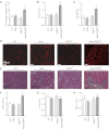
TIMP3 deficiency increases oxidative stress in 6-mo-old Akita diabetic kidneys. Shown are representative images of glomerular Dihydroethidium (DHE) staining (A) and nitrotyrosin staining (red) with nuclear DAPI staining (blue; B), with quantification of superoxide (C) and nitrotyrosin levels (D) showing increased superoxide levels in the Akita/TIMP3−/− kidneys. Also shown are increased NADPH oxidase activity (E), p47phox (F) and NOX2 (G) mRNA subunit expression in Akita/TIMP3−/− kidneys; n = 8/group. Gp91-tat is a specific peptide inhibitor of NOX2, gp91phox ds-tat; Scr tat is an inactive scrambled form of gp91phox ds-tat. Values are means ± SE. *P < 0.05 compared with WT. #P < 0.05 compared with all other groups. @P < 0.05 compared with scrambled (Scr) tat peptide group (using unpaired Student's t-test).
Fig. 8.
Fig. 8.
Differential alterations in TNF-α-converting enzyme (TACE) and matrix metalloproteinase 2 (MMP2) and MMP9 levels in kidneys of different genotypes. A: TACE mRNA expression (i) and activity (ii) in all groups; n = 5/group. B: representative gelatin zymography (i) and quantification of the band intensity for pro-MMP9 (ii), MMP9 (iii), pro-MMP2 (iv), and MMP 2 (v) in each genotype; n = 6/group. +ve, Positive control for MMP2 and MMP9. *P < 0.05 compared with WT. #P < 0.05 compared with all other groups.

References

    1. Amour A, Slocombe PM, Webster A, Butler M, Knight CG, Smith BJ, Stephens PE, Shelley C, Hutton M, Knauper V, Docherty AJ, Murphy G. TNF-alpha converting enzyme (TACE) is inhibited by TIMP-3. FEBS Lett 435: 39–44, 1998 - PubMed
    1. Awad AE, Kandalam V, Chakrabarti S, Wang X, Penninger JM, Davidge ST, Oudit GY, Kassiri Z. Tumor necrosis factor induces matrix metalloproteinases in cardiomyocytes and cardiofibroblasts differentially via superoxide production in a PI3K γ-dependent manner. Am J Physiol Cell Physiol 298: C679–C692, 2010 - PubMed
    1. Babu D, Soenen SJ, Raemdonck K, Leclercq G, De Backer O, Motterlini R, Lefebvre RA. TNF-alpha/cycloheximide-induced oxidative stress and apoptosis in murine intestinal epithelial MODE-K cells. Curr Pharm Des. [Epub ahead of print] - PubMed
    1. Basu R, Oudit GY, Wang X, Zhang L, Ussher JR, Lopaschuk GD, Kassiri Z. Type 1 diabetic cardiomyopathy in the Akita (Ins2WT/C96Y) mouse model is characterized by lipotoxicity and diastolic dysfunction with preserved systolic function. Am J Physiol Heart Circ Physiol 297: H2096–H2108, 2009 - PubMed
    1. Bodiga S, Gruenloh SK, Gao Y, Manthati VL, Dubasi N, Falck JR, Medhora M, Jacobs ER. 20-HETE-induced nitric oxide production in pulmonary artery endothelial cells is mediated by NADPH oxidase, H2O2, and PI3-kinase/Akt. Am J Physiol Lung Cell Mol Physiol 298: L564–L574, 2010 - PMC - PubMed

Publication types

MeSH terms