Comparative Study
. 2012 Nov 7;9(76):3140-55.
doi: 10.1098/rsif.2012.0479. Epub 2012 Aug 15.

Epidermis architecture and material properties of the skin of four snake species

Affiliations
Comparative Study

Epidermis architecture and material properties of the skin of four snake species

Marie-Christin G Klein et al. J R Soc Interface. .

Abstract

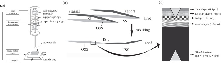
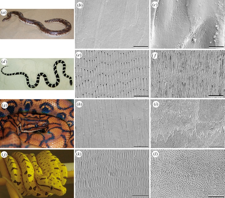
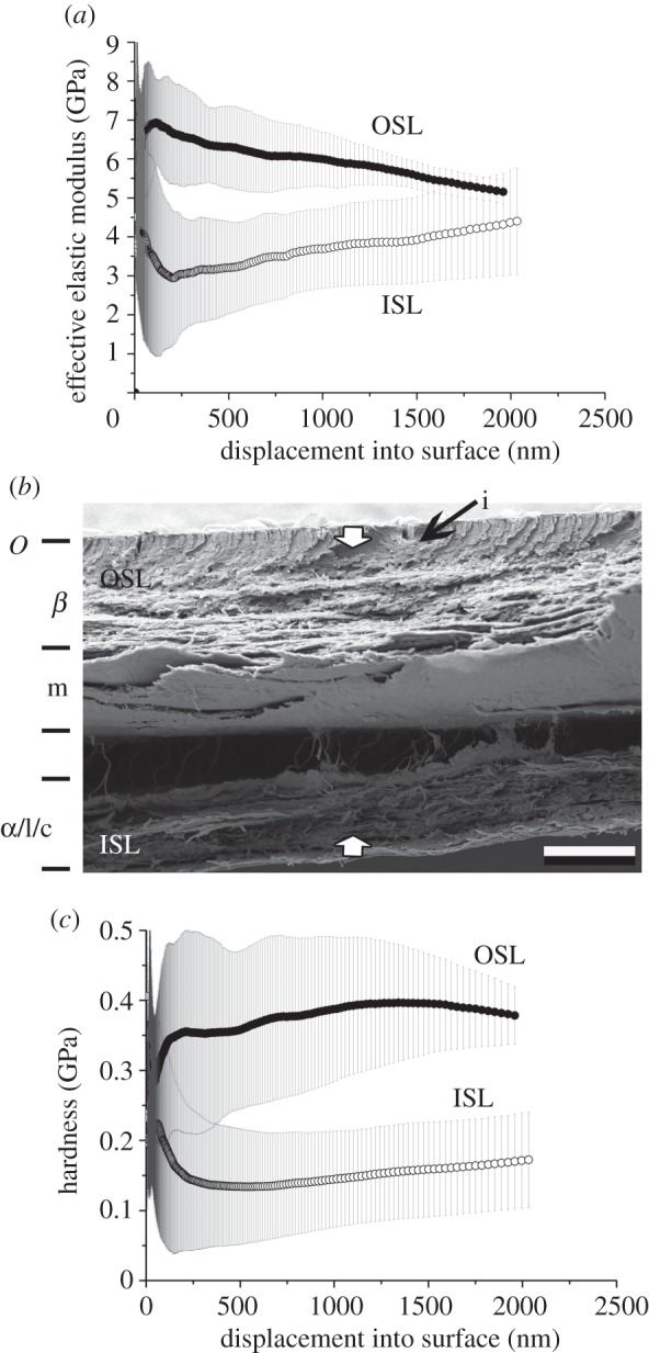
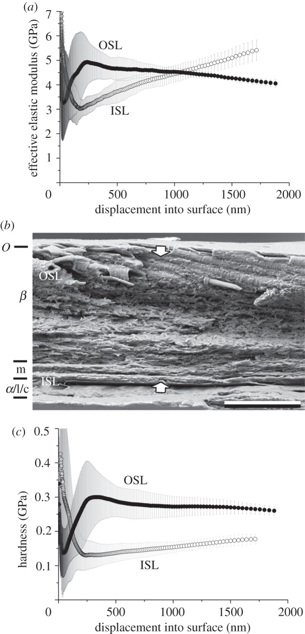
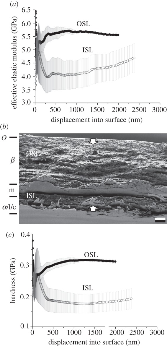
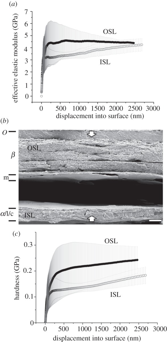
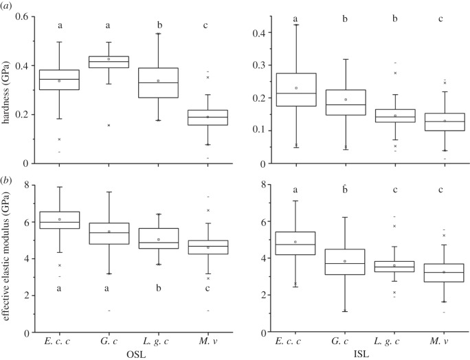
On the basis of structural and experimental data, it was previously demonstrated that the snake integument consists of a hard, robust, inflexible outer surface (Oberhäutchen and β-layer) and softer, flexible inner layers (α-layers). It is not clear whether this phenomenon is a general adaptation of snakes to limbless locomotion or only to specific conditions, such as habitat and locomotion. The aim of the present study was to compare the structure and material properties of the outer scale layers (OSLs) and inner scale layers (ISLs) of the exuvium epidermis in four snake species specialized to live in different habitats: Lampropeltis getula californiae (terrestrial), Epicrates cenchria cenchria (generalist), Morelia viridis (arboreal) and Gongylophis colubrinus (sand-burrowing). Scanning electron microscopy (SEM) of skin cross sections revealed a strong variation in the epidermis structure between species. The nanoindentation experiments clearly demonstrated a gradient of material properties along the epidermis in the integument of all the species studied. The presence of such a gradient is a possible adaptation to locomotion and wear minimization on natural substrates. In general, the difference in both the effective elastic modulus and hardness of the OSL and ISL between species was not large compared with the difference in epidermis thickness and architecture.


Figures

Figure 1.
Figure 1.
(a) Schematic diagram of the indentation system set-up with the coil-magnet assembly for load application, the capacitive displacement sensor and the indenter shaft support springs [5]. (b) Schematic drawing of the epidermis on scale level. The black square illustrates the skin part that was tested. (c) Diagram of the cross section of the snake integument (after [–8], and the present study). Triangles indicate the indentation depth undertaken in this study. ISL, inner scale layers; ISS, inner scale surface; OSS, outer scale surface. Figure 1a was adapted with permission from Barbakadze et al. [5]. Adapted with permission from Klein et al. [1].
Figure 2.
Figure 2.
Overview of the four model snake species studied. (a,d,g,j): animals. (b,c,e,f,h,i,k,l): SEM images illustrating the microstructure of the Oberhäutchen on the caudal outer scale surface of the ventral (b,e,h,k) and dorsal (c,f,i,l) scales. (ac) G. colubrinus; (df) L. g. californiae; (gi) E. c. cenchria; (jl) M. viridis. (Online version in colour.)
Figure 3.
Figure 3.
Gongylophis colubrinus. Results of nanoindentation measurements for both the OSL and ISL in relation with the cross-section architecture. (a) EEM versus displacement, (b) cross-section and (c) HD versus displacement. The error bars denote standard deviations. OSL, outer scale layers; ISL, inner scale layers; i, inclusions. The arrows indicate the indentation depth of 2 µm. O, Oberhäutchen; β, β-layer; m, mesos-layer; α, α-layer; l, lacunar tissue; c, clear-layer.
Figure 4.
Figure 4.
Lampropeltis getula californiae. Results of nanoindentation measurements for both the OSL and ISL in relation with the cross-section architecture. (a) EEM versus displacement, (b) cross-section and (c) HD versus displacement. The error bars denote s.d. OSL, outer scale layers; ISL, inner scale layers. The arrows indicate the indentation depth of 2 µm. O, Oberhäutchen; β, β-layer; m, mesos-layer; α, α-layer; l, lacunar tissue; c, clear-layer.
Figure 5.
Figure 5.
Epicrates cenchria cenchria. Results of nanoindentation measurements for both the OSL and ISL in relation with the cross-section architecture. (a) EEM versus displacement, (b) cross-section and (c) HD versus displacement. The error bars denote standard deviations. OSL, outer scale layers; ISL, inner scale layers. The arrows indicate the indentation depth of 2 µm. O, Oberhäutchen; β, β-layer; m, mesos-layer; α, α-layer; l, lacunar tissue; c, clear-layer.
Figure 6.
Figure 6.
Morelia viridis. Results of nanoindentation measurements for both the OSL and ISL in relation with the cross-section architecture. (a) EEM versus displacement, (b) cross-section and (c) HD versus displacement. The error bars denote s.d. OSL, outer scale layers; ISL, inner scale layers. The arrows indicate the indentation depth of 2 µm. O, Oberhäutchen; β, β-layer; m, mesos-layer; α, α-layer; l, lacunar tissue; c, clear-layer.
Figure 7.
Figure 7.
Summary of the (a) HD and (b) EEM results for penetration depths of 300–1800 nm for G. colubrinus (G. c.), 300–1200 nm for L. g. californiae (L. g. c.), 300 nm- minimum of 1700 nm for E. c. cenchria (E. c. c.) and 300–1500 nm for M. viridis (M. v.) of the nanoindentation measurements of all four snake species. Statistics: Bonferroni t-test.

Similar articles

Cited by

References

    1. Klein M.-C., Deuschle J., Gorb S. 2010. Material properties of the skin of the Kenyan sand boa Gongylophis colubrinus (Squamata, Boidae). J. Comp. Physiol. A 196, 659–668 10.1007/s00359-010-0556-y (doi:10.1007/s00359-010-0556-y) - DOI - DOI - PubMed
    1. Mattison C. 1995. The encyclopedia of snakes. London, UK: Cassel & Co
    1. Baden H. P., Maderson P. F. A. 1970. Morphological and biophysical identification of fibrous proteins in the amniote epidermis. J. Exp. Zool. 174, 225–232 10.1002/jez.1401740211 (doi:10.1002/jez.1401740211) - DOI - DOI - PubMed
    1. Landmann L. 1979. Keratin formation and barrier mechanisms in the epidermis of Natrix natrix (Reptilia: Serpentes): an ultrastructural study. J. Morphol. 162, 93–126 10.1002/jmor.1051620107 (doi:10.1002/jmor.1051620107) - DOI - DOI - PubMed
    1. Barbakadze N., Enders S., Gorb S., Arzt E. 2006. Local mechanical properties of the head articulation cuticle in the beetle Pachnoda marginata (Coleoptera, Scarabaeidae). J. Exp. Biol. 209, 722–730 10.1242/jeb.02065 (doi:10.1242/jeb.02065) - DOI - DOI - PubMed

Publication types

LinkOut - more resources