. 2012 Dec 7;9(77):3490-502.
doi: 10.1098/rsif.2012.0498. Epub 2012 Aug 15.

Aggregation by depletion attraction in cultures of bacteria producing exopolysaccharide

Affiliations

Aggregation by depletion attraction in cultures of bacteria producing exopolysaccharide

Gary Dorken et al. J R Soc Interface. .

Abstract

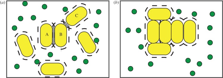
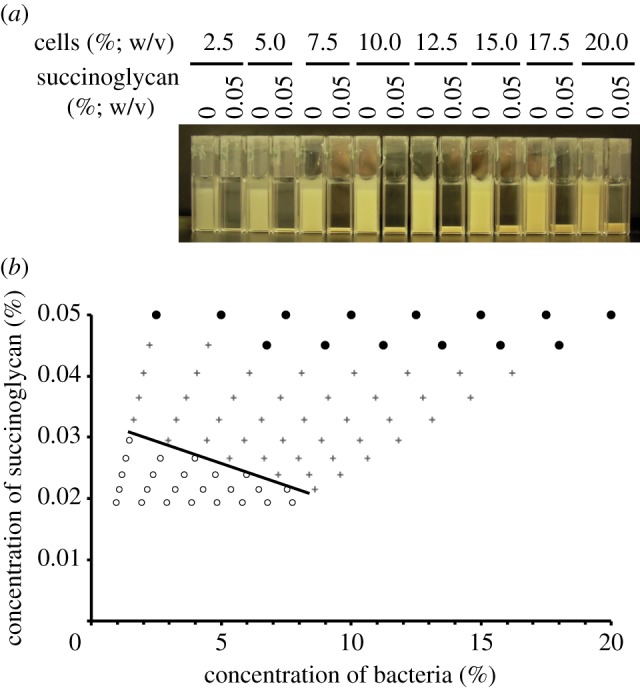
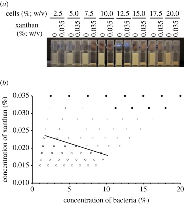
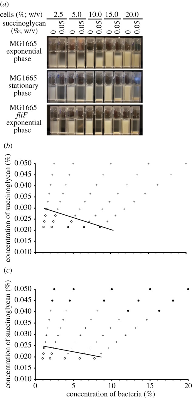
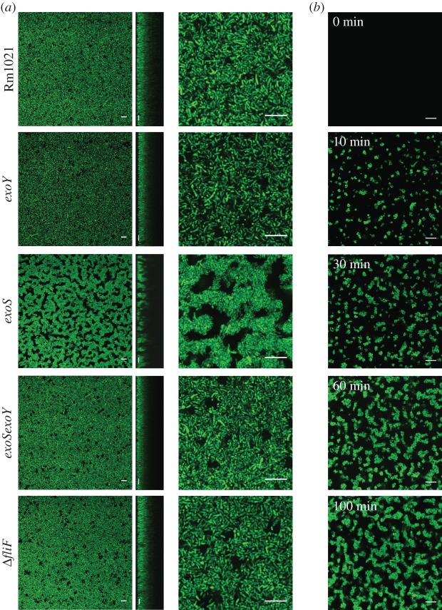
In bacteria, the production of exopolysaccharides--polysaccharides secreted by the cells into their growth medium--is integral to the formation of aggregates and biofilms. These exopolysaccharides often form part of a matrix that holds the cells together. Investigating the bacterium Sinorhizobium meliloti, we found that a mutant that overproduces the exopolysaccharide succinoglycan showed enhanced aggregation, resulting in phase separation of the cultures. However, the aggregates did not appear to be covered in polysaccharides. Succinoglycan purified from cultures was applied to different concentrations of cells, and observation of the phase behaviour showed that the limiting polymer concentration for aggregation and phase separation to occur decreased with increasing cell concentration, suggesting a 'crowding mechanism' was occurring. We suggest that, as found in colloidal dispersions, the presence of a non-adsorbing polymer in the form of the exopolysaccharide succinoglycan drives aggregation of S. meliloti by depletion attraction. This force leads to self-organization of the bacteria into small clusters of laterally aligned cells, and, furthermore, leads to aggregates clustering into biofilm-like structures on a surface.


Figures

Figure 1.
Figure 1.
(a,b) Schematic of how depletion attraction operates in a culture of bacteria producing exopolysaccharide. Bacteria, large spherocylinders; exopolysaccharide, small spheres. Dashed lines around the bacteria are zones of depletion or excluded volumes and represent the volume that is not accessible to the centre of mass of a given polymer after it has been secreted into the medium. As bacteria come closer together, these excluded volumes overlap (as shown in grey shaded zones; (a)). When bacteria aggregate, this volume then becomes available to the succinoglycan, increasing its entropy and maximizing the entropy of the system. As shown in (a), a greater volume is made available to the succinoglycan if cells align laterally (cells A and B) rather than end-on (cells B and C), so lateral alignment of cells would be expected to be favoured if a crowding mechanism is occurring. (Online version in colour.)
Figure 2.
Figure 2.
Screening of exopolysaccharide mutants for differences in rate of sedimentation. Rm1021 and the indicated succinoglycan biosynthesis mutants were grown to late-exponential phase in LBMC, transferred into cuvettes and incubated without shaking for 24 h. For each mutant strain, the gene location of the transposon is indicated, along with the relevant phenotype relative to Rm1021, which is motile and can make succinoglycan: EPS, succinoglycan; Mot, motility; +/−, gain or loss of that phenotype. ExoK and ExsH are extracellular glycanases; mutants have a decrease (down arrow) of low-molecular-weight (LMW) succinoglycan. Repeated experiments gave the same result. (Online version in colour.)
Figure 3.
Figure 3.
The exoS mutant has enhanced aggregation and lateral stacking of cells. (a) Sedimentation screen of Rm1021 and the indicated mutants. Strains were grown to late-exponential phase in LBMC, transferred into cuvettes and incubated without shaking for 24 h. (b) Percentage of aggregation as quantified from the change in OD600 over 24 h from the experiment performed as in (a). Data are means ± s.d., n = 3. (c) Phase contrast microscopy images of Rm1021 (i) and the exoS mutant (ii). The exoY, exoSexoY and ΔfliF mutants all looked the same as Rm1021. Strains were grown to late-exponential phase, 1 μl of culture was placed on a microscope slide and, after sealing on a coverslip, the slide was immediately imaged. The exoS mutant forms small aggregates, where the cells are aligned laterally. (c) Scale bars, 10 μm. (Online version in colour.)
Figure 4.
Figure 4.
Addition of the exoS supernatant to S. meliloti results in aggregation. (a) Left: cells of the indicated strains were grown to late-exponential phase, transferred to cuvettes and incubated without shaking for 24 h. Right: cultures of the indicated strains dispersed in exoS supernatant. An exoS culture was grown to late-exponential phase, centrifuged and the supernatant was filter sterilized to remove any remaining cells. The indicated strains were grown to late-exponential phase, and centrifuged to pellet the cells. The pelleted cells were then dispersed in the filter-sterilized exoS supernatant, transferred to cuvettes and incubated without shaking for 24 h. (b) The indicated strains were grown in nitrogen-free M9. After 30 h, cells were transferred to cuvettes and left without shaking for 24 h. (c) The indicated strains were grown to late-exponential phase, transferred to cuvettes and incubated without shaking for 24 h. (d) Rm1021 and the exoS mutant were grown to late-exponential phase. Heat-treated cultures were incubated at 65°C for 2 h before preparation. Rm1021 centrifuged, Rm1021 that were centrifuged and the pellet then re-dispersed without changing the supernatant. Rm1021 cells dispersed in exoS supernatant were prepared as in (a). All experiments were performed at least three times with the same result. (Online version in colour.)
Figure 5.
Figure 5.
Application of succinoglycan to the Rm1021 exoY mutant leads to aggregation, which is stronger under crowded conditions. (a) Cultures of the exoY mutant were grown to late-exponential phase and then diluted to a range of concentrations with the addition of 0.05% (w/v) succinoglycan where indicated. exoY cultures with no succinoglycan were used as controls. Cultures were incubated for 24 h without shaking. (b) Cultures as shown in (a) were successively diluted. After each dilution, cultures containing succinoglycan were visually scored for extent of aggregation and compared with control cuvettes, after 24 h without shaking. Top row indicates results for cuvettes containing 0.05% succinoglycan as shown in (a). Filled circles indicate cultures that are aggregating and leave a clear upper phase; plus symbols indicate cultures where a sediment is still visible but the upper phase is now turbid; open circles indicate cultures that are now equal turbidity to the relevant control. Line is drawn as a guide to the eye. n = 3. (Online version in colour.)
Figure 6.
Figure 6.
Application of xanthan also results in aggregation consistent with a crowding mechanism. (a) Cultures of the exoY mutant were grown to late-exponential phase and then diluted to a range of concentrations with the addition of 0.035% (w/v) xanthan where indicated. exoY cultures with no xanthan were used as controls. Cultures were incubated for 24 h without shaking. (b) Cultures as shown in (a) were successively diluted. After each dilution, cultures containing xanthan were visually scored for extent of aggregation and compared with control cuvettes, after 24 h without shaking. Top row indicates results for cuvettes as shown in (a). Filled circles indicate cultures that are aggregating and leave a clear upper phase; plus symbols indicate cultures where a sediment is still visible but the upper phase is now turbid; open circles indicate cultures that are now equal turbidity to the relevant control. Line is drawn as a guide to the eye. n = 3. (Online version in colour.)
Figure 7.
Figure 7.
Addition of succinoglycan to E. coli results in aggregation consistent with a crowding mechanism. (a) Cultures of E. coli MG1655 or a MG1655 fliF mutant were grown to late-exponential phase or stationary phase as indicated and then diluted to a range of concentrations with the addition of 0.05% (w/v) succinoglycan where indicated. Cultures with no succinoglycan were used as controls. Cultures were incubated for 24 h without shaking. (b,c) Escherichia coli MG1655 stationary phase cultures (b) and MG1655 fliF mutant late exponential phase cultures (c) were prepared as shown in (a) and successively diluted. After each dilution, cultures containing succinoglycan were visually scored for extent of aggregation and compared with control cuvettes, after 24 h without shaking. Top row indicates results for cuvettes as shown in (a). Filled circles indicate cultures that are aggregating and leave a clear upper phase; plus symbols indicate cultures where a sediment is still visible but the upper phase is now turbid; open circles indicate cultures that are now equal turbidity to the relevant control. Lines are guides to the eye. (Online version in colour.)
Figure 8.
Figure 8.
The sediment formed by aggregation of the exoS mutant has a biofilm-like structure. (a) The indicated strains, expressing GFP, were grown to late-exponential phase in LBMC, transferred to chambered coverslides, incubated without shaking for 24 h and then imaged by confocal microscopy. As the coverslip forms the base of the incubation chamber, this allows imaging of the sediment that forms through aggregation. Left and right, xy images; middle, xz. (b) Cultures of the exoS mutant were grown to late-exponential phase in LBMC and then transferred to chambered coverslides on a confocal microscope. Images were taken from the base of the chamber at the indicated times. (a,b) Scale bars, 10 μm. (Online version in colour.)

Similar articles

Cited by

References

    1. Deinema M. H., Zevenhuizen L. P. 1971. Formation of cellulose fibrils by gram-negative bacteria and their role in bacterial flocculation. Arch. Mikrobiol. 78, 42–51 10.1007/BF00409087 (doi:10.1007/BF00409087) - DOI - DOI - PubMed
    1. Friedman B. A., Dugan P. R. 1968. Identification of Zoogloea species and the relationship to zoogloeal matrix and floc formation. J. Bacteriol. 95, 1903–1909 - PMC - PubMed
    1. Friedman B. A., Dugan P. R., Pfister R. M., Remsen C. C. 1969. Structure of exocellular polymers and their relationship to bacterial flocculation. J. Bacteriol. 98, 1328–1334 - PMC - PubMed
    1. McKinney R. E. 1953. Staining bacterial polysaccharides. J. Bacteriol. 66, 453–454 - PMC - PubMed
    1. McKinney R. E., Weichlein R. G. 1953. Isolation of floc-producing bacteria from activated sludge. Appl. Microbiol. 1, 259–261 - PMC - PubMed

Publication types

MeSH terms

LinkOut - more resources