. 2012 Nov;86(21):11633-44.
doi: 10.1128/JVI.01479-12. Epub 2012 Aug 15.

The cytoplasmic tail domain of influenza B virus hemagglutinin is important for its incorporation into virions but is not essential for virus replication in cell culture in the presence of compensatory mutations

Affiliations

The cytoplasmic tail domain of influenza B virus hemagglutinin is important for its incorporation into virions but is not essential for virus replication in cell culture in the presence of compensatory mutations

Masaki Imai et al. J Virol. 2012 Nov.

Abstract

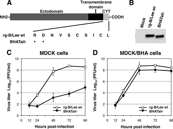
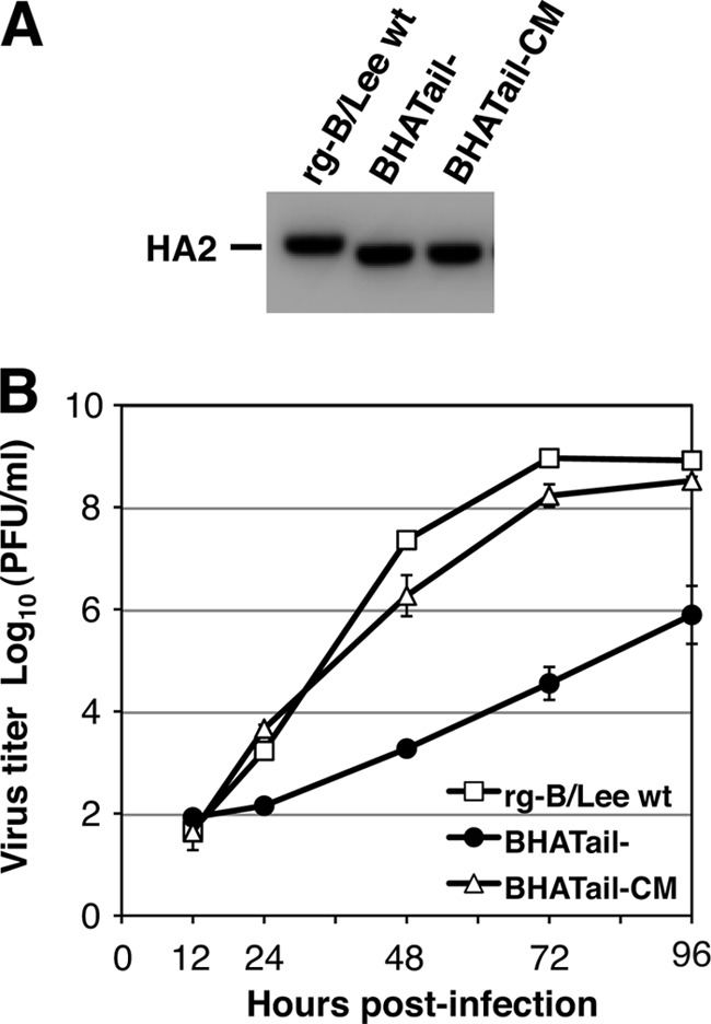
Influenza B virus hemagglutinin (BHA) contains a predicted cytoplasmic tail of 10 amino acids that are highly conserved among influenza B viruses. To understand the role of this cytoplasmic tail in infectious virus production, we used reverse genetics to generate a recombinant influenza B virus lacking the BHA cytoplasmic tail domain. The resulting virus, designated BHATail(-), had a titer approximately 5 log units lower than that of wild-type virus but grew normally when BHA was supplemented in trans by BHA-expressing cells. Although the levels of BHA cell surface expression were indistinguishable between truncated and wild-type BHA, the BHATail(-) virus produced particles containing dramatically less BHA. Moreover, removal of the cytoplasmic tail abrogated the association of BHA with Triton X-100-insoluble lipid rafts. Interestingly, long-term culture of a virus lacking the BHA cytoplasmic tail in Madin-Darby canine kidney (MDCK) cells yielded a mutant with infectivities somewhat similar to that of wild-type virus. Sequencing revealed that the mutant virus retained the original cytoplasmic tail deletion but acquired additional mutations in its BHA, neuraminidase (NA), and M1 proteins. Viral growth kinetic analysis showed that replication of BHA cytoplasmic tailless viruses could be improved by compensatory mutations in the NA and M1 proteins. These findings indicate that the cytoplasmic tail domain of BHA is important for efficient incorporation of BHA into virions and tight lipid raft association. They also demonstrate that the domain is not absolutely required for virus viability in cell culture in the presence of compensatory mutations.


Figures

Fig 1
Fig 1
(A) Schematic diagram of wild-type and mutant BHA proteins carrying a carboxy-terminal truncation in BHA. The amino acid sequence of the BHA cytoplasmic tail (CYT; residues 560 to 569) is shown. An asterisk indicates a stop codon. (B) Detection of BHA protein in BHA truncation mutant virus-infected MDCK cells in the presence of trypsin. MDCK cells were infected with rg-B/Lee wt and BHATail viruses in the presence of trypsin. BHA proteins in cell lysates were detected by Western blotting using an anti-BHA antibody. (C and D) Growth properties of BHATail mutant virus. MDCK (C) and MDCK/BHA (D) cells were infected at an MOI of 0.001 PFU, and supernatants of infected cells were harvested at the indicated times. Virus titers in the supernatant were determined by plaque assay on MDCK/BHA cells. The data points represent the means ± standard deviations from triplicate experiments.
Fig 2
Fig 2
BHA protein-induced RBC-cell fusion. MDCK cells were infected with rg-B/Lee wt and BHATail viruses at an MOI of 5 PFU. At 14 h p.i., R18/calcein-labeled RBCs were adsorbed to BHA expressed on the cell surface. Fusion was triggered by transiently shifting the pH to 5.2. (A and B) Time-lapse sequences of fluorescence images of RBCs and virus-infected cells during membrane fusion. Fluorescence images of the RBCs bound to virus-infected cells were captured before and after low-pH treatment at pH 5.2 and 23°C. (A) Time-lapse sequence of red fluorescence (R18) images. (B) Time-lapse sequence of green fluorescence (calcein) images. Both red and green fluorescence was initially located only in the RBC regions but became diffusely distributed over regions of the infected cells after the induction of fusion. (C) Changes in intensity of red (R18) and green (calcein) fluorescence at a single RBC-cell complex during membrane fusion. Fluorescence intensities at the single RBC region (panels A and B, circled in yellow) were plotted. Time zero corresponds to the pH shift. The arrows indicate the time points at which lipid mixing or content mixing started, that is, the response time for single-RBC fusion. (D) Response time distributions of red (R18) and green (calcein) fluorescence increase in wild-type and BHATail virus-infected cells. Red and green fluorescence intensities at individual RBCs were measured as in panel C to determine the response times for RBC-cell fusion. “n” in each box denotes the number of RBCs examined, with the number of fused RBCs/number of nonfused RBCs within 176 s in parentheses. The vertical axis represents the frequency of fusion for one RBC per 4 s (fusion/RBC/4s). (E and F) Kinetics of fusion in the wild-type and BHATail virus-infected cells. The kinetics of RBC-cell fusion were obtained by cumulative summation of the data in Fig. 2D.
Fig 3
Fig 3
Characterizations of BHA cytoplasmic tailless viruses with additional mutations. (A) Detection of BHA protein in BHA truncation mutant virus-infected MDCK cells. MDCK cells were infected with rg-B/Lee wt, BHATail, or BHATail-CM virus in the presence of trypsin. BHA proteins in cell lysates were detected by Western blotting using an anti-BHA antibody. (B) Multiple-step growth curve of BHA mutant viruses. MDCK cells were infected at an MOI of 0.001 PFU, and culture medium was harvested at the indicated times. Virus yields from the supernatants were determined by plaque assay on MDCK/BHA cells. The data points represent the means ± standard deviations from triplicate experiments.
Fig 4
Fig 4
Analysis of the virions of BHA cytoplasmic tailless mutants. (A and B) Protein composition of BHA mutant virions. MDCK cells were infected at an MOI of 5 PFU, and virions were purified by centrifugation through 30% sucrose. (A) Proteins of purified viruses were analyzed by using Coomassie brilliant blue staining (top) and Western blotting using an anti-BM2 antibody (bottom). (B) Relative amounts of viral proteins. Viral proteins were quantified by using Image J, and the relative staining intensity of each protein was normalized to that of NP for each virus. *, P < 0.05 compared with the value for rg-B/Lee wt. The error bars indicate standard deviations. (C) Morphology of mutants observed under negative-staining electron microscopy. Virions were grown in MDCK cells and purified by centrifugation through 30% sucrose. The virions were negatively stained with 1% uranyl acetate and observed under a transmission electron microscope. Bars, 500 nm.
Fig 5
Fig 5
Comparison of the amounts of viral glycoproteins expressed in virus-infected MDCK cells. (A) Surface expression of BHA and NA proteins. Infected MDCK cells were fixed with paraformaldehyde at the indicated times p.i. and permeabilized with TX-100 for the detection of NP or left untreated for the detection of BHA and NA on the cell surface. Viral glycoprotein levels were analyzed by means of a cell ELISA using anti-BHA and anti-NA antibodies. The amounts of BHA and NA proteins were normalized to that of the NP protein of each mutant virus. The amounts of BHA and NA in the wild-type virus were set to 100%, and the amounts of both proteins in the mutants are shown relative to that of the wild-type virus. The error bars indicate standard deviations. (B) Total expression of BHA and NA proteins. Infected MDCK cells were lysed at the indicated times p.i., and viral glycoprotein in cell lysates was detected by Western blotting using anti-BHA and anti-NA antibodies. Viral proteins were quantified by using Image J, and the relative staining intensity of each protein was normalized to that of the NP protein of each virus.
Fig 6
Fig 6
Effect of the cytoplasmic tail deletion on BHA protein-lipid raft association. (A) TX-100 solubility of the wild-type and mutant BHA proteins in virus-infected cells. MDCK cells were infected at an MOI of 5 PFU. After 15 h, the cells were surface biotinylated and then incubated with lysis buffer (0.2% TX-100, 50 mM Tris-HCl [pH 7.4], 150 mM NaCl, 1 mM EDTA) on ice for 10 min. The soluble (S) and insoluble (I) fractions were separated by centrifugation, and the insoluble fraction was homogenized by passage through a 25-gauge needle 20 times. Both fractions were immunoprecipitated and analyzed by use of SDS-PAGE. (B) The relative intensities of the viral proteins found in the insoluble and soluble fractions were quantified by using Image J. The error bars indicate standard deviations.
Fig 7
Fig 7
(A) Identification of compensatory mutations that contribute to the efficient growth of BHATail-CM mutant virus. Multicycle replication kinetics was determined as described in the legend to Fig. 3B. (B) Protein compositions of mutant virions. Proteins of purified viruses were analyzed by use of Coomassie brilliant blue staining as described in the legend to Fig. 4.

Similar articles

Cited by

References

    1. Ali A, Avalos RT, Ponimaskin E, Nayak DP. 2000. Influenza virus assembly: effect of influenza virus glycoproteins on the membrane association of M1 protein. J. Virol. 74:8709–8719 - PMC - PubMed
    1. Baigent SJ, McCauley JW. 2001. Glycosylation of haemagglutinin and stalk-length of neuraminidase combine to regulate the growth of avian influenza viruses in tissue culture. Virus Res. 79:177–185 - PubMed
    1. Barman S, et al. 2004. Role of transmembrane domain and cytoplasmic tail amino acid sequences of influenza A virus neuraminidase in raft association and virus budding. J. Virol. 78:5258–5269 - PMC - PubMed
    1. Bilsel P, Castrucci MR, Kawaoka Y. 1993. Mutations in the cytoplasmic tail of influenza A virus neuraminidase affect incorporation into virions. J. Virol. 67:6762–6767 - PMC - PubMed
    1. Bourmakina SV, Garcia-Sastre A. 2003. Reverse genetics studies on the filamentous morphology of influenza A virus. J. Gen. Virol. 84:517–527 - PubMed

Publication types

MeSH terms

LinkOut - more resources