. 2012 Sep 15;189(6):3150-8.
doi: 10.4049/jimmunol.1201140. Epub 2012 Aug 15.

Semaphorin 7A contributes to West Nile virus pathogenesis through TGF-β1/Smad6 signaling

Affiliations

Semaphorin 7A contributes to West Nile virus pathogenesis through TGF-β1/Smad6 signaling

Hameeda Sultana et al. J Immunol. .

Abstract

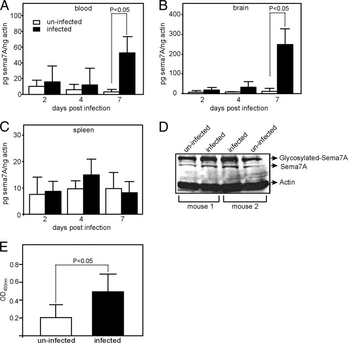
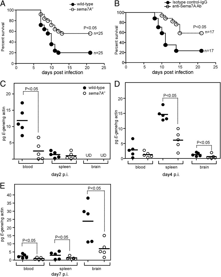
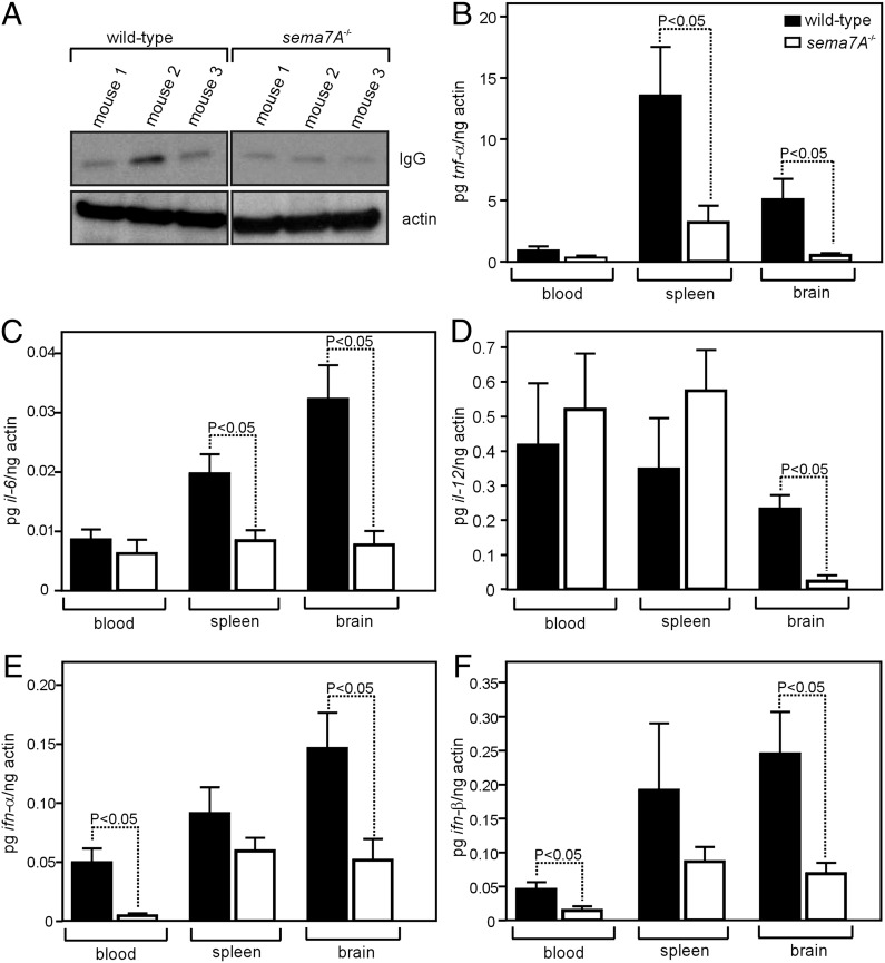
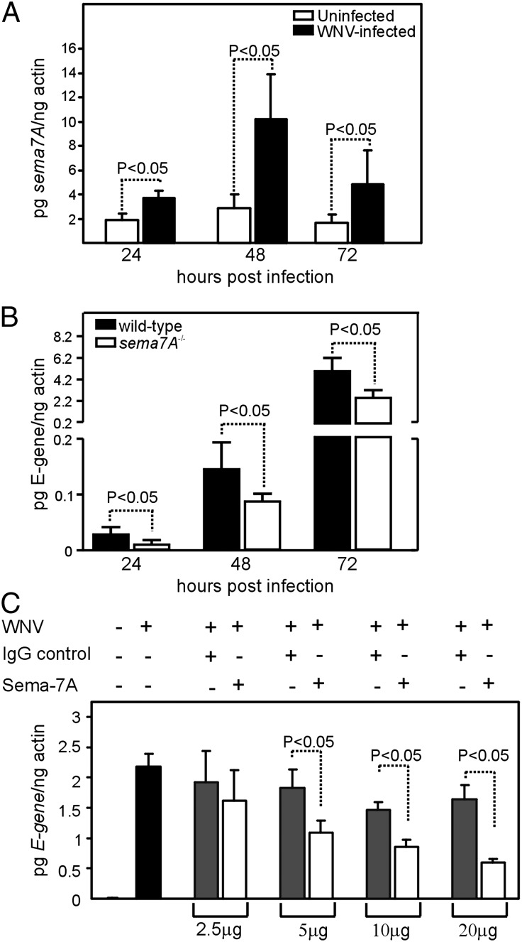
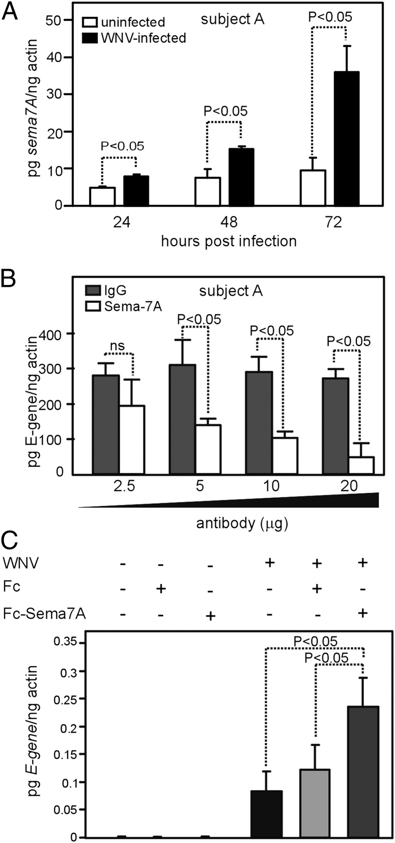
Semaphorin 7A (Sema7A) is a membrane-associated/secreted protein that plays an essential role in connecting the vertebrate neuronal and immune systems. However, the role of Sema7A has not been elucidated in viral pathogenesis. In this study, we show that abrogation of Sema7A protects mice from lethal West Nile virus (WNV) infection. Mice lacking Sema7A showed increased survival, reduced viral burden, and less blood-brain barrier permeability upon WNV infection. Increased Sema7A levels were evident in murine tissues, as well as in murine cortical neurons and primary human macrophages upon WNV infection. Treatment with Sema7A Ab blocked WNV infection in both of these cell types. Furthermore, Sema7A positively regulates the production of TGF-β1 and Smad6 to facilitate WNV pathogenesis in mice. Collectively, these data elucidate the role of Sema7A in shared signaling pathways used by the immune and nervous systems during viral pathogenesis that may lead to the development of Sema7A-blocking therapies for WNV and possibly other flaviviral infections.


Figures

FIGURE 1.
FIGURE 1.
WNV induces Sema7A expression in mice. Q-PCR results showing expression levels of sema7A mRNA in blood (A), brain (B), and spleen (C) on days 2, 4, and 7 in uninfected and WNV-infected mice (n = 5 mice per group and per data point). Levels of sema7A mRNA were normalized to the levels of actin mRNA. Error bars represent + SD from the mean value. Statistical significance was calculated using the Student t test. (D) Immunoblotting results showing expression of Sema7A in brain tissue lysates from uninfected and infected (day-7 p.i.) mice. Levels of total actin were used as loading control. Representative gel images from two independent experiments are shown. (E) ELISA to quantify total amount of Sema7A in uninfected and infected (day 7 p.i.) mice brain tissue lysates used in (D). Data from two independent experiments are shown in (A–C) and (E); Q-PCR was performed in triplicate.
FIGURE 2.
FIGURE 2.
Abrogation of Sema7A renders resistance to WNV-induced lethality in mice. (A) Survival of wild-type and sema7A−/− mice challenged i.p. with 103 PFU of wild-type WNV. Survival was recorded daily until day 21 p.i.; data were pooled from two independent experiments and represent a total of 25 animals/group. (B) Survival of C57BL/6 wild-type mice inoculated with WNV and treated with anti-Sema7A mAb or isotype-matched negative control Ab on days −1 and +2. Data are pooled from two independent experiments and represent a total of 17 animals/group. Statistical significance (p < 0.05) was calculated using Prism 4 software. WNV is less neuroinvasive in sema7A−/− mice than in wild-type mice. Viral loads in selected tissues (blood, spleen, and brain) of wild-type and sema7A−/− mice inoculated i.p. with 103 PFU of WNV. Each group contained fie animals examined at day 2 (C), day 4 (D), or day 7 (E) after viral challenge. Data are representative of results obtained in two independent experiments, performed in triplicate. Horizontal bars represent mean of the values. Statistical significance was calculated using the Student t test. UD, Undetectable.
FIGURE 3.
FIGURE 3.
sema7A−/− mice have reduced cytokine responses that are associated with inflammation during WNV infection. (A) Immunoblot of IgG (H chain) in WNV-infected wild-type or sema7A−/− mice brain tissue lysates. Actin served as the loading control. Representative gel images from two independent experiments are shown. TNF-α (B)), IL-6 (C), IL-12 (D), IFN-α (E), and IFN-β (F) mRNA levels in the blood, spleen, and brain of WNV-infected wild-type or sema7A−/− mice at day 7 p.i. Both groups of mice were infected with 103 PFU of WNV. Results are from five mice/group and two independent experiments, performed in triplicate. Statistical significance (p < 0.05) was calculated using the Student t test. Error bars represent + SD from the mean value.
FIGURE 4.
FIGURE 4.
Abrogation of Sema7A blocks WNV infection of cortical neurons in vitro. (A) Q-PCR analysis showing expression of sema7A mRNA levels at 24, 48, and 72 h p.i. with WNV in cortical neurons isolated from wild-type mice. Uninfected cortical neurons were used as controls. Levels of sema7A mRNA were normalized to the levels of actin mRNA. (B) Kinetics of WNV infection in cortical neuronal cells isolated from wild-type or sema7A−/− mice at 24, 48, and 72 h p.i. (C) Q-PCR analysis showing WNV burden in Sema7A mAb-treated or isotype-matched negative control Ab-treated cortical neurons at 48 p.i. All results are from three independent experiments, performed in triplicate. Error bars represent + SD from the mean value. Statistical significance was calculated using the Student t test.
FIGURE 5.
FIGURE 5.
Abrogation of Sema7A blocks WNV infection of human macrophages. (A) Q-PCR analysis showing expression of sema7A mRNA levels at 24, 48, and 72 h p.i. with WNV in macrophages isolated from a representative healthy human volunteer (Subject A). Uninfected macrophages were used as controls. Levels of sema7A mRNA were normalized to the levels of actin mRNA. (B) Q-PCR analysis showing WNV burden in Sema7A mAb-treated or isotype-matched negative control Ab-treated human macrophages from Subject A at 48 h p.i. (C) Q-PCR analysis showing viral burden in Fc or Fc-Sema7A–transfected HEK 293 cells. Infected, but untransfected, cells were used as controls. Data are representative of results performed in triplicates. Error bars represent + SD from the mean value. Statistical significance was calculated using the Student t test.
FIGURE 6.
FIGURE 6.
Sema7A facilitates WNV pathogenesis through TGF-β1/Smad6 signaling. (A) Q-PCR results showing expression of tgf-beta1 mRNA levels at 24, 48, and 72 h p.i. with WNV in cortical neurons isolated from wild-type mice. Levels of tgf-beta1 mRNA were normalized to the levels of actin mRNA. (B) Expression of tgf-beta1 mRNA levels at 24, 48, and 72 h p.i. with WNV in cortical neurons isolated from wild-type or sema7A−/− mice. (C) Expression of tgf-beta1 mRNA at 24, 48, and 72 h p.i. with WNV in HEK 293 cells transfected with tgf-beta1 or control siRNA. (D) Kinetics of WNV infection in HEK 293 cells transfected with tgf-beta1 or control siRNA at 24, 48, and 72 h p.i. (E) Q-PCR results showing smad6 mRNA levels in cortical neurons isolated from wild-type or sema7A−/− mice at 48 h p.i. with WNV. (F) Q-PCR results showing expression of smad6 mRNA levels at 72 h p.i. with WNV in HEK 293 cells transfected with tgf-beta1 or control siRNA. (G) Immunoblot showing levels of Smad6 and Smad2 in HEK 293 cells transfected with tgf-beta1 siRNA at 48 and 72 h p.i. with WNV. Actin levels served as loading control. Representative gel images from two independent experiments are shown. Q-PCR results showing expression of smad6 mRNA (H) or the kinetics of WNV infection (I) at 48 and 72 h p.i. in HEK 293 cells transfected with smad6 or control siRNA. All data are representative of results obtained from two independent experiments, performed in triplicate. Error bars represent + SD from the mean value. Statistical significance was calculated using the Student t test.

Similar articles

Cited by

References

    1. Klee A. L., Maidin B., Edwin B., Poshni I., Mostashari F., Fine A., Layton M., Nash D. 2004. Long-term prognosis for clinical West Nile virus infection. Emerg. Infect. Dis. 10: 1405–1411 - PMC - PubMed
    1. Petersen L. R., Marfin A. A. 2002. West Nile virus: a primer for the clinician. Ann. Intern. Med. 137: 173–179 - PubMed
    1. Diamond M. S., Klein R. S. 2004. West Nile virus: crossing the blood-brain barrier. Nat. Med. 10: 1294–1295 - PubMed
    1. Lustig S., Olshevsky U., Ben-Nathan D., Lachmi B. E., Malkinson M., Kobiler D., Halevy M. 2000. A live attenuated West Nile virus strain as a potential veterinary vaccine. Viral Immunol. 13: 401–410 - PubMed
    1. Samuel M. A., Diamond M. S. 2006. Pathogenesis of West Nile Virus infection: a balance between virulence, innate and adaptive immunity, and viral evasion. J. Virol. 80: 9349–9360 - PMC - PubMed

Publication types

MeSH terms