. 2012 Sep 15;189(6):2702-6.
doi: 10.4049/jimmunol.1201682. Epub 2012 Aug 15.

Cutting edge: intravascular staining redefines lung CD8 T cell responses

Affiliations

Cutting edge: intravascular staining redefines lung CD8 T cell responses

Kristin G Anderson et al. J Immunol. .

Abstract

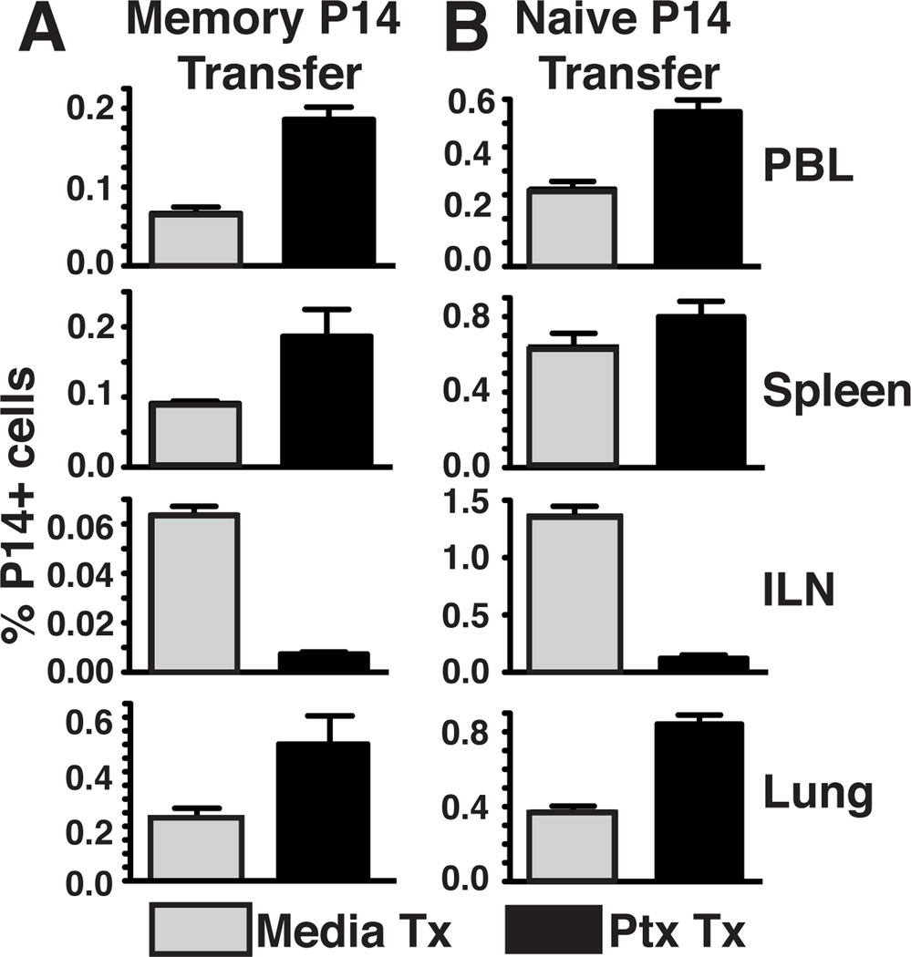
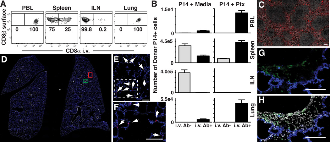
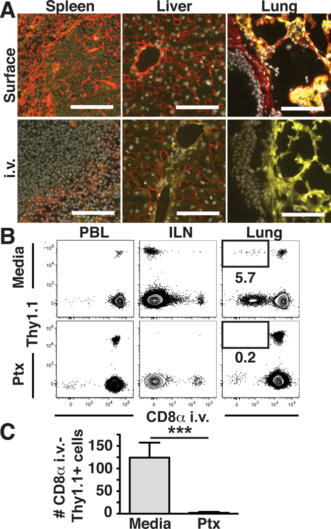
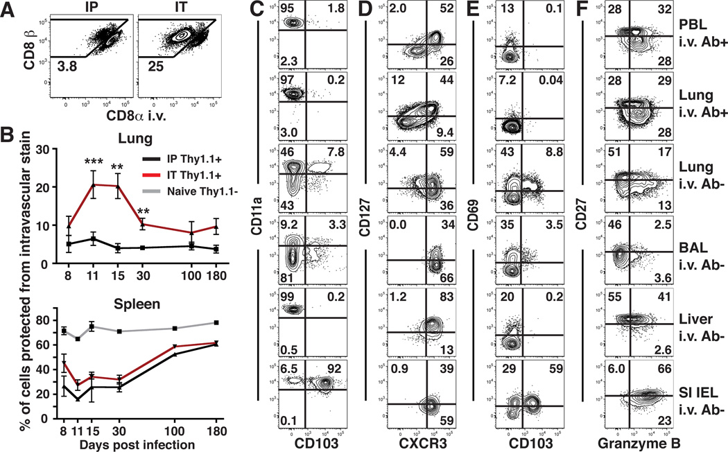
Nonlymphoid T cell populations control local infections and contribute to inflammatory diseases, thus driving efforts to understand the regulation of their migration, differentiation, and maintenance. Numerous observations indicate that T cell trafficking and differentiation within the lung are starkly different from what has been described in most nonlymphoid tissues, including intestine and skin. After systemic infection, we found that >95% of memory CD8 T cells isolated from mouse lung via standard methods were actually confined to the pulmonary vasculature, despite perfusion. A respiratory route of challenge increased virus-specific T cell localization within lung tissue, although only transiently. Removing blood-borne cells from analysis by the simple technique of intravascular staining revealed distinct phenotypic signatures and chemokine-dependent trafficking restricted to Ag-experienced T cells. These results precipitate a revised model for pulmonary T cell trafficking and differentiation and a re-evaluation of studies examining the contributions of pulmonary T cells to protection and disease.


Conflict of interest statement

This study was funded by R01AI084913 (DM), the Beckman Young Investigator Award (DM), and NIH Immunology grant T32-AI07313 (CNS). The authors have no conflicting financial interests.

Figures

Figure 1
Figure 1. Pertussis toxin treatment of transferred T cells yields increased recovery from lung
Naïve or memory P14 splenocytes were treated with pertussis toxin (PTx) or media control and transferred i.v. into naïve recipients. Tissues were harvested 1 (naïve) or 3 (memory) days post-transfer. Frequency of memory (A) or naïve (B) P14 cells in PBL, spleen, ILN, or lung. Data are representative of 3 independent experiments totaling >12 mice/condition. Error bars indicate SEM.
Figure 2
Figure 2. In vivo staining distinguishes between anatomic compartments
(A) Naïve Thy1.1+ P14 splenocytes were transferred i.v. into C57Bl/6J mice. Anti-CD8α was injected i.v. Three minutes later, tissues were harvested. Cells were isolated and then stained ex vivo with anti-CD8β. Plots gated on Thy1.1+ lymphocytes. (B) Number of i.v. Ab+ and i.v. Ab− naïve P14 cells, ± PTx treatment, isolated from tissues after transfer. (C) Spleen after anti-CD8α-PE i.v. injection. (D–H) Whole lung sections of P14 immune chimeras were imaged. Panels E and F or G and H represent enlarged images defined by red or green boxes, respectively, in panel D. CD8 T cells that stain with i.v. injected Ab (red) co-localize with blood vessels (CD31, blue, see E and enlarged inset F), whereas those protected from i.v. Ab (green surface stain) are spatially distinct (G and H, ± DAPI, respectively). Arrows designate red i.v. Ab+ cells. (A and B) are representative of 3 independent experiments totaling ≥12 mice/condition. Error bars indicate SEM. Images are representative of 2 independent experiments totaling 6 mice. Scale bars in C, E–H represent 100µm.
Figure 3
Figure 3. Migration of memory CD8 T cells to uninflamed lung tissue is chemokine dependent
(A) Anti-collagen IV Ab (red) was injected i.v. prior to tissue harvest or tissue sections were surface stained ex vivo along with anti-CD31 (yellow) and DAPI (gray). (B&C) Thy1.1+ memory P14 splenocytes were treated with PTx or media and transferred i.v. into naïve C57Bl/6J mice. Intravascular anti-CD8α staining and tissue harvest occurred 3 days later. (B) Representative flow cytometric analysis of the indicated recipient tissues. Plots gated on CD8β+ lymphocytes. (C) Number of donor memory P14 cells recovered from lung that were protected from intravascular staining (intravascular anti-CD8α negative). Images are representative of 2 independent experiments totaling 4 mice. Scale bars represent 100µm. Plots in (B) and (C) are representative of 3 independent experiments totaling ≥12 mice/condition. ***, P=0.006 unpaired Student’s t test. Error bars represent SEM.
Figure 4
Figure 4. Most lung memory CD8 T cells are capillary-associated after infection
(A) P14 immune chimeras were infected with LCMV via the i.p. or i.t. route. 15 days later, mice were subject to intravascular staining (as in Fig. 2), and lymphocytes were isolated from perfused lung. Plots are gated on Thy1.1+ P14 cells. (B) Frequency of P14 cells that were protected from intravascular staining isolated from lung or spleen after i.p (black) or i.t. (red) infection. Proportion of naïve (CD44lo) CD8 T cells (gray) that were protected from intravascular staining was also determined in spleen. (C–F) The phenotype of P14 cells was evaluated in the indicated tissues 15 days after i.t. LCMV infection. Data are representative of 2–3 independent experiments per time point totaling ≥8 mice per condition. ***, P=0.0003; **, P<0.006 unpaired Student’s t test. Error bars represent SEM.

References

    1. WHO. Geneva: 2004. The World Health Report: 2004, Changing History.
    1. Holgate ST. Innate and adaptive immune responses in asthma. Nat. Med. 2012;18:673–683. - PubMed
    1. Masopust D, Picker LJ. Hidden memories: frontline memory T cells and early pathogen interception. J. Immunol. 2012;188:5811–5817. - PMC - PubMed
    1. Gebhardt T, Wakim LM, Eidsmo L, Reading PC, Heath WR, Carbone FR. Memory T cells in nonlymphoid tissue that provide enhanced local immunity during infection with herpes simplex virus. Nat. Immunol. 2009;10:524–530. - PubMed
    1. Jiang X, Clark RA, Liu L, Wagers AJ, Fuhlbrigge RC, Kupper TS. Skin infection generates non-migratory memory CD8+ T(RM) cells providing global skin immunity. Nature. 2012;483:227–231. - PMC - PubMed

Publication types

MeSH terms