. 2012 Sep 15;189(6):2727-34.
doi: 10.4049/jimmunol.1200380. Epub 2012 Aug 15.

The adaptor 3BP2 is required for early and late events in FcεRI signaling in human mast cells

Affiliations

The adaptor 3BP2 is required for early and late events in FcεRI signaling in human mast cells

Erola Ainsua-Enrich et al. J Immunol. .

Abstract

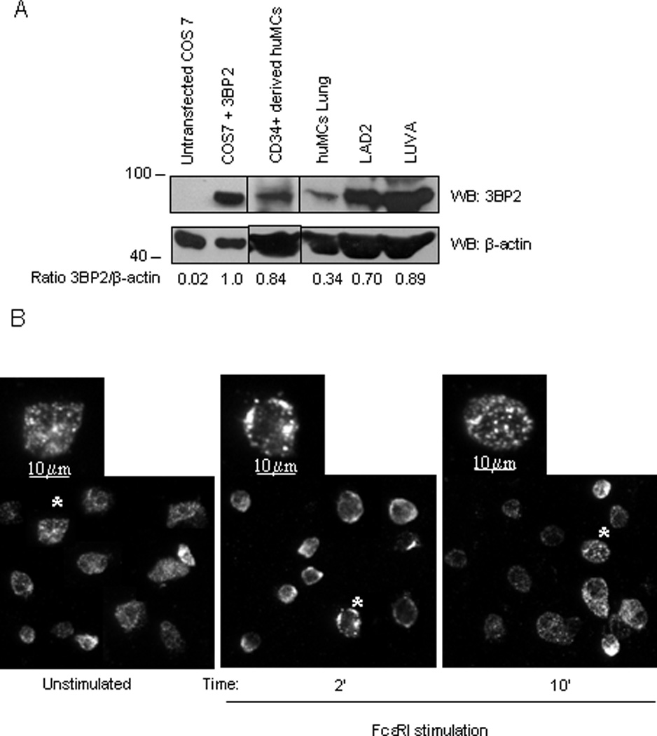
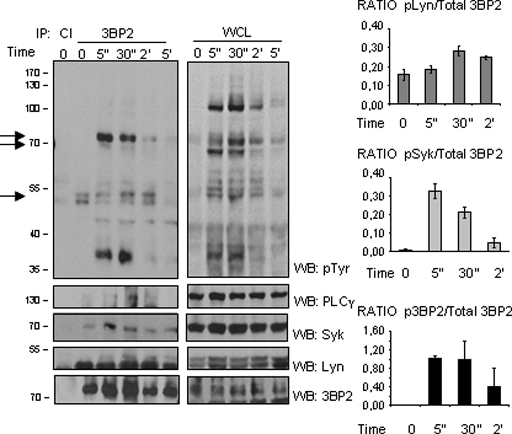
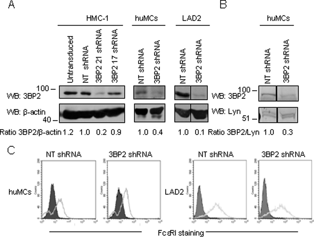
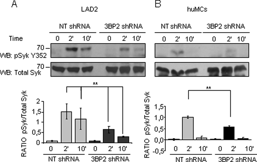
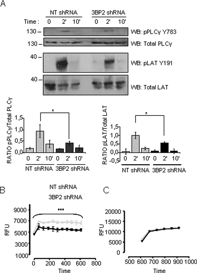
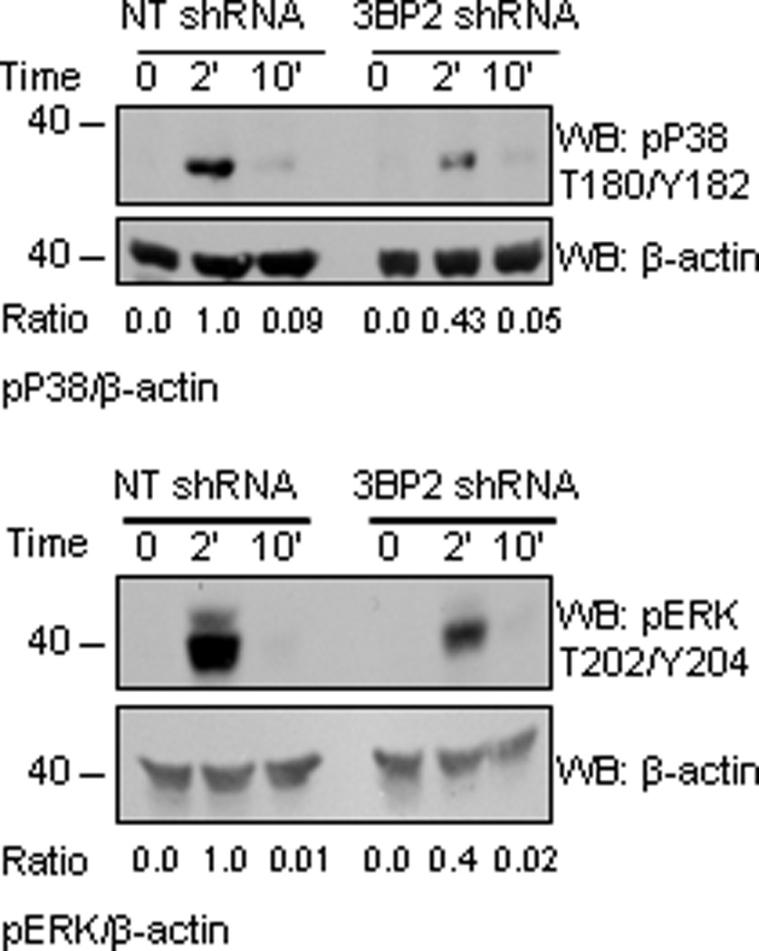
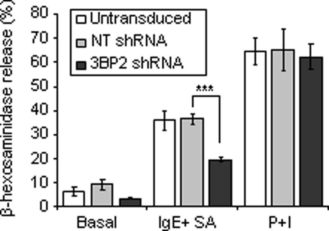
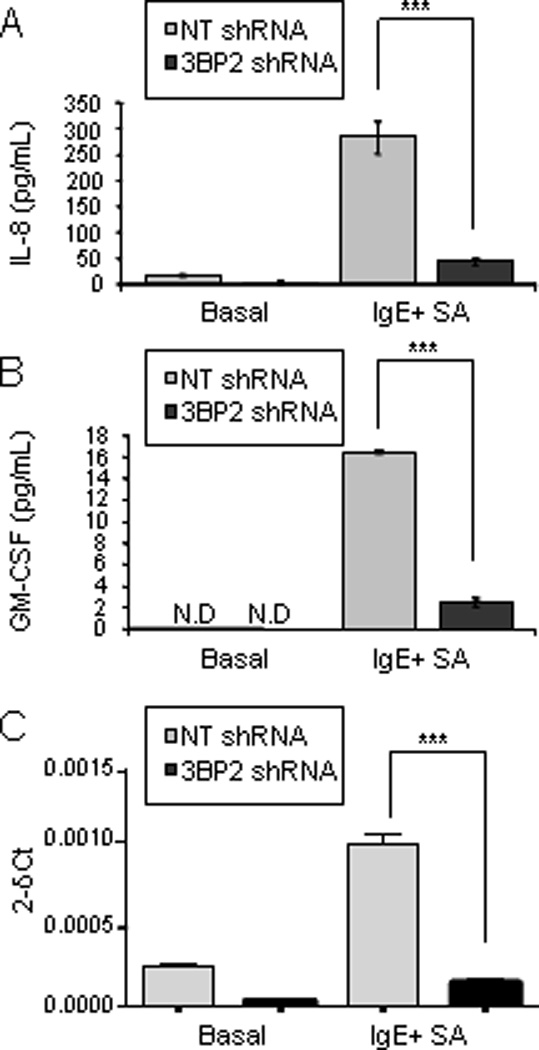
Adaptor molecules are essential in organizing signaling molecules and in coordinating and compartmentalizing their activity. SH3-binding protein 2 (3BP2) is a cytoplasmic adaptor protein mainly expressed by hematopoietic cells that has been shown to act as a positive regulator in T, B, and NK cell signal transduction. 3BP2 is an important regulator of cytotoxic granule release in NK cells. Mast cells (MCs) similarly degranulate following Ag-dependent aggregation of the FcεRI on the cell surface. Activation of these cells induces the release of preformed inflammatory mediators and the de novo synthesis and secretion of cytokines and chemokines. Thus, MCs participate in both innate and acquired responses. We observed that 3BP2 is expressed in human MCs (huMCs) from diverse origins. Moreover, 3BP2 coimmunoprecipitates with essential MC signaling mediators such as Lyn, Syk, and phospholipase C γ; thus, a role for this adaptor in MC function was postulated. In the present work, we used the short hairpin RNA lentiviral targeting approach to silence 3BP2 expression in huMCs. Our findings point to a requirement for 3BP2 in optimal immediate and late MCs responses such as degranulation and IL-8 or GM-CSF secretion. 3BP2 was determined to be necessary for optimal phosphorylation of Syk, linker for activation of T cells, and phospholipase C γ(1), critical signals for calcium release from intracellular stores. Taken together, our results show that by participating in FcεRI- mediated signal transduction 3BP2 is an important regulator of huMC activation. Thus, 3BP2 could be a potential therapeutic target for IgE-dependent MC-mediated inflammatory disease.


Figures

Figure 1
Figure 1. 3BP2 is expressed in human mast cells
Anti-3BP2 western blot analysis was performed on non-transfected and 3BP2- transfected COS 7, LAD2 and CD34+ peripheral blood-derived huMCs whole cell lysates, LUVA and lung mast cells. Membrane was reprobed with anti-β-actin, to calculate 3BP2/total protein ration and to check loading levels per lane. (A). IgE-bound LAD2 cells were activated with streptavidin (SA) at 2 and 10 min. Non-stimulated and activated cells were fixed, permeabilized and stained with anti-3BP2 and isotype control antibodies (data not shown) and further stained with anti-mouse-Cy3 as indicated in Materials and Methods section (B). Bars equal to 10µm
Figure 2
Figure 2. 3BP2 coprecipitates with Lyn, Syk and PLCγ in human mast cells
Biotinylated IgE-bound LAD2 cells (25 million cells per point) were activated with streptavidin (SA) at the indicated times and lysed as discussed in Materials and Methods. The lysates were immunoprecipitated using control Ig (CI) and 3BP2 antibodies. Western blot was performed with the following antibodies: anti-phosphotyrosine, anti-PLCγ, anti-Lyn and anti-Syk. The immunoprecipitates and WCL (one million and a half) were loaded in the same gel. WCL stands for whole cell lysate. The experiment has been performed various times and the graphics comparing phosphorylation of Lyn, Syk and 3BP2 from 3BP2 immunoprecipitation versus total 3BP2 immunoprecipitated are the mean of three independent experiments
Figure 3
Figure 3. Selective knock down of 3BP2 in human mast cells
3BP2 expression was analyzed in HMC-1 cells by western blot following treatment of the cells with control (NT shRNA) and 2 different 3BP2-targetting shRNAs. The sequence corresponding to shRNA clone 21 was chosen for further assays in CD34+ peripheral blood-derived huMCs and LAD2 cells (A). Quantitation of the band intensity was performed by densitometry. Lyn (B) and FcεRI expression (C) were similar in 3BP2 shRNA and control- transduced huMCs and LAD2 cells. FcεRI expression was determined by FACS using PE-conjugated α-FcεRI. The isotype control is grey filled. Data are representative of 3 experiments.
Figure 4
Figure 4. Silencing of 3BP2 reduces FcεRI-induced Syk Y352 phosphorylation
Specific antibodies recognizing phosphorylated Syk Y352 and total Syk were used to immunoprobe for these proteins in huMC lysates of 3BP2 shRNA- and NT shRNA- transduced cells following biotinylated IgE crosslinking with 100 ng/ml SA at indicated times. Western blots correspond to LAD2 cells (A) and CD34+ peripheral blood-derived huMCs (B). The band intensity quantitation was performed by densitometry. Densitometric analysis and phosphorylated/total ratio for each molecule from 3 independent experiments is represented in the bar charts on the bottom (C). Statistical significance (**p< 0.001,) is relative to NT shRNA.
Figure 5
Figure 5. FcεRI-mediated phosphorylation of PLCγ and LAT and resulting calcium signal are reduced in 3BP2 shRNA- silenced cells
Specific anti-phosphorylated LATY191 and PLCγ Y783 antibodies were used to blot membranes with 3BP2 knockdown versus control shRNA huMCs cell lysates upon biotinylated IgE crosslinking with 100ng/ml SA for various times as indicated. Densitometric analysis and phosphorylated/total ratio for each molecule from 3 independent experiments is represented in the bar charts next to the blot (A). Calcium fluxes following FcεRI stimulation were measured in biotinylated IgE sensitized 3BP2 shRNA silenced and NT shRNA transduced LAD2 (B) cells loaded with Fluo4. Cells were stimulated with 100 ng/ml streptavidin (SA) and analyzed by fluorimetric measure of free calcium concentrations. Results are expressed as mean +/− SD of triplicates and it is representive of 6 independent experiments. 3BP2 shRNA and NT shRNA transduced LAD2 cells responses to ionomycin, respectively are shown in (C). Statistical significance (*p< 0.005, ***p< 0.0001) is relative to NT shRNA. RFU means relative fluorescence units.
Figure 6
Figure 6. ERK and p38 MAPK phosphorylation is reduced in 3BP2 silenced huMCs
Biotinylated IgE-sensitized 3BP2 shRNA- and NT shRNA-transduced huMCs were stimulated with 100 ng/ml SA for 2 and 10 min. Cells were lysed and total cell lysates were analyzed for ERK1/2 and p38 MAPK phosphorylation. Actin was used as loading control. Band intensity quantitation for each blot was performed by densitometry. The experiment is representative of 3 independent assays.
Figure 7
Figure 7. 3BP2 silencing impairs FcεRI mediated degranulation in huMCs
Biotinylated-IgE-sensitized CD34+ peripheral blood-derived huMCs with normal or reduced levels of 3BP2 (shRNA silenced) were stimulated with 100 ng/ml SA for 30 min. β-hexosaminidase release was measured in collected supernatants. Results are expressed as a percent of total β-hexosaminidase content reported as mean +/− SE. Data represent the mean of 3 independent experiments. Statistical significance (***p< 0.0001) is relative to NT shRNA.
Figure 8
Figure 8. 3BP2 is required for normal huMC cytokine production
CD34+ peripheral blood-derived huMCs with normal or reduced levels of 3BP2 (NT shRNA and 3BP2 shRNA respectively) were sensitized O/N with biotinylated IgE and stimulated for 6 h with 1 µg/ml streptavidin (SA). IL-8 and GM-CSF production were measured by ELISA (A and B) and IL-8 transcripts relative to a control RNA were measured by real time PCR (C). Results are expressed as the mean +/− SD. Significant (***p<0.0001) difference was found between NT shRNA and 3BP2 shRNA. Data are the mean of triplicates and it is representative of 3 independent experiments from 3 different donors. N.D. means not detectable.

Similar articles

Cited by

References

    1. Galli SJ, Kalesnikoff J, Grimbaldeston MA, Piliponsky AM, Williams CM, Tsai M. Mast cells as "tunable" effector and immunoregulatory cells: recent advances. Annu Rev Immunol. 2005;23:749–786. - PubMed
    1. Galli SJ, Grimbaldeston M, Tsai M. Immunomodulatory mast cells: negative, as well as positive, regulators of immunity. Nat Rev Immunol. 2008;8:478–486. - PMC - PubMed
    1. Mekori YA, Oh CK, Metcalfe DD. The role of c-Kit and its ligand, stem cell factor, in mast cell apoptosis. Int Arch Allergy Immunol. 1995;107:136–138. - PubMed
    1. Ronnstrand L. Signal transduction via the stem cell factor receptor/c-Kit. Cellular and molecular life sciences : CMLS. 2004;61:2535–2548. - PMC - PubMed
    1. Alvarez-Errico D, Lessmann E, Rivera J. Adapters in the organization of mast cell signaling. Immunol Rev. 2009;232:195–217. - PMC - PubMed

Publication types

Substances