. 2012 Oct 1;206(7):1050-8.
doi: 10.1093/infdis/jis433. Epub 2012 Aug 14.

Hereditary hemochromatosis restores the virulence of plague vaccine strains

Affiliations

Hereditary hemochromatosis restores the virulence of plague vaccine strains

Lauriane E Quenee et al. J Infect Dis. .

Abstract

Nonpigmented Yersinia pestis (pgm) strains are defective in scavenging host iron and have been used in live-attenuated vaccines to combat plague epidemics. Recently, a Y. pestis pgm strain was isolated from a researcher with hereditary hemochromatosis who died from laboratory-acquired plague. We used hemojuvelin-knockout (Hjv(-/-)) mice to examine whether iron-storage disease restores the virulence defects of nonpigmented Y. pestis. Unlike wild-type mice, Hjv(-/-) mice developed lethal plague when challenged with Y. pestis pgm strains. Immunization of Hjv(-/-) mice with a subunit vaccine that blocks Y. pestis type III secretion generated protection against plague. Thus, individuals with hereditary hemochromatosis may be protected with subunit vaccines but should not be exposed to live-attenuated plague vaccines.


Figures
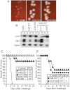
Figure 1.
Figure 1.
Yersinia pestis KIM D27 and UC91309 are attenuated for virulence due to a defect in iron scavenging. A, Y. pestis strains CO92 (virulent wild-type plague isolate), KIM D27 (nonpigmented vaccine type strain), and UC91309 (clinical isolate from a case of fatal plague septicemia) were spread on Congo red agar plates and incubated at 26°C for 3 days, after which colonies were photographed. B, Culture medium (supernatant [S]) and bacterial extract (pellet [P]) of Y. pestis strains CO92, KIM D27, and UC91309 were analyzed by immunoblotting with antibodies specific for LcrV (V antigen), Caf1 (F1 or capsular fraction 1 antigen) and RNA polymerase subunit A (RpoA), as a loading control. KIM D27 variants with a mutation in the lcrV gene (lcrV1) or a deletion of caf1caf1) were included as controls. C, Cohorts of Swiss Webster mice (n = 10) were challenged by subcutaneous injection with suspensions of Y. pestis strains CO92 (100 colony-forming units [CFUs]), KIM D27 (105 or 106 CFUs), or UC91309 (105 or 10CFUs). For each challenge experiment, inocula were verified by plating aliquots of bacterial suspensions on agar plates and enumerating CFUs. Animals were monitored over 14 days after challenge for survival. Results are representative of at least 3 independent experimental determinations. D, Mice were treated 24 hours before challenge with 0.1 mL 50% (v/v) iron dextran (100 mg/mL) and 50 mg/mL desferrioxamine B mesylate into the peritoneal cavity (+). Cohorts of Swiss Webster mice (n = 10) were challenged by subcutaneous injection with 105 CFUs of Y. pestis KIM D27 or UC91309. For each challenge experiment, inocula were verified by plating aliquots of bacterial suspensions on agar plates and enumerating CFUs. Animals were monitored for 14 days after challenge. Results are representative of 2 independent experimental determinations.
Figure 2.
Figure 2.
Histopathologic analysis of iron dextran–treated mice following infection with nonpigmented plague strains. Mice were treated 24 hours before challenge with 0.1 mL 50% (v/v) iron dextran (100 mg/mL) and 50 mg/mL desferrioxamine B mesylate into the peritoneal cavity. Cohorts of Swiss Webster mice (n = 10) were challenged by subcutaneous injection with 105 colony-forming units of Yersinia pestis KIM D27 or UC91309. Animals that succumbed to subcutaneous challenge underwent necropsy, and livers and spleens were removed and fixed. Tissues were embedded, thin sectioned, stained with hematoxylin-eosin, and analyzed by light microscopy with a 40 × objective. Iron-treated animals developed hepatic granulomas with histiocytes (black arrows) and neutrophils (blue arrows; A and B). Histiocytes harbored hemosiderin deposits, a brown-yellow pigment (red arrows). Spleen tissues displayed massive coagulative necrosis in the sinusoids with fibrin and neutrophil debris (blue arrows; C and D).
Figure 3.
Figure 3.
Mice with hereditary hemochromatosis are susceptible to bubonic and pneumonic plague challenge. A, Spleen and liver from naive 8-week-old wild-type mice (WT; 129S1/SvImJ) and hemojuvelin mutant (Hjv−/−) mice (129S6/SvEvTac) were removed during necropsy, fixed in 10% neutral buffered formalin, and embedded. Blocks were sectioned in 5-µm intervals, stained with Prussian blue, and viewed by microscopy with a 40 × objective. B, Blood from 8-week-old mice (n = 10) was collected, and serum was evaluated to determine the total iron level, using the serum iron/UIBC kit (Roche Diagnostics). Error bars indicate standard error of the means. C, WT or Hjv−/− mice aged 6–8 weeks (n = 10) were infected with Yersinia pestis CO92 via subcutaneous injection. D, WT or Hjv−/− mice aged 6–8 weeks (n = 10) were infected with Y. pestis CO92 via intranasal instillation. Infected animals were observed over 14 days for morbidity, mortality, or recovery. Results of bubonic plague (C) and pneumonic plague (D) challenge studies with Hjv−/− mice are representative of 2 independent experimental determinations.
Figure 4.
Figure 4.
Hemojuvelin-knockout (Hjv−/−) mice display increased susceptibility to challenge with nonpigmented Yersinia pestis. A and B, Wild-type (WT) or Hjv−/− mice aged 6–8 weeks (n = 10) were infected with Y. pestis KIM D27 (A) or Y. pestis UC91309 (B) via subcutaneous injection. Infected animals were observed over 14 days for morbidity, mortality, or recovery. Data were averaged from 2 (A) or 3 (B) independent experiments. Error bars indicate standard error of the means. Statistical significance was examined with the log-rank test: Hjv−/− 107 colony-forming units (CFUs) versus WT 107 CFUs, < .0001 (A); Hjv−/− 105 CFUs versus WT 105 CFUs, < .0001 (A); Hjv−/− 107 CFUs versus WT 107 CFUs, < .0001 (B); Hjv−/− 105 CFUs versus WT 105 CFUs, < .0001 (B). C and D, WT mice (C) or Hjv−/− mice (D) were infected with 1 × 106 CFUs of Y. pestis KIM D27 (pgm) via subcutaneous injection. At timed intervals, mice (n = 5) were euthanized and subjected to necropsy to determine the load of nonpigmented plague bacteria in tissue homogenates of the inguinal lymph node (LN; proximal to the injection site), the spleen, and the liver. Homogenates were spread on agar media and incubated for colony formation. Results are representative of 3 independent experimental determinations.
Figure 5.
Figure 5.
Histopathologic analysis of mice with hemochromatosis that were infected with nonpigmented Yersinia pestis. Wild-type (WT) mice or hemojuvelin-knockout (Hjv−/−) mice (n = 5) were infected by subcutaneous injection with 1 × 106 colony-forming units (CFUs) of Yersinia pestis KIM D27 (pgm). On day 4 (for Hjv−/− mice) or day 7 (for wild-type mice), animals were euthanized, and necropsy was performed. Liver and spleen were removed, and the organs were fixed, embedded, thin sectioned, and stained with hematoxylin-eosin and analyzed by light microscopy with a 40 × objective. WT animals displayed the physiological architecture of spleen tissues with lymphoid follicles (LF) and sinusoids (SI; A) or liver tissues with healthy liver parenchyma (LP) and blood vessels (BV; C). Hjv−/− mice harbored focal inflammatory infiltrates composed of histocytes (black arrows) and neutrophils (blue arrows) in both liver and spleen tissues.
Figure 6.
Figure 6.
Plague vaccine protection of mice with hereditary hemochromatosis. Hemojuvelin-knockout (Hjv−/−) mice aged 6–8 weeks were immunized at day 0 and day 21 via intramuscular injections with 25 µg of rV10-2 protein adsorbed to 250 µg of Alhydrogel. As a control, animals received intramuscular injections of phosphate-buffered saline (PBS) with 250 µg of Alhydrogel. A, Blood was collected from mice (n = 5) on day 0 (prior to the first immunization) and on day 42 (21 days following the booster), and serum was analyzed for rV10-2 antigen–specific immunoglobulin G (IgG). B, Forty-two days after immunization, mice (n = 10) were challenged by subcutaneous injection with either 500 colony-forming units (CFUs) of the fully virulent plague isolate Yersinia pestis CO92 or 5 × 106 CFUs of the attenuated vaccine-type strain Y. pestis KIM D27 (pgm) via subcutaneous inoculation. Infected animals were observed over 14 days for morbidity and mortality.

Similar articles

Cited by

References

    1. Yersin A. La peste bubonique à Hong-Kong. Ann Inst Pasteur. 1894;2:428–30.
    1. Bos KI, Schuenemann VJ, Golding GB, et al. A draft genome of Yersinia pestis from victims of the black death. Nature. 2011;478:506–10. - PMC - PubMed
    1. Simond PL. La propagation de la peste. Ann Inst Pasteur. 1898;12:625–87.
    1. Meyer KF. Effectiveness of live or killed plague vaccines in man. Bull World Health Organ. 1970;42:653–66. - PMC - PubMed
    1. Girard G. Plague. Annu Rev Microbiol. 1955;9:253–77. - PubMed

Publication types

MeSH terms