. 2012;2(7):723-33.
doi: 10.7150/thno.4275. Epub 2012 Aug 1.

PEGylated Phospholipid Micelle-Encapsulated Near-Infrared PbS Quantum Dots for in vitro and in vivo Bioimaging

Affiliations

PEGylated Phospholipid Micelle-Encapsulated Near-Infrared PbS Quantum Dots for in vitro and in vivo Bioimaging

Rui Hu et al. Theranostics. 2012.

Abstract

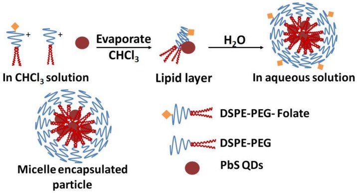
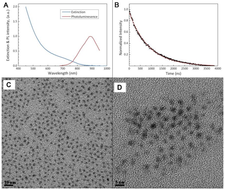
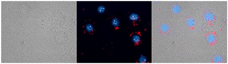
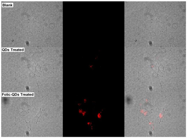
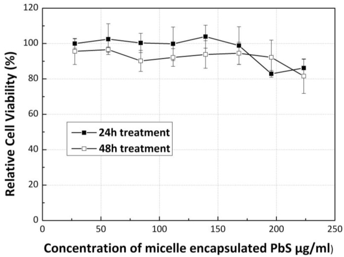
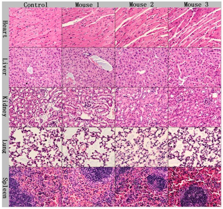
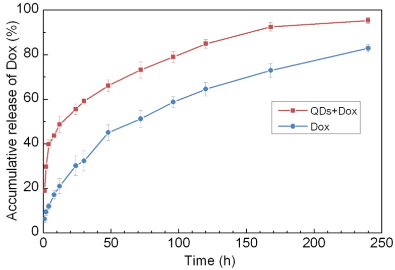
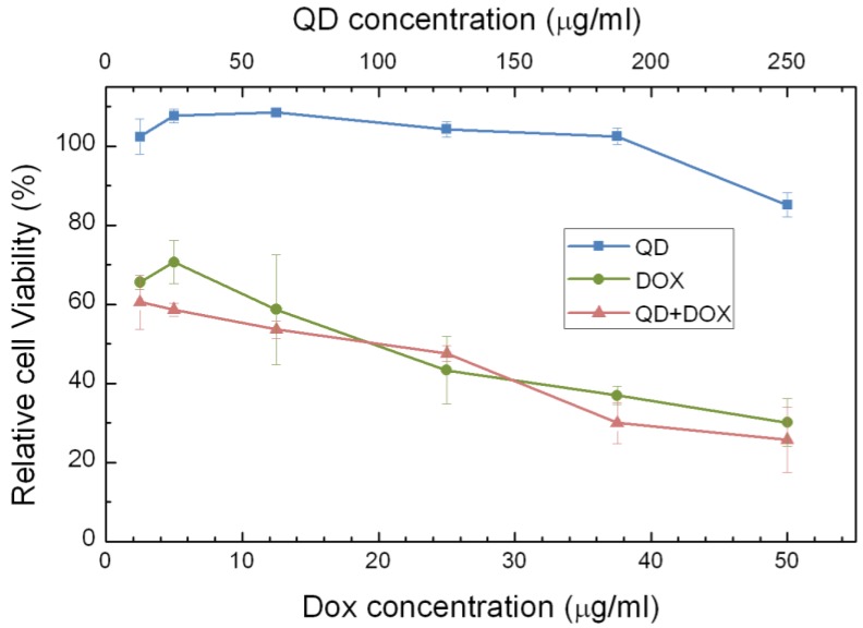
Surface modification and functionalization of bioconjugated quantum dots (QDs) has drawn great attention for the past few years due to their wide applications in biomedical research. In this contribution, we demonstrate the use of PEGylated phospholipid micelles to encapsulate near infrared emitting ultra-small lead sulfide (PbS) QDs for in vitro and in vivo imaging. The cytotoxicity of the micelle-encapsulated QDs formulation was evaluated using MTS assay and histological analysis studies. We have found that upon encapsulating the QDs with phospholipid micelle, the toxicity of the PbS QDs is reduced, from which we envision that the PEGylated phospholipid micelle-encapsulated PbS QDs formulation can be used as theranostics probes for some selected applications in cell imaging and small animals study.

Keywords: Micelle encapsulation; PEG.; Phospholipid; bioimaging; quantum dots.


Conflict of interest statement

Competing Interests: The authors have declared that no competing interest exists.

Figures

Figure 1
Figure 1
Schematic illustration of the PEGylated phospholipid micelle encapsulation of PbS QDs.
Figure 2
Figure 2
Characterization on the physical properties of the PbS QD nanoparticles. (a) Extinction & photolumination (PL) profile of the QDs after micelle encapsulation. (b) Temporal decay of the photoluminescence of PbS QDs after excited by a pulse laser. Solid line shows the fitting result using a single-exponential function. (c) and (d), TEM images of the PbS QDs before and after micelle encapsulation, respectively.
Figure 3
Figure 3
RAW264.7 macrophages labeled with micelle-encapsulated PbS QDs (red), the cell nucleus is stained with Hoechst 33342 (blue).
Figure 4
Figure 4
Imaging of live pancreatic cancer cells (Blank) as a comparison with which treated with folic acid conjugated phospholipid micelle encapsulated PbS QDs (Folic-QDs Treated) or non-conjugated QDs (QDs Treated). Left panel shows the bright-field image, middle panel is the fluorescent signal pseudo-colored in red and the right panel is the merged images.
Figure 5
Figure 5
Normalized characterization spectra from the QDs sample and the background signal of an untreated mouse, both under deep-red illumination. Inset in the upper left is the QDs sample in a centrifuge tube and the inset in the lower right shows the back of a mouse with its tail labeled. The two insets are pseudo-colored in red (QDs) and green (background).
Figure 6
Figure 6
In vivo fluorescent images of a nude mouse tail vein injected with PEGylated phospholipid micelle encapsulated PbS QDs at 1h, 3h, 6h and 42h after injection. Left panel shows the original unprocessed images, middle panel is the QDs signal channel and the panel to the right shows the pseudo-colored images of the spectrally separated signals, with background in green and the QDs signals in red.
Figure 7
Figure 7
Relative cell viability of cells treated with PEGylated phospholipid micelle encapsulated PbS QDs of different concentrations, after 24h and 48h.
Figure 8
Figure 8
Histological studies on the major organs of the QDs injected mice at a dosage of 25mg/kg after 4 weeks. No abnormal pathological changes were observable.
Figure 9
Figure 9
Accumulative release of Doxorubicin (Dox) from phospholipid micelle encapsulated formulations, with and without PbS QDs co-encapsulation.
Figure 10
Figure 10
Relative cell viabilities of Panc-1 celles treated with different formulations of phospholipid micelles, i.e., QDs encapsulated, Dox only encapsulated and QDs-Dox co-encapsulated, as a function of drug/QDs concentration 72 h following the treatment.

Similar articles

Cited by

References

    1. Zachariasse KA, Vanphuc N, Kozankiewicz B. Investigation of Micelles, Micro-Emulsions, and Phospholipid-Bilayers With the Pyridinium N-Phenolbetaine Et(30), A Polarity Probe for Aqueous Interfaces. J Phys Chem. 1981;85:2676–83. doi:10.1021/j150618a022.
    1. Wendoloski JJ, Kimatian SJ, Schutt CE, Salemme FR. Molecular-Dynamics Simulation of a Phospholipid Micelle. Science. 1989;243:636–8. doi:10.1126/science.2916118. - PubMed
    1. Bangham AD, Horne RW. Negative staining of phospholipids and their structural modification by surface-active agents as observed in the electron microscope. Journal of Molecular Biology. 1964;8:660–IN10. doi:10.1016/s0022-2836(64)80115-7. - PubMed
    1. Gabizon A, Shmeeda H, Horowitz AT, Zalipsky S. Tumor cell targeting of liposome-entrapped drugs with phospholipid-anchored folic acid-PEG conjugates. Adv Drug Deliv Rev. 2004;56:1177–92. doi:10.1016/j.addr.2004.01.011. - PubMed
    1. Walsh TJ, Finberg RW, Arndt C, Hiemenz J, Schwartz C, Bodensteiner D. et al. Liposomal Amphotericin B for Empirical Therapy in Patients with Persistent Fever and Neutropenia. New England Journal of Medicine. 1999;340:764–71. doi:doi:10.1056/NEJM199903113401004. - PubMed