Review
. 2012 Jul 18;3(7):494-504.
doi: 10.1021/cn300013p. Epub 2012 Apr 16.

Neurochemical and neurostructural plasticity in alcoholism

Affiliations
Review

Neurochemical and neurostructural plasticity in alcoholism

Justin T Gass et al. ACS Chem Neurosci. .

Abstract

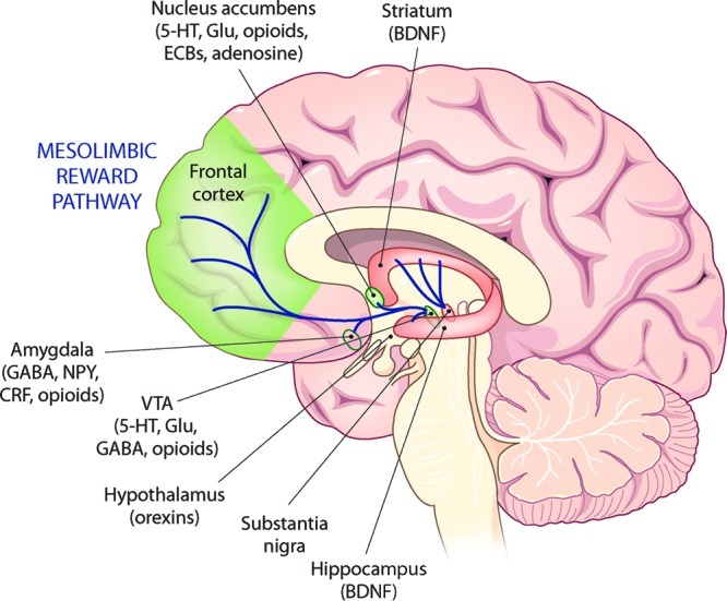
The behavioral manifestations of alcoholism are primarily attributable to the numerous and lasting adaptations that occur in the brain as a result of chronic heavy alcohol consumption. As will be reviewed here, these adaptations include alcohol-induced plasticity in chemical neurotransmission, density and morphology of dendritic spines, as well as neurodegeneration and cerebral atrophy. Within the context of these neuroadaptations that have been observed in both human and animal studies, we will discuss how these changes potentially contribute to the cognitive and behavioral dysfunctions that are hallmark features of alcoholism, as well as how they reveal novel potential pharmacological targets for the treatment of this disorder.


Figures

Figure 1
Figure 1
Schematic of the mesolimbic reward pathway in the human brain and neurochemical influences on this pathway that are modulated by alcohol. The reinforcing effects of alcohol are believed to be mediated by the activation of dopamine-containing cell bodies in the ventral tegmental area (VTA) that project rostrally to the nucleus accumbens and frontal cortex. This activation occurs primarily at the level of the VTA, where alcohol promotes the release of endogenous opioid peptides which hyperpolarize local inhibitory GABAergic neurons, thereby disinhibiting mesolimbic dopamine neurons. In addition, alcohol promotes the release of the excitatory amino acid glutamate in the VTA and also affects various ionic currents intrinsic to VTA dopamine neurons that regulate the excitability of these cells. Dopaminoceptive terminal field regions targeted by mesolimbic dopamine projections are modulated by various neurotransmitters and neuromodulators as detailed in this review. Abbreviations: Glu, glutamate; 5-HT, 5-hydroxytrypamine (serotonin), ECBs, endocannabinoids; NPY, neuropeptide Y; CRF, corticotropin releasing factor, BDNF, brain-derived neurotrophic factor.
Figure 2
Figure 2
Voluntary ethanol consumption increases extracellular levels of glutamate in the VTA as measured by enzyme-coated glutamate biosensors. See text for details of experimental procedures. (a) Principles of amperometric detection of changes in extracellular glutamate in near real-time by glutamate biosensors (Pinnacle Technologies, Lawrence, KS). Biosensors are equipped with a 1 mm sensor cavity (176 μm O.D.) consisting of a Pt–Ir electrode coated with Nafion (to repel anionic interferants) and an enzyme layer containing immobilized glutamate oxidase (GluOx, which catalyzes the breakdown of glutamate to α-ketoglutarate and electroactive H2O2) and ascorbate oxidase (AscOx, which catalyzes the breakdown of ascorbate to neutral dehydroascorbate and H2O). An amperometric current of +600 mV applied to the electrode oxidizes H2O2 to form electrons (e−) that provide a near real-time (∼1 s resolution) indirect measurement of changes in extracellular levels of glutamate. Image couresty of Jim Urich (Pinnacle Technologies). (b) Histological photograph of a rat brain showing the location of glutamate biosensor placement in the VTA. (c) Sample biosensor current tracings obtained during the consumption of sweetened alcohol (10% v/v ethanol + 3% w/v glucose + 0.125% w/v saccharin, n = 5) or control (3% w/v glucose + 0.125% w/v saccharin, n = 5) solutions. Consumption of the solution, as determined from video recordings, occurred during the time indicated by the shaded gray bar. In animals consuming the sweetened alcohol solution, the amount of alcohol consumed during the 30 min session was 0.45 ± 0.07 g/kg (mean ± SEM), resulting in blood alcohol levels assessed immediately following the session of 25.4 ± 2.5 mg/dL (mean ± SEM). (d) Cumulative changes in extracellular glutamate during the 30 min consumption session as determined by cumulative changes in GluOx-mediated currents (in nA) converted to changes in extracellular glutamate (in μM) based on preimplantation calibration curves. Animals consuming the sweetened alcohol solution exhibited significantly greater changes in glutamate in the VTA as compared to animals consuming the control solution (p < 0.05).
Figure 3
Figure 3
Analysis of structural plasticity of dendritic spines. (a) Diolistically labeled neuron in the prefrontal cortex. (b) 3-D reconstruction of a labeled dendrite showing spine morphology based on the classification of spine shape as filopodia, long, stubby, or mushroom (bottom row).

Similar articles

Cited by

References

    1. Breese G. R.; Sinha R.; Heilig M. (2011) Chronic alcohol neuroadaptation and stress contribute to susceptibility for alcohol craving and relapse. Pharmacol. Ther. 129, 149–171. - PMC - PubMed
    1. Spanagel R. (2009) Alcoholism: a systems approach from molecular physiology to addictive behavior. Physiol. Rev. 89, 649–705. - PubMed
    1. Kiefer F.; Mann K. (2010) Acamprosate: how, where, and for whom does it work? Mechanism of action, treatment targets, and individualized therapy. Curr. Pharm. Des. 16, 2098–2102. - PubMed
    1. Ripley T. L.; Stephens D. N. (2011) Critical thoughts on current rodent models for evaluating potential treatments of alcohol addiction and withdrawal. Br. J. Pharmacol. 164, 1335–1356. - PMC - PubMed
    1. Crabbe J. C.; Phillips T. J.; Belknap J. K. (2010) The complexity of alcohol drinking: studies in rodent genetic models. Behav. Genet. 40, 737–750. - PMC - PubMed

Publication types