. 2012 Dec;14(12):1892-903.
doi: 10.1111/cmi.12008. Epub 2012 Sep 25.

In silico directed mutagenesis identifies the CD81/claudin-1 hepatitis C virus receptor interface

Affiliations
Free PMC article

In silico directed mutagenesis identifies the CD81/claudin-1 hepatitis C virus receptor interface

Christopher Davis et al. Cell Microbiol. 2012 Dec.
Free PMC article

Abstract

Hepatitis C virus (HCV) entry is dependent on host cell molecules tetraspanin CD81, scavenger receptor BI and tight junction proteins claudin-1 and occludin. We previously reported a role for CD81/claudin-1 receptor complexes in HCV entry; however, the molecular mechanism(s) driving association between the receptors is unknown. We explored the molecular interface between CD81 and claudin-1 using a combination of bioinformatic sequence-based modelling, site-directed mutagenesis and Fluorescent Resonance Energy Transfer (FRET) imaging methodologies. Structural modelling predicts the first extracellular loop of claudin-1 to have a flexible beta conformation and identifies a motif between amino acids 62-66 that interacts with CD81 residues T149, E152 and T153. FRET studies confirm a role for these CD81 residues in claudin-1 association and HCV infection. Importantly, mutation of these CD81 residues has minimal impact on protein conformation or HCV glycoprotein binding, highlighting a new functional domain of CD81 that is essential for virus entry.


Figures

Fig. 1
Fig. 1
Structural modelling of claudin-CD81 association. Ribbon models of the ECL1 domain of native claudin-1 (A) or I32M (B), E48K (C) and I32M/E48K (D) mutants with CD81 ECL2 (PDB: 1G8Q). Claudin-1 is depicted according to its predicted secondary structure (alpha helices in red, beta turn in white) with CD81 in green, key interacting residues are labelled. The interacting regions include claudin-1 residues 33–35 and 63–66, and CD81 residues K148, T149, E152 and T153. I32M and E48K claudin-1 mutations alter key inter-residue distances and interactions with CD81. Model of the ECL1 domain of native claudin-7 (E) or M32I/K48E mutant (F) with the CD81 ECL. Mutagenesis of claudin-7 relocates the loop around C64 to a position similar to that of claudin-1 and withdraws a prominent loop away from the region of claudin-CD81 interaction. Images were produced using the Chimera program (University of San Francisco).
Fig. 2
Fig. 2
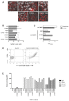
Effect of claudin-1 mutations on protein localization, CD81 association and HCV infection. A. 293-T cells were transfected to express wild-type (WT) AcGFP–claudin-1 and mutants and their localization assessed by confocal microscopy. B. Cell surface expression of AcGFP–claudin mutants was determined using the Zeiss profiling function to trace cell boundaries, the data shown is the average MFI of 10 cell profiles, the data are representative of three transfection experiments. C. %FRET between AcGFP–claudin-1 mutants and DsRED-CD81 in transfected 293-T cells. None of the mutants in group I (W30A, I32A, D38A, G49A and W51A) showed any significant interaction with CD81. The mean %FRET values are derived from triplicate estimates within a single experiment and are representative of two further experiments. The mean plus 2 SD of non-specific FRET values observed in parental non-transfected is indicated by the dashed line and represents the threshold for the assay. The diminution in %FRET signal for the members of group I relative to the other two groups was significant (Kruskal Wallis test, P < 0.01). D. Two-hybrid screening of interactions between mutant claudin-1 and WT CD81. None of the claudin-1 mutants in group I showed evidence for association in vitro. E and F. Infectivity of HCVpp (E) and MLVpp (F) in parental 293-T cells and those transfected to express AcGFP–claudin-1 WT and mutants. 293-T cells expressed comparable levels of WT and claudin-1 mutants. Data are expressed as specific infectivity where the value of an envelope deficient pseudoparticle is subtracted from both HCVpp and MLVpp relative light unit (RLU) signals. The dashed line represents the mean plus 2 SD of HCVpp infection of claudin-1-negative parental 293-T cells; levels of infection below this threshold are considered negative. The data presented are from a single experiment and are representative of two independent experiments.
Fig. 3
Fig. 3
Effect of CD81 mutations on protein localization, antigenicity and HCV E2 binding. HepG2 cells were transduced to express wild-type (WT) DsRED-CD81 and mutant proteins and their localization assessed by confocal microscopy (A). %FRET between DsRED-CD81 mutants and AcGFP–CD81 in transfected 293-T cells, The mean values are derived from triplicate estimates which are representative of three further experiments (B). Anti-CD81 mAbs and rabbit anti-MBP were tested at 5 μg ml−1 for their reactivity with monomeric and dimeric MBP–CD81 by ELISA, where the data are presented as optical density at 450 nm (C). Flow cytometric binding of anti-CD81 2s20 to parental HepG2 and cells transduced to express DsRED-CD81 (D). Anti-CD81 mAb binding to HepG2 cells expressing WT DsRED-CD81 and mutant proteins. mAb binding is expressed as mean fluorescence intensity (MFI) relative to DsRED CD81 signals (E). The data presented are from a single experiment and are representative of two independent experiments.
Fig. 4
Fig. 4
Effect of CD81 mutations on claudin-1 association and HCV infection. FRET between AcGFP–claudin-1 and DsRED-CD81 and mutant proteins expressed in 293-T cells (A). The mean plus 2SD of non-specific FRET values observed in parental non-transfected is indicated by the dashed line and represents the threshold for the assay. Flow cytometric binding of HCV E2 (strain H77) to parental CHO cells and those transduced to express DsRED-CD81 (B). HCV E2 strain H77 and JFH-1 binding to CHO cells expressing wild-type (WT) DsRED-CD81 and mutant proteins. HCV E2 binding is expressed as mean fluorescence intensity (MFI) relative to DsRED CD81 signals (C). HepG2 cells were transduced to express WT DsRED-CD81 and mutant proteins and evaluated for their ability to support HCVpp or MLVpp infection (D). Flow cytometry of transduced HepG2 cells confirmed comparable levels of CD81 expression. Data are expressed as specific infectivity where the value of an envelope deficient pseudoparticle is subtracted from both HCVpp and MLVpp relative light unit (RLU) signals. The dashed line represents the mean plus 2 SD of HCVpp infection of CD81-negative parental HepG2 cells; levels of infection below this threshold are considered negative. The data presented are from a single experiment and are representative of two independent experiments.

Similar articles

Cited by

References

    1. Altschul SF, Gish W, Miller W, Myers EW, Lipman DJ. Basic local alignment search tool. J Mol Biol. 1990;215:403–410. - PubMed
    1. Burlone ME, Budkowska A. Hepatitis C virus cell entry: role of lipoproteins and cellular receptors. J Gen Virol. 2009;90:1055–1070. - PubMed
    1. Chung SK, Vanbellinghen JF, Mullins JG, Robinson A, Hantke J, Hammond CL, et al. Pathophysiological mechanisms of dominant and recessive GLRA1 mutations in hyperekplexia. J Neurosci. 2010;30:9612–9620. - PMC - PubMed
    1. Coffey GP, Rajapaksa R, Liu R, Sharpe O, Kuo CC, Krauss SW, et al. Engagement of CD81 induces ezrin tyrosine phosphorylation and its cellular redistribution with filamentous actin. J Cell Sci. 2009;122:3137–3144. - PMC - PubMed
    1. Cukierman L, Meertens L, Bertaux C, Kajumo F, Dragic T. Residues in a highly conserved claudin-1 motif are required for hepatitis C virus entry and mediate the formation of cell–cell contacts. J Virol. 2009;83:5477–5484. - PMC - PubMed

Publication types