Review
. 2012 Sep;21(9):660-4.
doi: 10.1111/j.1600-0625.2012.01566.x.

Innate immunity and the regulation and mobilization of keratinocyte stem cells: are the old players playing a new game?

Affiliations
Review

Innate immunity and the regulation and mobilization of keratinocyte stem cells: are the old players playing a new game?

Ashok Singh et al. Exp Dermatol. 2012 Sep.

Abstract

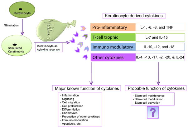
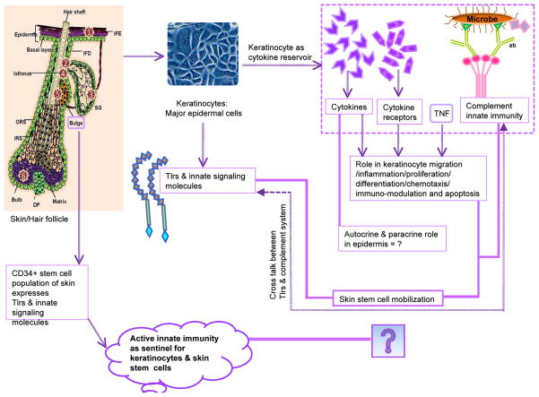
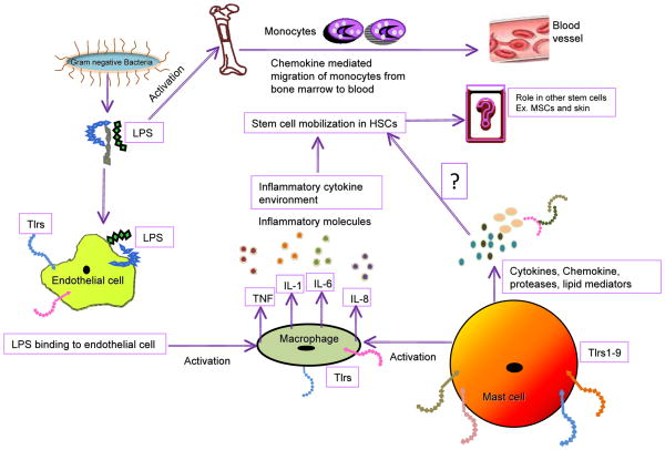
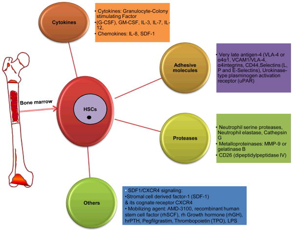
The skin provides an anatomical barrier to physical, chemical and biological agents. Hence, it is not surprising that it has well-developed innate immunity. What we find surprising is that the CD49f(+) /CD34(+) hair follicle stem cells should have an enriched expression profile of so many genes involved in innate immunity. Do these stem cells require extra protection from environmental insults? Or, could there be a new role for these genes? To probe these questions, we first summarize the roles of some key players in epidermal innate immunity. We next focus on their expression in CD49f(+) /CD34(+) hair follicle stem cells. Then, we consider recent data suggesting a new role for these 'old players' in the regulation and mobilization of haematopoietic and mesenchymal stem cells. Finally, we hypothesize that the 'old players' in these hair follicle stem cells may be playing a 'new game'.


Conflict of interest statement

Conflict of interests:

We declare no conflict of interest.

Figures

Figure 1
Figure 1. Keratinocytes as reservoir of cytokines
This figure illustrates some of the known and plausible unknown function of keratinocyte derived cytokines. The cartoon also depicts some of the various cytokines released from keratinocytes.
Figure 2
Figure 2. This figure illustrates the involvement of Tlrs, cytokines, and other innate signatures of stem cells use to boost the innate immunity of keratinocytes in mammalian skin
The left side of the figure depicts skin and its CD34+ stem cells (located in bulge region), and their connection to the innate immune system via Tlrs and other associated molecules. The numbers inside the stars indicates the locations of known stem cell populations within the hair follicle. The middle portion of the figure shows the presence of Tlrs in keratinocytes, whereas the right portions show cytokines and their receptors, and their role in keratinocytes (inside the dotted box). The far right of the figure illustrates the complement cascade and it’s connection in HSC mobilization. The bold lines represent the Viewpoint question related to paracrine or autocrine role of cytokines, and propose a role for innate immunity and its working arms such as Tlrs and the complement system in epidermal stem cell mobilization and stabilization. A divided double line indicates probable cross-talk between the Tlrs and the complement system. Abbreviations:(TNF= Tumor Necrosis Factor, c= complement molecule, ab = antibody, SG = Sebaceous gland, IM = Arrector pili muscle, DP = Dermal papillae, IFE = inter-follicular epidermis, IFD = Infundibulum, ORS =Outer root sheath, IRS = Inner root sheath).
Figure 3
Figure 3. This figure illustrates one of the probable mechanisms of HSC mobilization via involvement of endothelial cells, macrophages, cytokines and bacterial LPS
The LPS of the aerobic Gram-negative bacteria damages the endothelial cells and activates macrophages to releases pro-inflammatory cytokines such as TNF, IL-1 (Interleukin-1), IL-6, and IL-8 in response to LPS. LPS also induces monocyte migration from bone marrow to blood. It is noteworthy that the macrophage and endothelial cells express Tlrs. The mast cells also express Tlrs and releases cytokines, histamine, chemokines, proteases and lipid mediators against diverse signals; however, their involvement in stem cell mobilization has not yet been determined. Involvement of cytokines, interleukins and LPS induced HSCs activation is reported in literature.
Figure 4
Figure 4
This figure illustrates involvement of various cytokines, adhesive molecules, proteases and other factors during the process of HSC mobilization.

References

    1. Tizard I. Immunity at body surface. Philadelphia, PA: Saunders; 2000.
    1. Trempus CS, Morris RJ, Ehinger M, et al. CD34 expression by hair follicle stem cells is required for skin tumor development in mice. Cancer Res. 2007;67:4173–4181. - PMC - PubMed
    1. Ito M, Liu Y, Yang Z, et al. Stem cells in the hair follicle bulge contribute to wound repair but not to homeostasis of the epidermis. Nat Med. 2005;11:1351–1354. - PubMed
    1. Morris RJ, Liu Y, Marles L, et al. Capturing and profiling adult hair follicle stem cells. Nat Biotechnol. 2004;22:411–417. - PubMed
    1. Janeway CA., Jr Approaching the asymptote? Evolution and revolution in immunology. Cold Spring Harb Symp Quant Biol. 1989;54(Pt 1):1–13. - PubMed

Publication types

MeSH terms