

Protein kinase C epsilon is important in modulating organic-dust-induced airway inflammation
- PMID: 22897707
- PMCID: PMC4066446
- DOI: 10.3109/01902148.2012.714841
Protein kinase C epsilon is important in modulating organic-dust-induced airway inflammation
Abstract
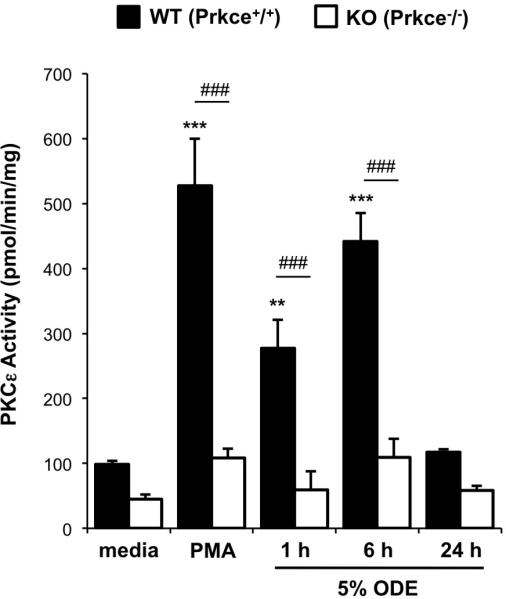
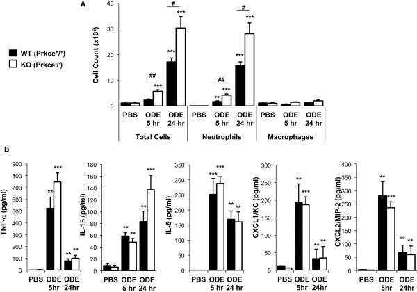
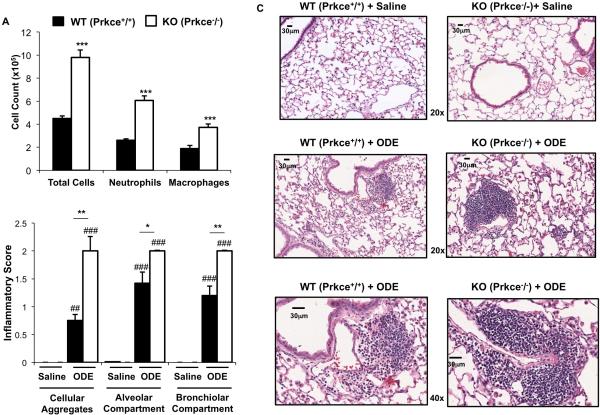
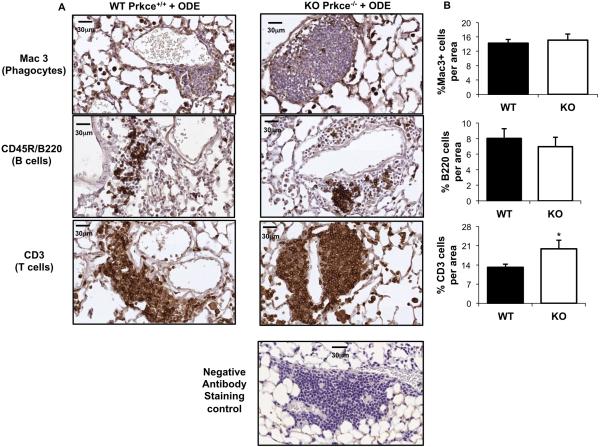
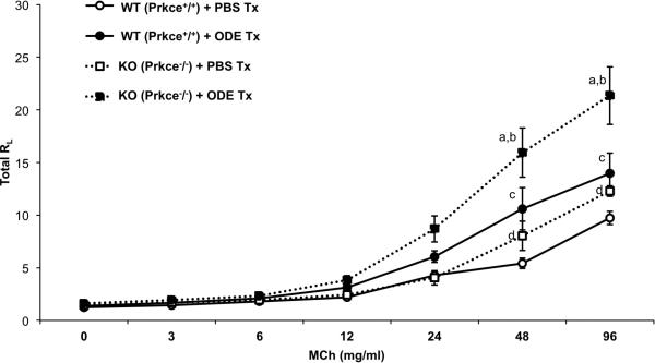
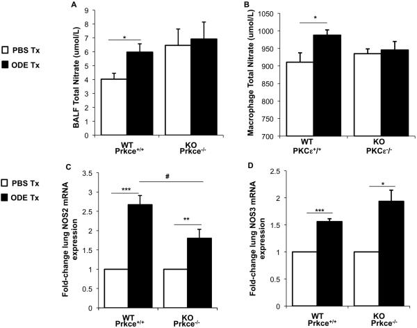
Organic dust samples from swine confinement facilities elicit pro-inflammatory cytokine/chemokine release from bronchial epithelial cells and monocytes, dependent, in part, upon dust-induced activation of the protein kinase C (PKC) isoform, PKCε. PKCε is also rapidly activated in murine tracheal epithelial cells following in vivo organic dust challenges, yet the functional role of PKCε in modulating dust-induced airway inflammatory outcomes is not defined. Utilizing an established intranasal inhalation animal model, experiments investigated the biologic and physiologic responses following organic dust extract (ODE) treatments in wild-type (WT) and PKCε knock-out (KO) mice. We found that neutrophil influx increased more than twofold in PKCε KO mice following both a one-time challenge and 3 weeks of daily challenges with ODE as compared with WT mice. Lung pathology revealed increased bronchiolar and alveolar inflammation, lymphoid aggregates, and T cell influx in ODE-treated PKCε KO mice. Airway hyperresponsiveness to methacholine increased in PKCε KO + ODE to a greater magnitude than WT + ODE animals. There were no significant differences in cytokine/chemokine release elicited by ODE treatment between groups. However, ODE-induced nitric oxide (NO) production differed in that ODE exposure increased nitrate levels in WT mice but not in PKCε KO mice. Moreover, ODE failed to upregulate NO from ex vivo stimulated PKCε KO lung macrophages. Collectively, these studies demonstrate that PKCε-deficient mice were hypersensitive to organic dust exposure and suggest that PKCε is important in the normative lung inflammatory response to ODE. Dampening of ODE-induced NO may contribute to these enhanced inflammatory findings.
Figures
Similar articles
-
The Effect of Inhalant Organic Dust on Bone Health.Curr Allergy Asthma Rep. 2018 Feb 22;18(3):16. doi: 10.1007/s11882-018-0773-y. Curr Allergy Asthma Rep. 2018. PMID: 29470660 Free PMC article. Review.
-
Myeloid differentiation factor 88-dependent signaling is critical for acute organic dust-induced airway inflammation in mice.Am J Respir Cell Mol Biol. 2013 Jun;48(6):781-9. doi: 10.1165/rcmb.2012-0479OC. Am J Respir Cell Mol Biol. 2013. PMID: 23492189 Free PMC article.
-
Toll-like receptor 2 regulates organic dust-induced airway inflammation.Am J Respir Cell Mol Biol. 2011 Oct;45(4):711-9. doi: 10.1165/rcmb.2010-0427OC. Epub 2011 Jan 28. Am J Respir Cell Mol Biol. 2011. PMID: 21278324 Free PMC article.
-
MyD88 in lung resident cells governs airway inflammatory and pulmonary function responses to organic dust treatment.Respir Res. 2015 Sep 16;16(1):111. doi: 10.1186/s12931-015-0272-9. Respir Res. 2015. PMID: 26376975 Free PMC article.
-
Ovalbumin-sensitized mice have altered airway inflammation to agriculture organic dust.Respir Res. 2019 Mar 7;20(1):51. doi: 10.1186/s12931-019-1015-0. Respir Res. 2019. PMID: 30845921 Free PMC article.
Cited by
-
The Effect of Inhalant Organic Dust on Bone Health.Curr Allergy Asthma Rep. 2018 Feb 22;18(3):16. doi: 10.1007/s11882-018-0773-y. Curr Allergy Asthma Rep. 2018. PMID: 29470660 Free PMC article. Review.
-
β2-Adrenergic agonists attenuate organic dust-induced lung inflammation.Am J Physiol Lung Cell Mol Physiol. 2016 Jul 1;311(1):L101-10. doi: 10.1152/ajplung.00125.2016. Epub 2016 May 17. Am J Physiol Lung Cell Mol Physiol. 2016. PMID: 27190062 Free PMC article.
-
Influence of farming exposure on the development of asthma and asthma-like symptoms.Int Immunopharmacol. 2014 Nov;23(1):356-63. doi: 10.1016/j.intimp.2014.07.014. Epub 2014 Jul 31. Int Immunopharmacol. 2014. PMID: 25086344 Free PMC article. Review.
-
Surface Modification of Biodegradable Microparticles with the Novel Host-Derived Immunostimulant CPDI-02 Significantly Increases Short-Term and Long-Term Mucosal and Systemic Antibodies against Encapsulated Protein Antigen in Young Naïve Mice after Respiratory Immunization.Pharmaceutics. 2022 Sep 1;14(9):1843. doi: 10.3390/pharmaceutics14091843. Pharmaceutics. 2022. PMID: 36145590 Free PMC article.
-
Effect of epithelial-specific MyD88 signaling pathway on airway inflammatory response to organic dust exposure.J Immunotoxicol. 2023 Dec;20(1):2148782. doi: 10.1080/1547691X.2022.2148782. J Immunotoxicol. 2023. PMID: 36538286 Free PMC article.
References
-
- Schwartz DA, Landas SK, Lassise DL, Burmeister LF, Hunninghake GW, Merchant JA. Airway injury in swine confinement workers. Ann Intern Med. 1992;116:630–5. - PubMed
-
- Sundblad BM, Larsson BM, Palmberg L, Larsson K. Exhaled nitric oxide and bronchial responsiveness in healthy subjects exposed to organic dust. Eur Respir J. 2002;20:426–31. - PubMed
-
- Zhiping W, Malmberg P, Larsson BM, Larsson K, Larsson L, Saraf A. Exposure to bacteria in swine-house dust and acute inflammatory reactions in humans. Am J Respir Crit Care Med. 1996;154:1261–6. - PubMed
-
- Palmberg L, Larssson BM, Malmberg P, Larsson K. Airway responses of healthy farmers and nonfarmers to exposure in a swine confinement building. Scand J Work Environ Health. 2002;28:256–63. - PubMed
Publication types
MeSH terms
Substances
Grants and funding
- R01 AA008769/AA/NIAAA NIH HHS/United States
- R01 AA017993/AA/NIAAA NIH HHS/United States
- ES015522-03S1/ES/NIEHS NIH HHS/United States
- R01 ES019325/ES/NIEHS NIH HHS/United States
- K08 ES015522/ES/NIEHS NIH HHS/United States
- U54 OH010162/OH/NIOSH CDC HHS/United States
- R01AA017993/AA/NIAAA NIH HHS/United States
- R01OH008539/OH/NIOSH CDC HHS/United States
- R01 OH008539/OH/NIOSH CDC HHS/United States
- P01OH010162/OH/NIOSH CDC HHS/United States
- K08 ES015522-01/ES/NIEHS NIH HHS/United States
- I01 BX000728/BX/BLRD VA/United States
LinkOut - more resources
Full Text Sources
Research Materials