Review
. 2013 Feb 1;18(4):432-68.
doi: 10.1089/ars.2011.4234. Epub 2012 Aug 16.

Redox control of cardiac excitability

Affiliations
Review

Redox control of cardiac excitability

Nitin T Aggarwal et al. Antioxid Redox Signal. .

Abstract

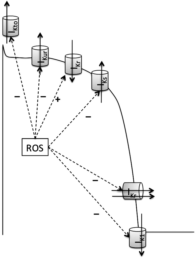
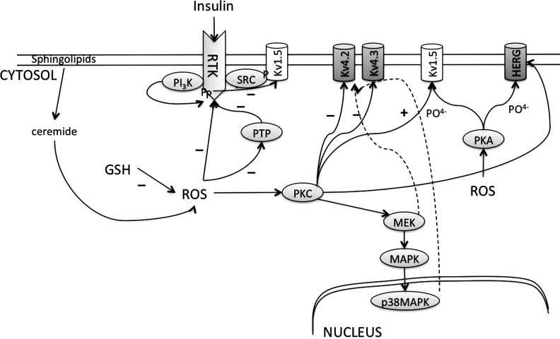
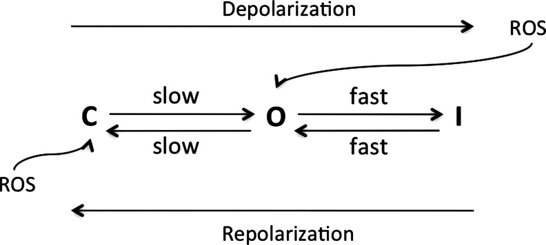
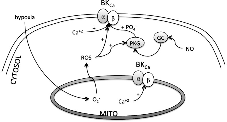
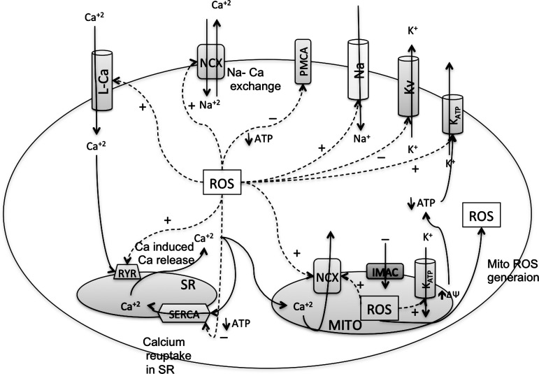
Reactive oxygen species (ROS) have been associated with various human diseases, and considerable attention has been paid to investigate their physiological effects. Various ROS are synthesized in the mitochondria and accumulate in the cytoplasm if the cellular antioxidant defense mechanism fails. The critical balance of this ROS synthesis and antioxidant defense systems is termed the redox system of the cell. Various cardiovascular diseases have also been affected by redox to different degrees. ROS have been indicated as both detrimental and protective, via different cellular pathways, for cardiac myocyte functions, electrophysiology, and pharmacology. Mostly, the ROS functions depend on the type and amount of ROS synthesized. While the literature clearly indicates ROS effects on cardiac contractility, their effects on cardiac excitability are relatively under appreciated. Cardiac excitability depends on the functions of various cardiac sarcolemal or mitochondrial ion channels carrying various depolarizing or repolarizing currents that also maintain cellular ionic homeostasis. ROS alter the functions of these ion channels to various degrees to determine excitability by affecting the cellular resting potential and the morphology of the cardiac action potential. Thus, redox balance regulates cardiac excitability, and under pathological regulation, may alter action potential propagation to cause arrhythmia. Understanding how redox affects cellular excitability may lead to potential prophylaxis or treatment for various arrhythmias. This review will focus on the studies of redox and cardiac excitation.


Figures

FIG. 1.
FIG. 1.
Examples of the effects of reactive oxygen species (ROS) on various channel currents. (A, B) Hydrogen peroxide (H2O2) increased the Ca+2 currents from the L-type Ca2+ channels (ICaL) and the persistent Na+ currents from the cardiac Na+ channels (INaP). (C) Dihydroxyfumarate (DHF), an oxidant, decreased the inward rectifier K+ currents (IKr) from the Kv11.1 channels. All these currents were measured in the guinea pig myocytes, and are taken from (44, 167, 249), with copywrite permissions, respectively. The arrows indicate differences in the currents.
FIG. 2.
FIG. 2.
Major ROS synthesis pathways in cardiac myocytes. In the mitochondria (MITO), a small percentage of electrons from the respiratory chain prematurely leak to O2 at complexes I, II, or III, resulting in the formation of the toxic superoxide radical ion (O2•−). O2•− is also generated by release of a free electron from the reactions where NADPH is oxidized to NADP by NADPH oxidase, and hypoxanthine is oxidized to xanthine (X) by xanthine oxidase (XO). The O2•− generated in the mitochondria can freely pass through the mitochondrial membrane into the cytosol. Cytosolic O2•− can interact with other intracellular molecules to generate (hydroxyl radical [OH], a neutral form of the OH) from H2O2. This H2O2, but not O2•−, is mainly generated in the peroxisomes of the cells. When peroxisomes are damaged, their H2O2-consuming enzymes are downregulated, and H2O2 releases into the cytosol (62). In a Haber–Wiess reaction, O2•− reacts with H2O2 to form OH and a hydroxyl anion (OH). Moreover, under conditions of metabolic stress, iron-containing molecules in a cell, such as 4Fe-4S cluster-containing enzymes, release free iron. This iron participates in a Fenton reaction generating OH radical. The Haber–Wiess and the Fenton reactions occur in conjunction, because the OH released from the Haber–Wiess reaction feeds into the Fenton reaction to form more OH. The reduction of Fe by O2•− yielding Fe2+ and O2 further propels the Fenton reaction. Other radicals derived from O2 that can be formed in living systems are lipid peroxyl radicals (RCOO) that are synthesized due to peroxidation of the membrane lipids (6). Overproduction of nitrogen species is called nitrosative stress. Reactive nitrogen species (RNS) are formed from a single nitrogen radical (NO) generated in cells by nitric oxide synthase (NOS), which metabolizes l-arginine (l-Arg) to l-citruline (l-Cit) with the formation of NO (145). NO and O2•− react together to produce a much more potent oxidative molecule peroxynitrite anion (ONOO). The figure was created by adapting information from (176, 195, 255), respectively.
FIG. 3.
FIG. 3.
Synthesis of superoxide radical in the mitochondria. Electron transfer occurs between the Complex I through Complex IV of the respiratory chain. At the mitochondrial inner membrane, electrons from NADH (at Complex I) and succinate (at Complex II) pass to the coenzyme Q (ubiquinone [UQ]). UQ passes electrons to Complex III (cytochrome bc1 complex), which then passes the electrons to the cytochrome c (cyt c). Cyt c passes electrons to Complex IV (cyt c oxidase), which uses the electrons and hydrogen ions to reduce molecular oxygen to water. This enzymatic series produces a proton gradient across the mitochondrial membrane, producing a thermodynamic state that has the potential to do work. However, in this respiratory chain, a small percentage of electrons prematurely leak to O2 at complexes I, II, or III, resulting in the formation of the toxic O2•−. Reverse electron flow from Complex I also generated superoxide radicals. Inhibitors of Complex I and III that enhance the generation of ROS in cardiac mitochondria are Rotenone and Antimycin, respectively. Mitochondrial membrane potential (ΔΨm) is determined by the proton gradient across the membrane. Mitochondrial ion channels such as inner membrane anionic channel (IMAC) and ATP-sensitive K+ (KATP) channels cause ionic flux through the membrane and contribute toward mitochondrial membrane potential. The figure was created by adapting the information from (106, 195, 215), respectively.
FIG. 4.
FIG. 4.
Antioxidants in the cardiac cells. The major antioxidants are glutathione (GSH)/oxidized glutathione (GSSG) along with the enzymes glutathione reductase (GRed) and glutathione peroxidase (GPx), catalase, superoxide dismutase (SOD), NADPH oxidase, and α-tocopherol and vitamin C. MITO, mitochondria; RCOO and RCOOH, lipid radical and lipid; α-toco and α-toco, α-tocopheroxyl radical and α-tocopherol.
FIG. 5.
FIG. 5.
Pathways affected by ROS in cardiac myocytes. Apart from the hypoxia/reoxygenation-mediated ROS-synthesis, ROS are also produced after activation of cell surface receptors such as the cytokine receptors (Cytokine-R), the G-protein-coupled receptors (GPCR), and the growth factor receptor (GF-R) such as TGF-β1 (135, 290). Cellular ROS, such as H2O2, activate the three subfamilies of mitogen-activated protein kinases (MAPKs), namely the stress-activated protein kinases/c-Jun N-terminal kinases (SAPKs/JNKs), the extracellularly responsive kinases (ERK1/2), and p38-MAPK (52). ROS activate ERK in a protein kinase C (PKC)-dependent manner because the activation of ERKs can be inhibited by PD98059 [which inhibits the activation of ERK kinases (MEKKs)], and also by the PKC inhibitor GF109203X. ROS also activates p38-MAPK that then activates the downstream MAPK-activated protein kinase 2 (MAPKAPK2) that in turn phosphorylates HSP25/27 (52). Free-radical trapping agents such as dimethyl sulfoxide (DMS) and N-t-butyl-a-phenyl nitrone (tBPN) inhibit the activation of MAPKAPK2 (52). ROS were estimated as lipid peroxides, activate apoptosis-regulating signal kinase-1 (ASK-1), and Rac/cdc42 pathways. Activation of ASK-1 and Rac/cdc42 leads to activation of other kinases such as MEKK, SAPK/JNK, p38MAPKs, and Jun kinases, and this is reversed by addition of the reducing agents (290). Activation of such kinases activates several downstream nuclear transcription factors as NFkβ, STATs, and HSP. Therefore, dominant negative mutant ASK-1 can attenuate the agonist-mediated activation of NFkβ in cardiac cells in response to increased ROS (225). Activation of such transcription factors as Jun and NFkβ will lead to an altered gene expression. Indeed, DNA microarray studies show induction of nearly 100 genes in response to oxidant stress (203). Various ion channels such as Ca2+, Na+, and K+ channels as well as Na+/Ca+2 exchanger (NCX) and Na-H exchanger (NHX), and ATP sensitive K+ channels in the MITO are affected by these pathways. Inhibitors of MEKK, PKC, and ROS are shown.
FIG. 6.
FIG. 6.
Effect of ROS on various Ca+2 handling channels in cardiac myocytes. Depolarization causes opening of the voltage-gated L-type Ca2+ channel (L-Ca) on sarcolemmal membranes, which produces the entry of a small amount of Ca+2 that triggers the opening of another Ca+2 channel on the sarcoplasmic reticulum (SR) called the ryanodine receptors (RyRs). This process, known as calcium-induced calcium release (CICR), increases the total Ca+2 content in the cytoplasm. Intracellular Ca+2 is decreased by reuptake into the SR by the sarcoendoplasmic reticulum Ca+2 ATPase (SERCA) pump and removed from the cell by NCX (114, 298). Each of these Ca+2 regulators is affected by the ROS in the cells to increase or decrease the cellular Ca+2. ROS induce the Ca+2 currents from the L-Ca, the Ca+2 release from RyR, and expulsion of Ca+2 out of the cytosol through the NCX on the sarcolemmal and mitochondrial membranes. ROS decrease the Ca+2 sequestering by SERCA or expulsion of Ca+2 by the plasma membrane Ca+2 ATPase (PMCA) pump.
FIG. 7.
FIG. 7.
ROS induces the activity of L-type Ca+2 channels. Activation of various receptors such as angiotensin (AII) receptor, adenosine receptor (A1R), or GPCR increases ROS in the myocytes. Cytosolic ROS activate PKC or PKA that activates the channels by phosphorylation. PKC also activates CRE-binding protein (CREB) that binds to cAMP-response elements (CRE) on DNA to induce the expression of α-1 subunit of the L-type Ca+2 channels. BtS, buthionine sulfoxime; MXT, myxothiazol; FCCP, carbonylcyanide p-trifluoromethoxyphenylhydrazone, a protonophore and uncoupler of mitochondrial respiratory chain; DIP, diphenylene iodonium; Apo, apocynin; 20-HETE, 20-hydroxyeicosatetraenoic acid; AA, arachidonic acid; NcA, N-acetylcysteine; Iso, isoproterenol.
FIG. 8.
FIG. 8.
RyR activity is enhanced, and SERCA pump activity is decreased by ROS/RNS. ROS/RNS enhance the RyR activity directly or by inhibiting the interaction with triadin (TRI) that alters the Ca+2 sensitivity of receptor in the SR. Triadin stabilizes binding of calsequestrin (CSQ) to the RyR. ROS also decrease the ability of RyR to bind with calmodulin (CAM), thereby decreasing the activity. ROS decrease the activity of SERCA by direct interaction or by decreasing the cellular ATP content necessary for the SERCA activity. XO, xanthine oxidase; HyXa, hypoxanthine; Xa, xanthine.
FIG. 9.
FIG. 9.
ROS modulate the activity of various voltage-dependent K+currents that contribute to the cardiac action potential. Various K+ currents such as the transient outward current (IKto), the ultrarapid (IKur), rapid (IKr), and slow (IKs) components of the delayed rectifier and the inward rectifier (IK1) are shown on the action potential when their activity is maximum. The outward currents IKto, IKur, IKs, and IKr and the inward IK1 are all inhibited, whereas the outward current by HERG, a depolarizing phase, is generally increased by ROS.
FIG. 10.
FIG. 10.
ROS effects on the voltage-gated K+channels. Phosphorylation of Kv1.5, Kv4.2, Kv4.3, or HERG decreases the currents from these channels. ROS activate PKC or PKA that are responsible for this phosphorylation. ROS also inactivate protein tyrosine phosphatase (PTP), causing increased phosphorylation of the receptor tyrosine kinases (RTK) that leads to activation of SRC-mediated phosphorylation of Kv1.5. ROS content is increased by cytosolic fatty acids such as ceramide and decreased by GSH.
FIG. 11.
FIG. 11.
Schematic of activation–inactivation kinetic model for HERG. ROS cause a depolarization shift in inactivation kinetics, and thus leads to more open channels to increase outward currents. At repolarizing potentials, most of the channels recover from inactivation to be active. However, ROS accelerate the deactivation at the repolarizing voltages, decreasing the inward currents. C, closed-deactivated; O, open; I, inactivation.
FIG. 12.
FIG. 12.
ROS enhance the activity of the large-conductance calcium-activated K-channels (BKCa). Activation by ROS occurs due to increase in cytosolic Ca+2, as well as directly by modulating channel subunits. Nitric oxide (NO) activates protein kinase G (PKG) that increases phosphorylation of the BKCa channels to increase the activity.
FIG. 13.
FIG. 13.
ROS activate KATP channels. KATP are formed by sulfonylurea receptors (SURs) and inward rectifier (Kir) on sarcolemma or mitochondrial membranes. ROS-mediated activation of Protein Kinase-C and G (PKC and PKG) activates KATP. ROS, such as H2O2, itself activate KATP directly. Hypoxia decreases mitochondrial ATP synthesis and thus cellular ATP content, which lead to increased opening of mitochondrial or sarcolemmal KATP, respectively.
FIG. 14.
FIG. 14.
Membrane excitability and mitochondrial energetics. (A) In-phase oscillations of the sarc-KATP currents correlate with the oscillations of the NADPH fluorescence in adult guinea pig myocytes. The NADPH fluorescence in shown in uncalibrated units, and the membrane currents were measured at −40 mV. This indicated that mitochondrial functions might be related to the cellular excitability. (B) Simultaneous, whole-cell recordings of the temporal evolution of the fluorescence intensity of TMRE (mito-membrane potential-sensitive), CM-DCF (ROS-sensitive), and the endogenous NADH signals (ROS-sensitive). Compounding these results, it has been suggested that the mitochondrial ROS production and mito-membrane potential influence the membrane sarc-KATP currents and thus membrane potential. Panels for this figure are taken from (54, 207), with copyright permission.
FIG. 15.
FIG. 15.
Characterization of cell-wide mitochondrial oscillations. (A) Images of a cardiomyocyte loaded with TMRE (for detection of mitochondrial membrane potential) and DCF (for detection of ROS generation at the frame interval of 7 s. The phasic increase in intensity of fluorescence is evident. (B) Time course of average whole-cell fluorescence of TMRE and NADH (as an indicator of ROS), and (C) DCF and the derivative of DCF signals (dF/dt, purple). The precise phase relationship between all signals can be clearly appreciated from the vertical reference line drawn in (B, C). The individual panels in the figure were originally published in Aon et al. (11) © The American Society for Biochemistry and Molecular Biology. Reprinted with permission.
FIG. 16.
FIG. 16.
ROS enhance persistent sodium currents (INaP), but decrease transient Na+ currents (INaT). Increased INaP increases cytosolic Na, which then increases the cytosolic Ca+2 via increase Na/Ca exchange by NCX. Decreased INaT decreases depolarization amplitude and may cause arrhythmia.
FIG. 17.
FIG. 17.
Effects of ROS on cardiac ion channels, cytosolic ionic balance, and excitation-contraction coupling. ROS increase (although controversial) the activation of L-type Ca2+ channels, RyR, and mitochondrial NCX, but decrease the activity of SERCA and PMCA. This increases cytosolic Ca+2 content. Increased cytosolic Ca+2 is expelled out of the cytosol by ROS-mediated increased activity of NCX that increases cytosolic Na+ content. ROS also increase Na+ content by enhancing persistent Na+ currents (INaP) from Na+ channels. The increase in positive charge is balanced by expulsion of cytosolic K+ content. However, ROS decrease K+ efflux from the cytosol by inactivating voltage-dependent K+ channels. ROS-mediated activation of the sarc-KATP and mito-KATP channels, which increases K-efflux from the cytosol or sequesteration into the mitochondria, respectively. This may compensate for some of the increased positive charge in the cytosol. Overall, increased positive charge in the cytosol may prolong the depolarization phase of the action potential and reduce the excitability.

Similar articles

Cited by

References

    1. Abriel H. Cardiac sodium channel Na(v)1.5 and interacting proteins: Physiology and pathophysiology. J Mol Cell Cardiol. 2010;48:2–11. - PubMed
    1. Adachi T. Weisbrod RM. Pimentel DR. Ying J. Sharov VS. Schoneich C. Cohen RA. S-Glutathiolation by peroxynitrite activates SERCA during arterial relaxation by nitric oxide. Nat Med. 2004;10:1200–1207. - PubMed
    1. Ahern GP. Hsu SF. Klyachko VA. Jackson MB. Induction of persistent sodium current by exogenous and endogenous nitric oxide. J Biol Chem. 2000;275:28810–28815. - PubMed
    1. Ahmmed GU. Xu Y. Hong Dong P. Zhang Z. Eiserich J. Chiamvimonvat N. Nitric oxide modulates cardiac Na(+) channel via protein kinase A and protein kinase G. Circ Res. 2001;89:1005–1013. - PubMed
    1. Aiba T. Hesketh GG. Liu T. Carlisle R. Villa-Abrille MC. O'Rourke B. Akar FG. Tomaselli GF. Na+ channel regulation by Ca2+/calmodulin and Ca2+/calmodulin-dependent protein kinase II in guinea-pig ventricular myocytes. Cardiovasc Res. 2010;85:454–463. - PMC - PubMed

Publication types

LinkOut - more resources