
Genetic alterations activating kinase and cytokine receptor signaling in high-risk acute lymphoblastic leukemia

Kathryn G Roberts et al. Cancer Cell. .

Abstract

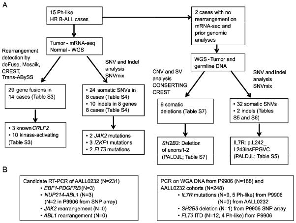
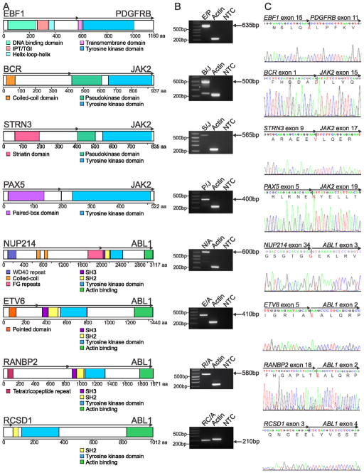
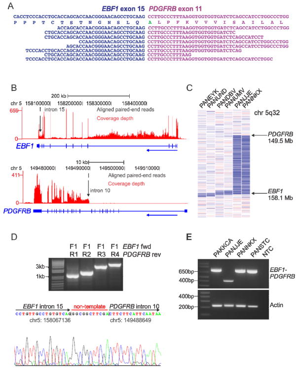
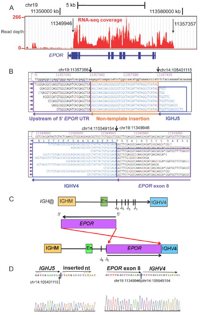
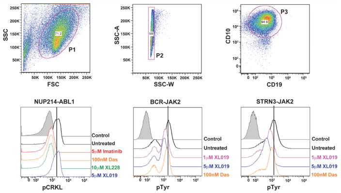
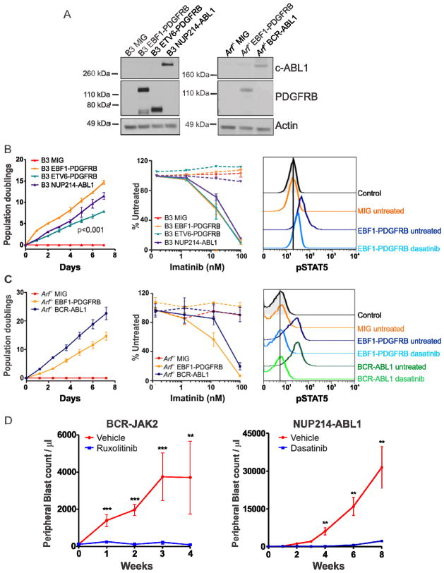
Genomic profiling has identified a subtype of high-risk B-progenitor acute lymphoblastic leukemia (B-ALL) with alteration of IKZF1, a gene expression profile similar to BCR-ABL1-positive ALL and poor outcome (Ph-like ALL). The genetic alterations that activate kinase signaling in Ph-like ALL are poorly understood. We performed transcriptome and whole genome sequencing on 15 cases of Ph-like ALL and identified rearrangements involving ABL1, JAK2, PDGFRB, CRLF2, and EPOR, activating mutations of IL7R and FLT3, and deletion of SH2B3, which encodes the JAK2-negative regulator LNK. Importantly, several of these alterations induce transformation that is attenuated with tyrosine kinase inhibitors, suggesting the treatment outcome of these patients may be improved with targeted therapy.


Figures

Figure 1
Figure 1
Flow chart of methodology. (A) 15 Ph-like high-risk (HR) ALL cases were subjected to mRNA-seq, with matched normal DNA subjected to whole genome sequencing (WGS). Two cases also had WGS of tumor DNA. (B) For recurrence testing of ABL1, JAK2 and PDGFRB fusions, cases with available RNA from AALL0232 were screened by RT-PCR. The two NUP214-ABL1 cases identified in P9906 showed gain of 9q34 between NUP214 and ABL1 on SNP array analysis, and the presence of NUP214-ABL1 was confirmed by RT-PCR. Whole genome amplified (WGA) leukemic DNA was used for recurrence of IL7R and SH2B3 mutations. *FLT3 mutations were reported previously (Zhang et al., 2011). See also Figure S1.
Figure 2
Figure 2
Rearrangements in Ph-like ALL. (A) Predicted domain structure of the in-frame fusions EBF1-PDGFRB (E/P), BCR-JAK2 (B/J), STRN3-JAK2 (S/J), PAX5-JAK2 (P/J), NUP214-ABL1 (N/A), ETV6-ABL1 (E/A), RANBP2-ABL1 (R/A) and RCSD1-ABL1 (RC/A) identified by mRNA-seq in eight Ph-like ALL cases. Confirmation of predicted fusions by RT-PCR (B) and Sanger sequencing (C). The two bands for BCR-JAK2 correspond to two different fusion breakpoints within JAK2 (exon 15 and 17), both of which were confirmed by Sanger sequencing. IPT/TIG, immunoglobulin-like fold, plexins, transcription factors/transcription factor immunoglobulin; FG, phenylalanine and glycine; SH, Src homology domain. See also Figure S2.
Figure 3
Figure 3
mRNA-seq data, recurrence screening and genomic mapping of the EBF1-PDGFRB fusion. (A) Split reads mapping across the EBF1-PDGFRB fusion point for case PAKKCA. Amino acid substitution from wild-type PDGFRB (Ser>Ala), is highlighted in green. (B) Coverage depth for all mRNA-seq reads at the EBF1 and PDGFRB locus in case PAKKCA, showing expression across the EBF1 locus and increased expression of PDGFRB at intron 10 (arrowed). The vertical height of the red bar indicates the number of reads covering the site. (C) SNP 6.0 microarray log2 ratio DNA copy number heatmap showing deletion (blue) between EBF1 and PDGFRB for two EBF1-PDGFRB cases (PANJJE and PANNKX), and four non-rearranged cases with focal EBF1 deletions (left). (D) Genomic mapping of the EBF1-PDGFRB rearrangement breakpoint by PCR (top) and sequencing (bottom), showing juxtaposition of EBF1 intron 15 (chr5:158067136) to PDGFRB intron 10 (chr5:149488649), with the addition of non-template nucleotides between the breakpoints. (E) RT-PCR confirmation of EBF1-PDGFRB fusion in four high-risk B-ALL cases with exon 14 (bottom band) or exon 15 (top band) of EBF1 fused to exon 11 of PDGFRB. See also Figure S3.
Figure 4
Figure 4
Schematic of the IGH@-EPOR rearrangement. (A) Plot of read depth obtained from mRNA-seq data showing increased read depth across the EPOR locus. The arrows correspond to the genomic breakpoints identified by genomic PCR and sequencing. (B) Bambino viewer of mRNA-seq reads showing IGHJ5 with 40bp of inserted sequence joined to ~1.3 kb upstream of EPOR 5′ untranslated region (UTR) on chromosome 19. Bottom view shows split reads spanning exon 8 of EPOR adjacent to IGHV4. (C) IGH@ contains an IGHM domain, enhancer (En), J domains (J1–J6) and the downstream IGHV4 gene segment. The EPOR locus is inverted so the 5′ end is adjacent to the J5 domain and the 3′ end is within the IGHV4 gene segment. (D) Sanger sequencing confirming the rearrangement at IGHJ5 (chr14:105401115) to non-template sequence (left), and exon 8 of EPOR (chr19:11349946) to IGHV4 (chr14:105549154). See also Figure S4.
Figure 5
Figure 5
IL7R activating mutations and SH2B3 deletion. (A) Protein domain structure of IL7R with location of sequence mutations identified in the transmembrane domain of nine B-ALL cases. (B) Copy number variant analysis of PALDJL comparing tumor and matched non-tumor DNA at the SH2B3 locus. (C) Transcript read depth data from mRNA-seq analysis comparing three cases with normal expression of SH2B3 to PALJDL. The vertical height of the red bar indicates the number of reads covering the site. (D) Sanger sequencing confirming the deletion from chr12:110325422 (first arrow) to chr12:110361365 (second arrow). See also Figure S5.
Figure 6
Figure 6
Phosphosignaling analysis of primary leukemic blasts harboring NUP214-ABL1, BCR-JAK2 and STRN3-JAK2 rearrangements. Cells were untreated or treated with indicated tyrosine kinase inhibitors for 1 hour and levels of phosphorylated CRKL or tyrosine were assessed by phosphoflow cytometric analysis. Viable (P1) and single cells (P2) were gated by expression of CD10 and CD19 (P3). Das, dasatinib. Control is secondary antibody alone. See also Figure S6.
Figure 7
Figure 7
Kinase-activating fusions induce growth factor-independence and show response to tyrosine kinase inhibitors. (A) Immunoblot of c-ABL1 and PDGFRB in Ba/F3 (B3) and Arf−/− pre-B cells expressing empty vector (MIG), EBF1-PDGFRB, ETV6-PDGFRB or NUP214-ABL1. Transduced Ba/F3 (B) or Arf−/− pre-B cells (C) were grown in the absence of cytokine and cell number was recorded as indicated (left). Ba/F3 or Arf−/− pre-B cells were grown in increasing concentrations of imatinib (middle). No cytokine (solid line) or cytokine (dotted line). Error bars represent mean ± SD of three independent experiments. Cells were untreated or treated with dasatinib for 1 hour and levels of phosphorylated STAT5 were assessed by phosphoflow cytometric analysis (right). (D) Xenograft model of BCR-JAK2 and NUP214-ABL1. Mice were randomized to receive vehicle (40% dimethyl acetamide, 60% propylene glycol; N=5), ruxolitinib (30 mg/kg/day; N=7) or dasatinib (20 mg/kg/day; N=5). Error bars represent mean ± SEM. **, p<0.001; ***, p<0.0001. See also Figure S7.

Comment in

References

    1. Apperley JF, Gardembas M, Melo JV, Russell-Jones R, Bain BJ, Baxter EJ, Chase A, Chessells JM, Colombat M, Dearden CE, et al. Response to imatinib mesylate in patients with chronic myeloproliferative diseases with rearrangements of the platelet-derived growth factor receptor beta. N Engl J Med. 2002;347:481–487. - PubMed
    1. Armstrong SA, Kung AL, Mabon ME, Silverman LB, Stam RW, Den Boer ML, Pieters R, Kersey JH, Sallan SE, Fletcher JA, et al. Inhibition of FLT3 in MLL. Validation of a therapeutic target identified by gene expression based classification. Cancer Cell. 2003;3:173–183. - PubMed
    1. Bernad R, van der Velde H, Fornerod M, Pickersgill H. Nup358/RanBP2 attaches to the nuclear pore complex via association with Nup88 and Nup214/CAN and plays a supporting role in CRM1-mediated nuclear protein export. Mol Cell Biol. 2004;24:2373–2384. - PMC - PubMed
    1. Bowman WP, Larsen EL, Devidas M, Linda SB, Blach L, Carroll AJ, Carroll WL, Pullen DJ, Shuster J, Willman CL, et al. Augmented therapy improves outcome for pediatric high risk acute lymphocytic leukemia: results of Children’s Oncology Group trial P9906. Pediatr Blood Cancer. 2011;57:569–577. - PMC - PubMed
    1. Carroll M, Tomasson MH, Barker GF, Golub TR, Gilliland DG. The TEL/platelet-derived growth factor beta receptor (PDGF beta R) fusion in chronic myelomonocytic leukemia is a transforming protein that self-associates and activates PDGF beta R kinase-dependent signaling pathways. Proc Natl Acad Sci U S A. 1996;93:14845–14850. - PMC - PubMed

Publication types

MeSH terms

Associated data