. 2012 Aug 14;22(2):180-93.
doi: 10.1016/j.ccr.2012.06.032.

ASXL1 mutations promote myeloid transformation through loss of PRC2-mediated gene repression

Affiliations

ASXL1 mutations promote myeloid transformation through loss of PRC2-mediated gene repression

Omar Abdel-Wahab et al. Cancer Cell. .

Abstract

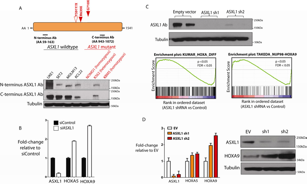
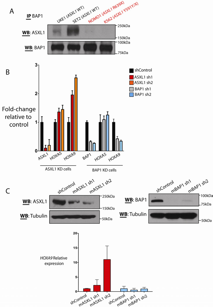
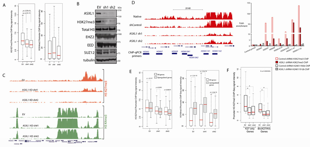
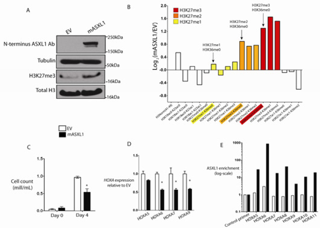
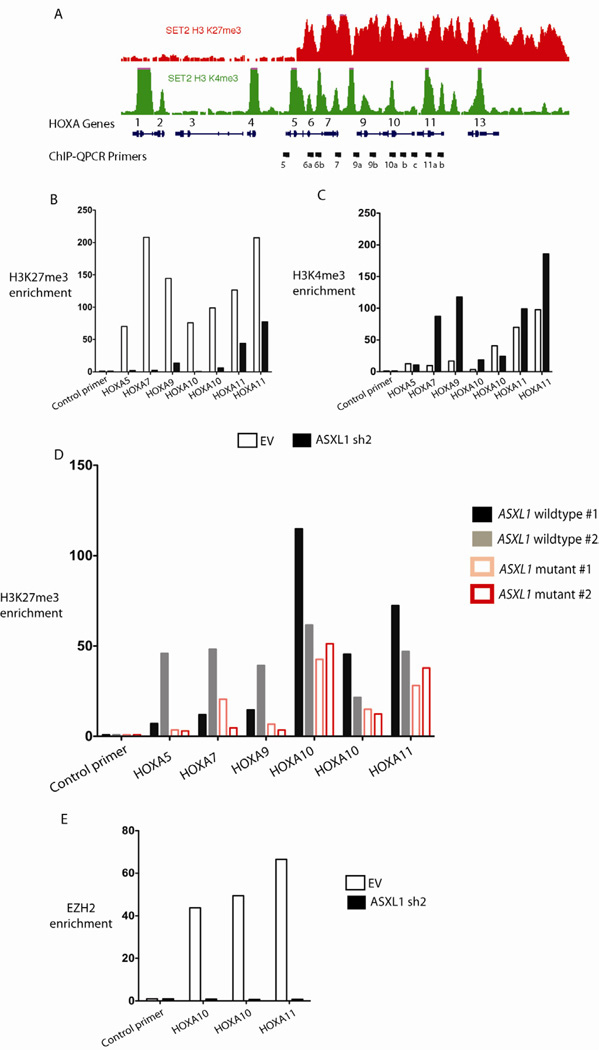
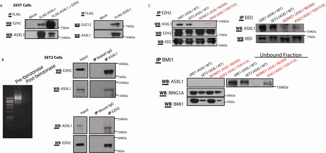
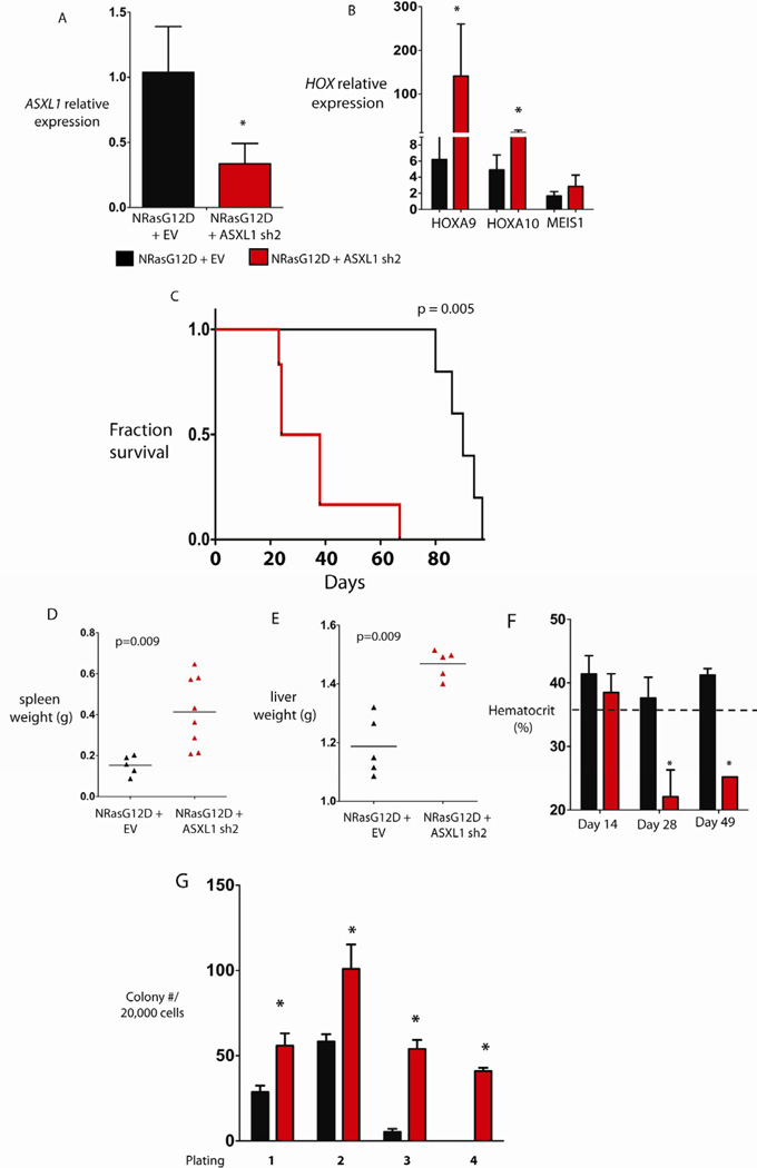
Recurrent somatic ASXL1 mutations occur in patients with myelodysplastic syndrome, myeloproliferative neoplasms, and acute myeloid leukemia, and are associated with adverse outcome. Despite the genetic and clinical data implicating ASXL1 mutations in myeloid malignancies, the mechanisms of transformation by ASXL1 mutations are not understood. Here, we identify that ASXL1 mutations result in loss of polycomb repressive complex 2 (PRC2)-mediated histone H3 lysine 27 (H3K27) tri-methylation. Through integration of microarray data with genome-wide histone modification ChIP-Seq data, we identify targets of ASXL1 repression, including the posterior HOXA cluster that is known to contribute to myeloid transformation. We demonstrate that ASXL1 associates with the PRC2, and that loss of ASXL1 in vivo collaborates with NRASG12D to promote myeloid leukemogenesis.


Figures

Figure 1
Figure 1. Leukemogeneic ASXL1 mutations are loss-of-function mutations and ASXL1 loss is associated with upregulation of HOXA gene expression
Characterization of ASXL1 expression in leukemia cells with nonsense mutations in ASXL1 reveals loss of ASXL1 expression at the protein level in cells with homozygous ASXL1 mutations as shown by Western blotting using N- and C-terminal anti-ASXL1 antibodies (A). Displayed are the ASXL1-mutant cell line lines NOMO1 (homozygous ASXL1 R639X), K562 (heterozygous ASXL1 Y591Y/X), and KBM5 (heterozygous ASXL1 G710X) and a panel of ASXL1-wildtype cell lines. ASXL1 siRNA in human primary CD34+ cells form cord blood results in upregulation of HOXA5 and HOXA9 with ASXL1 knockdown (KD) as revealed by quantitative real-time PCR (qRT-PCR analysis) (B). Stable KD of ASXL1 in ASXL1-wildype transformed human myeloid leukemia UKE1 cells (as shown by Western Blot) followed by Gene Set Enrichment Analysis reveals significant enrichment of gene sets characterized by upregulation of 5’ HOXA genes (C) as was confirmed by qRT-PCR (D). Statistical significance is indicated by the p-value and false-discovery rate (FDR). Similar upregulation of HOXA9 is seen by Western Blot following stable ASXL1 knockdown in the ASXL1-wildtype human leukemia SET2 cells (D). Error bars represent standard deviation of expression relative to control. See also Figure S1, Table S1, and Table S2.
Figure 2
Figure 2. ASXL1 and BAP1 physically interact in human hematopoietic cells but BAP1 loss does not result in increased HoxA gene expression
Immunoprecipitation of BAP1 in a panel of ASXL1-wildtype and mutant human myeloid leukemia cells reveals co-association of ASXL1 and BAP1 (A). Cells with heterozygous or homozygous mutations in ASXL1 with reduced or absent ASXL1 expression have minimal interaction with BAP1 in vitro. BAP1 knockdown in the ASXL1/BAP1 wildtype human leukemia cell line UKE1 fails to alter HOXA gene expression (B). In contrast, stable knockdown of ASXL1 in the same cell type results in a significant upregulation of HOXA9. Similar results are seen with knockdown of Asxl1 or Bap1 in murine precursor-B lymphoid Ba/F3 cells (C). Error bars represent standard deviation of expression relative to control. See also Figure S2.
Figure 3
Figure 3. ASXL1 loss is associated with loss of H3K27me3 and with increased expression of genes poised for transcription
ASXL1 loss is associated with a significant genome-wide decrease in H3K27me3 as illustrated by box plot showing the 25th, 50th, and 75th percentiles for H3K27me3 and H3K4me3 enrichment at transcription start sites in UKE1 cells treated with an empty vector or shRNAs directed against ASXL1 (A). The whiskers indicate the most extreme data point less than 1.5 interquartile range from box and red bar represents the median. Loss of ASXL1 is associated with a global loss of H3K27me3 without affecting PRC2 component expression as shown by Western blot of purified histones from cells with UKE1 knockdown and Western blot for core PRC2 component in whole cell lysates from ASXL1 knockdown UKE1 cells (B). Loss of H3K27me3 is evident at the HOXA locus as shown by ChIP-Seq promoter track signals across the HOXA locus in UKE1 cells treated with an EV or shRNA knockdown of ASXL1 (C). H3K27me3 ChIP-Seq promoter track signals from HOXA5 to HOXA13 in UKE1 cells treated with shRNA control or one of 2 anti-ASXL1 shRNAs is shown in (D) with location of primers used in ChIP-quantitative PCR (ChIP-qPCR) validation. ChIP for H3K27me3 and H2AK119Ub followed by ChIP-qPCR in cells treated with control or ASXL1 knockdown confirms a significant decrease in H3K27me3 at the HOXA locus with ASXL1 KD but minimal effects of ASXL1 KD on H2AK119Ub levels at the same primer locations (D). Integrating gene-expression data with H3K27me3/H3K4me3 ChIP-Seq identifies a significant correlation between alterations in chromatin state and increases in gene expression following ASXL1 loss at loci normally marked by the simultaneous presence of H3K27me3 and H3K4me3 in control cells (E). Loss of H3K27me3 is seen at promoters normally marked by the presence of H3K27me3 alone or at promoters co-occupied by H3K27me3 and H3K4me3 in the control state (F). See also Figure S3.
Figure 4
Figure 4. Expression of ASXL1 in ASXL1-null leukemic cells results in global increase in H3K27me2/3, growth suppression, and suppression of HoxA gene expression
ASXL1 expression in ASXL1-null NOMO1 cells is associated with a global increase in H3K27me3 as detected by Western blot of purified histones (A) as well as by quantitative liquid-chromatography/mass spectrometry of H3 peptides from amino acids 18–40 (B) (arrows indicate quantification of H3K27me1/2/3). ASXL1 overexpression results in growth suppression in 7-day growth assay performed in triplicate (C). Overexpression of ASXL1 was associated with a decrement in posterior HoxA gene expression in NOMO1 cells as shown by qRT-PCR for HOXA5, 6, 7, and 9 (D). This downregulation in HOXA gene expression was concomitant with a strong enrichment of ASXL1 at the loci of these genes as shown by chromatin immunoprecipitation of ASXL1 followed by quantitative PCR with BCRRP1 as a control locus. (E). Error bars represent standard deviation of target gene expression relative to control. See also Figure S4.
Figure 5
Figure 5. ASXL1 loss is associated with loss of PRC2 recruitment at the HOXA locus
Chromatin-immunoprecipitation (ChIP) for H3K27me3 and H3K4me3 followed by next-generation sequencing reveals the abundance and localization of H3K27me3 and H3K4me3 at the HoxA locus in SET2 cells (A). ChIP for H3K27me3 (B) and H3K4me3 (C) followed by quantitative PCR (qPCR) across the 5’ HOXA locus in SET2 cells treated with an empty vector or stable knockdown of ASXL1 reveals a consistent downregulation of H3K27me3 across the 5’ HOXA locus following ASXL1 loss and a modest increase in H3K4me3 at the promoters of 5’ HOXA genes with ASXL1 loss (primer locations are shown in A). Similar ChIP for H3K27me3 followed by qPCR across the HOXA locus in primary leukemic blasts from 2 patients with ASXL1 mutations versus 2 without ASXL1 mutations reveals H3K27me3 loss across the HOXA locus in ASXL1 mutant cells (D). ChIP for EZH2 followed by qPCR at the 5’ end of HOXA locus in SET2 cells reveals loss of EZH2 enrichment with ASXL1 loss in SET2 cells (E). CHIP-qPCR was performed in biologic duplicates and ChIP-qPCR data is displayed as enrichment relative to input. qPCR at the gene body of RRP1, a region devoid of H3K4me3 or H3K27me3, is utilized as a control locus. The error bars represent standard deviation.
Figure 6
Figure 6. ASXL1 interacts with the Polycomb Repressive Complex 2 (PRC2) in hematopoietic cells
Physical interaction between ASXL1 and EZH2 is demonstrated by transient transfection of HEK 293T cells with FLAG-hASXL1 cDNA with or without hEZH2 cDNA followed by immunoprecipitation (IP) of FLAG epitope and Western blotting for EZH2 and ASXL1 (A). HEK293T cells were transiently transfected with FLAG-hASXL1 cDNA followed by IP of FLAG epitope and Western blotting for SUZ12 and ASXL1. Endogenous interaction of ASXL1 with PRC2 members was also demonstrated by IP of endogenous EZH2 and ASXL1 followed by Western blotting of the other proteins in whole cell lysates from SET2 cells (B). Lysates from the experiment shown in Figure 6B were treated with benzonase to ensure nucleic acid free conditions in the lysates prior to IP as shown by ethidium bromide staining of an agarose gel before and after benzonase treatment. IP of endogenous EZH2 and EED in a panel of ASXL-wildtype and mutant human leukemia cells reveals a specific interaction between ASXL1 and PRC2 members in ASXL1-wildype human myeloid leukemia cells (C). In contrast, IP of the PRC1 member BMI1 failed to pulldown ASXL1. See also Figure S5.
Figure 7
Figure 7. Asxl1 silencing cooperates with NRasG12D in vivo
Retroviral bone marrow transplantation of NRasG12D with or without an shRNA for Asxl1 resulted in decreased Asxl1 mRNA expression as shown by qRT-PCR results in nucleated peripheral blood cells from transplanted mice at 14 days following transplant (A). qRT-PCR revealed an increased expression of HOXA9 and HOXA10 but not MEIS1 in the bone marrow of mice sacrificed 19 days following transplantation (B). Transplantation of bone marrow cells bearing overexpression of NRasG12D in combination with downregulation of Asxl1 led to a significant hastening of death compared to mice transplanted with NRasG12D/EV (C). Mice transplanted with NRasG12D/ASXL1 shRNA experienced increased splenomegaly (D) and hepatomegaly (E), and progressive anemia (F) compared with mice transplanted with NRasG12D + an empty vector (EV). Bone marrow cells from mice with combined NRasG12D overexpression/Asxl1 knockdown revealed increased serial replating compared with cells from NRasG12D/EV mice (G). Error bars represent standard deviation relative to control. Asterisk indicates p<0.05 (two-tailed, Mann Whitney U test). See also Figure S6.

Similar articles

  • Loss of BAP1 function leads to EZH2-dependent transformation.
    LaFave LM, Béguelin W, Koche R, Teater M, Spitzer B, Chramiec A, Papalexi E, Keller MD, Hricik T, Konstantinoff K, Micol JB, Durham B, Knutson SK, Campbell JE, Blum G, Shi X, Doud EH, Krivtsov AV, Chung YR, Khodos I, de Stanchina E, Ouerfelli O, Adusumilli PS, Thomas PM, Kelleher NL, Luo M, Keilhack H, Abdel-Wahab O, Melnick A, Armstrong SA, Levine RL. LaFave LM, et al. Nat Med. 2015 Nov;21(11):1344-9. doi: 10.1038/nm.3947. Epub 2015 Oct 5. Nat Med. 2015. PMID: 26437366 Free PMC article.
  • HHEX promotes myeloid transformation in cooperation with mutant ASXL1.
    Takeda R, Asada S, Park SJ, Yokoyama A, Becker HJ, Kanai A, Visconte V, Hershberger C, Hayashi Y, Yonezawa T, Tamura M, Fukushima T, Tanaka Y, Fukuyama T, Matsumoto A, Yamasaki S, Nakai K, Yamazaki S, Inaba T, Shibata T, Inoue D, Honda H, Goyama S, Maciejewski JP, Kitamura T. Takeda R, et al. Blood. 2020 Oct 1;136(14):1670-1684. doi: 10.1182/blood.2019004613. Blood. 2020. PMID: 32492700
  • Aberrant histone modifications induced by mutant ASXL1 in myeloid neoplasms.
    Asada S, Kitamura T. Asada S, et al. Int J Hematol. 2019 Aug;110(2):179-186. doi: 10.1007/s12185-018-2563-7. Epub 2018 Dec 5. Int J Hematol. 2019. PMID: 30515738 Review.
  • Tumor-derived neomorphic mutations in ASXL1 impairs the BAP1-ASXL1-FOXK1/K2 transcription network.
    Xia YK, Zeng YR, Zhang ML, Liu P, Liu F, Zhang H, He CX, Sun YP, Zhang JY, Zhang C, Song L, Ding C, Tang YJ, Yang Z, Yang C, Wang P, Guan KL, Xiong Y, Ye D. Xia YK, et al. Protein Cell. 2021 Jul;12(7):557-577. doi: 10.1007/s13238-020-00754-2. Epub 2020 Jul 18. Protein Cell. 2021. PMID: 32683582 Free PMC article.
  • Deregulated Polycomb functions in myeloproliferative neoplasms.
    Sashida G, Oshima M, Iwama A. Sashida G, et al. Int J Hematol. 2019 Aug;110(2):170-178. doi: 10.1007/s12185-019-02600-6. Epub 2019 Jan 31. Int J Hematol. 2019. PMID: 30706327 Review.

Cited by

References

    1. Abdel-Wahab O, Pardanani A, Patel J, Wadleigh M, Lasho T, Heguy A, Beran M, Gilliland DG, Levine RL, Tefferi A. Concomitant analysis of EZH2 and ASXL1 mutations in myelofibrosis, chronic myelomonocytic leukemia and blastphase myeloproliferative neoplasms. Leukemia. 2011;25:1200–1202. - PMC - PubMed
    1. Aravind L, Iyer LM. The HARE-HTH and associated domains: Novel modules in the coordination of epigenetic DNA and protein modifications. Cell Cycle. 2012;11 - PMC - PubMed
    1. Bejar R, Stevenson K, Abdel-Wahab O, Galili N, Nilsson B, Garcia-Manero G, Kantarjian H, Raza A, Levine RL, Neuberg D, Ebert BL. Clinical effect of point mutations in myelodysplastic syndromes. N Engl J Med. 2011;364:2496–2506. - PMC - PubMed
    1. Bernstein BE, Mikkelsen TS, Xie X, Kamal M, Huebert DJ, Cuff J, Fry B, Meissner A, Wernig M, Plath K, et al. A bivalent chromatin structure marks key developmental genes in embryonic stem cells. Cell. 2006;125:315–326. - PubMed
    1. Bott M, Brevet M, Taylor BS, Shimizu S, Ito T, Wang L, Creaney J, Lake RA, Zakowski MF, Reva B, et al. The nuclear deubiquitinase BAP1 is commonly inactivated by somatic mutations and 3p21.1 losses in malignant pleural mesothelioma. Nature genetics. 2011;43:668–672. - PMC - PubMed

Publication types

MeSH terms

Associated data