. 2012 Aug 16:13:46.
doi: 10.1186/1471-2172-13-46.

Regulation of in vitro human T cell development through interleukin-7 deprivation and anti-CD3 stimulation

Affiliations

Regulation of in vitro human T cell development through interleukin-7 deprivation and anti-CD3 stimulation

Ekta S Patel et al. BMC Immunol. .

Abstract

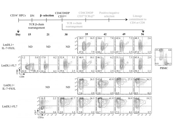
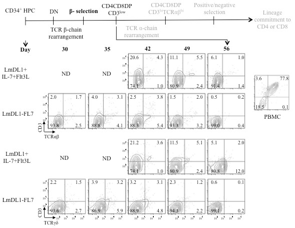
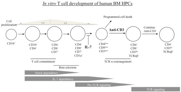
Background: The role of IL-7 and pre-TCR signaling during T cell development has been well characterized in murine but not in human system. We and others have reported that human BM hematopoietic progenitor cells (HPCs) display poor proliferation, inefficient double negative (DN) to double positive (DP) transition and no functional maturation in the in vitro OP9-Delta-like 1 (DL1) culture system.

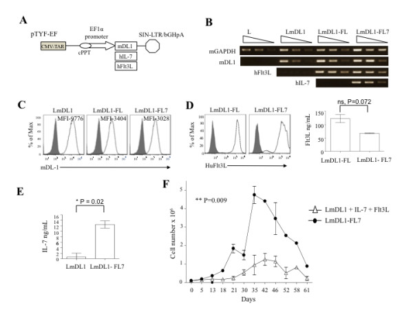
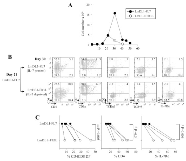
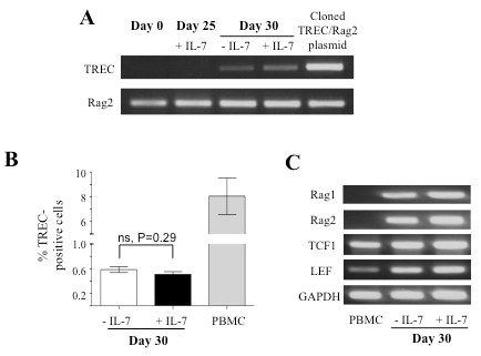
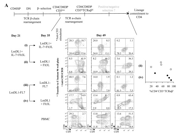
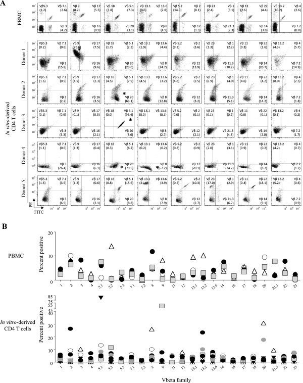
Results: In this study, we investigated the importance of optimal IL-7 and pre-TCR signaling during adult human T cell development. Using a modified OP9-DL1 culture ectopically expressing IL-7 and Fms-like tyrosine kinase 3 ligand (Flt3L), we demonstrated enhanced T cell precursor expansion. IL-7 removal at various time points during T cell development promoted a slight increase of DP cells; however, these cells did not differentiate further and underwent cell death. As pre-TCR signaling rescues DN cells from programmed cell death, we treated the culture with anti-CD3 antibody. Upon pre-TCR stimulation, the IL-7 deprived T precursors differentiated into CD3+TCRαβ+DP cells and further matured into functional CD4 T cells, albeit displayed a skewed TCR Vβ repertoire.

Conclusions: Our study establishes for the first time a critical control for differentiation and maturation of adult human T cells from HPCs by concomitant regulation of IL-7 and pre-TCR signaling.


Figures

Figure 1
Figure 1
Enhanced proliferation of T cell precursors on LmDL1-FL7.A, Lentivector constructs expressing mouse DL1, human IL-7 and human Flt3L. B, qRT-PCR analysis for DL-1, Flt3L, and IL-7. C &D, Flow cytometry analysis of mDL-1 (C) and Flt3L (D) surface expression in the various OP9 cell lines. ELISA analyses for Flt3L (D) and IL-7 (E) production. F, Growth kinetics of developing T cells from adult human BM-derived CD34+ HPCs on LmDL + IL-7 + Flt3L versus LmDL1-FL7 with p value indicated.
Figure 2
Figure 2
Flow cytometry analyses of expression kinetics of CD8, CD4, CD7, and CD1a of the developing T cells under LmDL1 + IL-7 + Flt3L or LmDL1-FL7 co-culture condition. Schematic illustration of key stages of T cell development is shown at top. Adult human BM CD34+ HPCs were plated on LmDL1-FL7 or LmDL1 + IL-7 + Flt3L and surface expression of CD8, CD4, CD7 and CD1a was examined over time as depicted. The percentages of stained cells are indicated in the flow graph quadrons. ND, not done, due to insufficient amount of developing T cells from the early time points of the LmDL1 + IL-7 + Flt3L coculture.
Figure 3
Figure 3
Flow cytometry analyses of expression kinetics of CD3, TCRαβ and TCRγδ of the developing T cells under LmDL1 + IL-7 + Flt3L or LmDL1-FL7 co-culture condition. Schematic illustration of key stages of T cell development is shown at top. T cell markers CD3, TCRαβ, and TCRγδ were analyzed for the developing T cells under the two different co-culture conditions up to 56 days. ND, not done, due to insufficient amount of developing T cells from the early time points of the LmDL1 + IL-7 + Flt3L coculture.
Figure 4
Figure 4
The effect of IL-7 withdrawal on T cell differentiation. Adult BM CD34+ HPCs were co-cultured on LmDL1-FL7 for 21 days and continued on LmDL-FL7 (IL-7 present) or transferred to LmDL-Flt3L (IL-7 deprived) for additional nine days. A, Growth curves for the developing T cells in the presence or absence of IL-7 after day 21. The cell growth declined markedly upon IL-7 removal at day 21 (depicted by open circles, data represents 4 independent experiments). B, Analysis of T cell markers in the T cell development cultures with or without IL-7 withdrawal after day 21. C, Summary of flow cytometry analysis of surface marker CD4CD8DP, CD4, and IL-7Ra in the developing T cells with (LmDL1-FL7) or without IL-7 (LmDL1-Flt3L), and p values were determined as shown.
Figure 5
Figure 5
TCR rearrangement in developing T cell precursors in LmDL1-FL7 co-culture.A, TREC analysis in developing T cells. PCR analysis for TREC and Rag2 was performed using genomic DNA harvested at the indicated time points from co-cultures under different IL-7 conditions. B, Quantitative PCR analysis of TREC-positive cells in developing T cells. SYBR Green quantitative PCR analysis of TREC was performed using genomic DNA isolated from T cell precursors under indicated culture conditions. C, Analyses of Rag1, Rag2, TCF1 and LEF RNA expression. The expression of the indicated genes was detected by RT-PCR using 1ug of mRNA harvested from day 30 of the developing T cell precursors.
Figure 6
Figure 6
Increased proliferation and TCR rearrangement of developing T cells upon concomitant IL-7 removal and anti-CD3/CD28 co-stimulation. Adult human HPCs were co-cultured on LmDL1-FL7 for 21 days and continued on LmDL-FL7 (IL-7 present) or transferred to LmDL-Flt3 (IL-7 deprived) for an additional nine days. A, Diminished precursor T cell growth upon continued presence of IL-7. Day 21 T precursor cells under IL-7 present or deprived conditions were stimulated using anti-CD3/CD28 beads for an additional two weeks. The fold increases in cell number were determined and significant difference was observed between the two groups (n =5, P = 0.006). B, Intracellular staining for Ki67 of the developing T cells with or without IL-7 and stimulated by anti-CD3/CD28 Ab beads. Un-stimulated and stimulated PBMCs from healthy donors were included as controls. C, Quantitative PCR analysis of TREC positive cells in the developing T cell population after IL-7 deprivation and anti-CD3/28 co-stimulation, as compared with control PBMC from healthy donors.
Figure 7
Figure 7
In vitromaturation and functional analysis of CD4 SP T cells upon IL-7 deprivation and anti-CD3/CD28 co-stimulation in the absence of stromal cell environment.A, Analysis of T cell development kinetics upon IL-7 removal. Diagram of key events and markers for precursor T cell development is illustrated at top. Adult HPCs were co-cultured with stromal cells for 21 days and continued on with IL-7 (IL-7 present, i and iii) or transferred to culture without IL-7 (IL-7 deprived, ii and iv) for an additional 17 days. The cells were then transferred to 96 well U bottom plates and stimulated with anti-CD3/CD28 beads for an additional 14 days. Representative results of analysis of T cell surface markers two weeks after stimulation are shown with percentage of cells indicated in the flow graph quadrons. The percentages of CD4+CD3+TCRαβ+ cell population of four independent T cell development experiments are shown. B, Analysis of effector functions of the in vitro-derived CD4 T cells. PBMCs of healthy donors and the in vitro-derived CD4 T cells were stimulated using anti-CD3/CD28 beads in the presence of IL-2, IL-7 and IL-15 for two weeks. Expression of intracellular effector cytokine (IFNγ) and T helper functional markers (IL-4, IL-17) was detected after Brefeldin A treatment; unstimulated (left panel) or PMA and ionomycin stimulated (right panel) cells were analyzed by antibody staining and flow cytometry. Note that the small percentage of in vitro-derived CD8+ cells were not CD3+ or viable propagating cells.
Figure 8
Figure 8
Vβ repertoire analysis of thein vitro-derived CD4 T cells. After anti-CD3/28 stimulation, the in vitro-derived CD4 T cells were surface stained for 24 Vβ families using the IOTest® Beta Mark TCR Vβ Repertoire Kit. CD3-gated population was evaluated for the expression of Vβ families of protein. A, TCR Vβ analysis of in vitro developed CD4 T cells of five CD34+ HPC donors versus a control PBMC. B, Summary analysis of Vβ distributions of four control PBMCs versus five in vitro derived CD4 T cells.
Figure 9
Figure 9
Schematic illustration ofin vitroT cell developmental program of adult human CD34+BM HPCs. The key events, known surface markers and critical checkpoints of the in vitro developing T cells from adult human BM CD34+ HPCs under the stromal cell co-culture condition are illustrated.

Similar articles

Cited by

References

    1. Storek J, Gooley T, Witherspoon RP, Sullivan KM, Storb R. Infectious morbidity in long-term survivors of allogeneic marrow transplantation is associated with low CD4 T cell counts. Am J Hematol. 1997;54:131–138. doi: 10.1002/(SICI)1096-8652(199702)54:2<131::AID-AJH6>3.0.CO;2-Y. - DOI - PubMed
    1. Douek DC, Vescio RA, Betts MR, Brenchley JM, Hill BJ, Zhang L, Berenson JR, Collins RH, Koup RA. Assessment of thymic output in adults after haematopoietic stemcell transplantation and prediction of T-cell reconstitution. Lancet. 2000;355:1875–1881. doi: 10.1016/S0140-6736(00)02293-5. - DOI - PubMed
    1. van den Brink MRM, Alpdogan O, Boyd RL. Strategies to enhance T-cell reconstitution in immunocompromised patients. Nat Rev Immunol. 2004;4:856–867. doi: 10.1038/nri1484. - DOI - PubMed
    1. Heng TSP, Chidgey AP, Boyd RL. Getting back at nature: understanding thymic development and overcoming its atrophy. Curr Opin Pharmacol. 2010;10:425–433. doi: 10.1016/j.coph.2010.04.006. - DOI - PubMed
    1. Schmitt TM, Zuniga-Pflucker JC. T-cell development, doing it in a dish. Immunol Rev. 2006;209:95–102. doi: 10.1111/j.0105-2896.2006.00353.x. - DOI - PubMed

Publication types

MeSH terms