. 2012 Aug 16;10(1):23.
doi: 10.1186/1546-0096-10-23.

Comparison of ultrasonography with Doppler and MRI for assessment of disease activity in juvenile idiopathic arthritis: a pilot study

Affiliations

Comparison of ultrasonography with Doppler and MRI for assessment of disease activity in juvenile idiopathic arthritis: a pilot study

Louise Laurell et al. Pediatr Rheumatol Online J. .

Abstract

Background: In juvenile idiopathic arthritis (JIA), the trend towards early therapeutic intervention and the development of new highly effective treatments have increased the need for sensitive and specific imaging. Numerous studies have demonstrated the important role of MRI and US in adult rheumatology. However, investigations of imaging in JIA are rare, and no previous study has been comparing MRI with Doppler ultrasonography (US) for assessment of arthritis. The aim of the present study was to compare the two imaging methods regarding their usefulness for evaluating disease activity in JIA, and to compare the results with those obtained in healthy controls.

Methods: In 10 JIA patients (median age 14 years, range 11-18), 11 joints (six wrists, three knees, two ankles) with arthritis were assessed by color Doppler US and MRI. The same imaging modalities were used to evaluate eight joints (three wrists, three knees, two ankles) in six healthy age- and sex-matched controls. The US examinations of both the patients and controls were compared with the MRI findings.

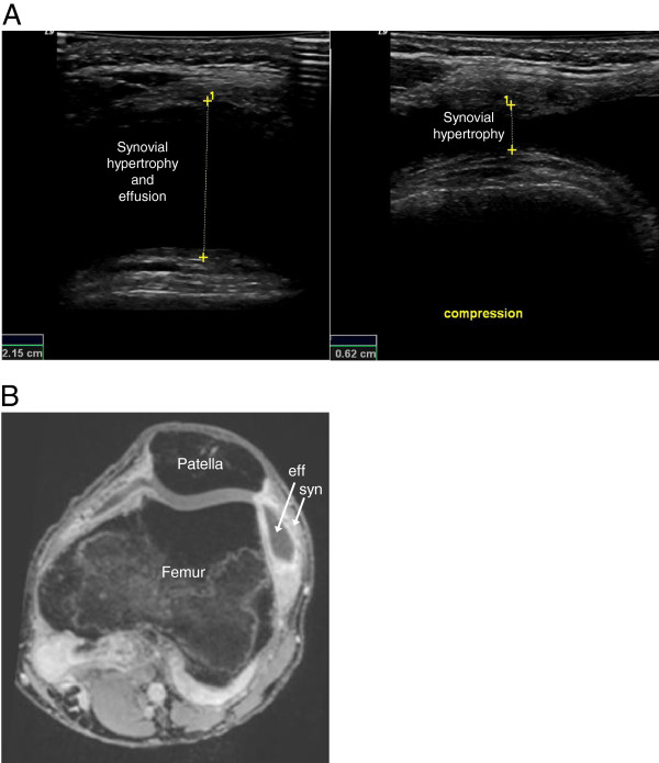
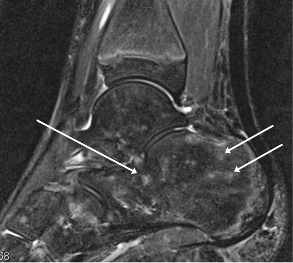
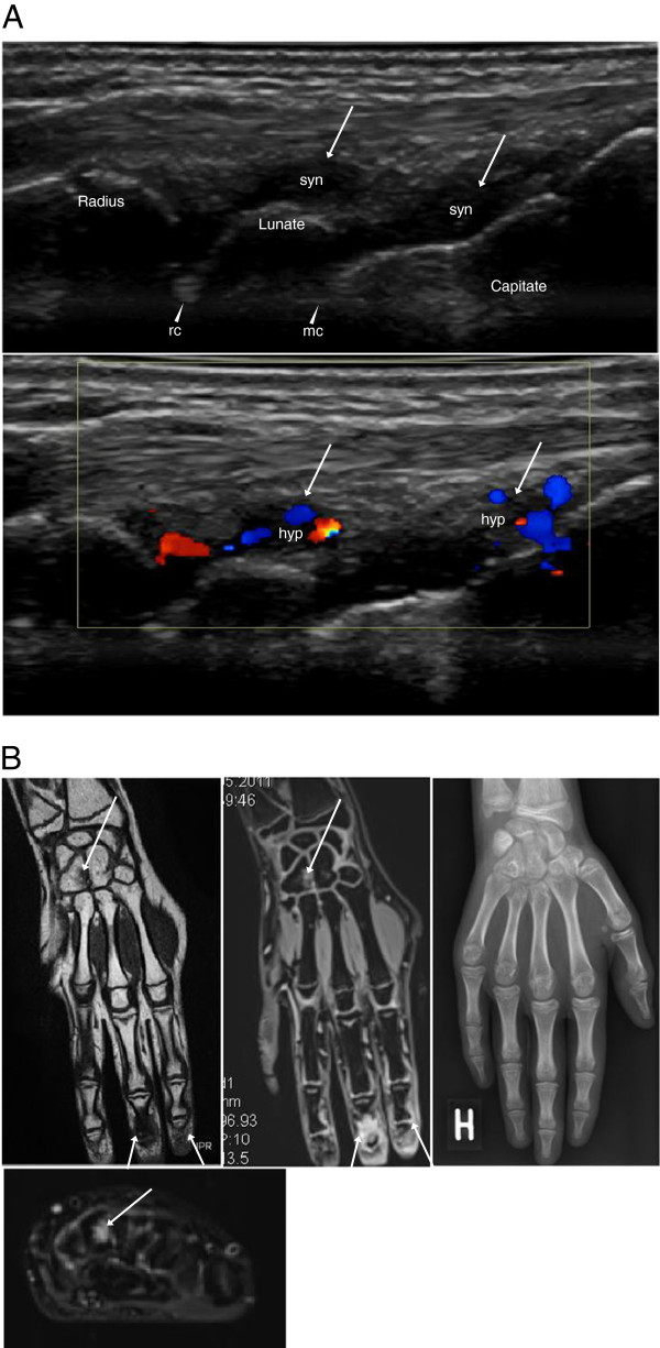
Results: In 10 JIA patients, US detected synovial hypertrophy in 22 areas of 11 joints, 86% of which had synovial hyperemia, and MRI revealed synovitis in 36 areas of the same 11 joints. Erosions were identified by US in two areas of two joints and by MRI in six areas of four joints. Effusion was shown by US in nine areas of six joints and by MRI in 17 areas of five joints. MRI detected juxta-articular bone marrow edema in 16 areas of eight joints.

Conclusions: The results of this pilot study indicate that both MRI and US provide valuable imaging information on disease activity in JIA. Importantly, the two techniques seem to complement each other and give partly different information. Although MRI is considered to be the reference standard for advanced imaging in adult rheumatology, US seems to provide useful imaging information that could make it an option in daily clinical practice, in JIA as well as in adult rheumatology. However, the current work represents a pilot study, and thus our results need to be confirmed in a larger prospective clinical investigation.


Figures

Figure 1
Figure 1
Imaging of the symptomatic ankle region of an 11-year-old girl with JIA. (A) Sagittal US scanning of the anterior talo-crural joint showing synovial hypertrophy (syn) to the left and hyperemia (hyp) on color Doppler to the right. (B) T1-weighted sagittal MRI images before (left) and after (middle) contrast injection. The left image reveals a small bony erosion at the neck of the talus (arrowhead). The erosion is surrounded by bone marrow edema (arrowhead, middle image). In the same image synovial hypertrophy with contrast enhancement (syn) and effusion (eff) are visualized in the anterior and posterior recesses of the talo-crural (tc) and posterior subtalar (pst) joints. On the STIR image to the right, high signal intensity depicts the location of synovitis (syn + eff) and the bone marrow edema surrounding the erosion (arrowhead, right image).
Figure 2
Figure 2
Imaging of the symptomatic and swollen knee of a 17-year-old boy with JIA. (A) Axial US scanning lateral to the patella. The left image shows an anechoic recess composed of synovial thickening and effusion, measured (1) before compression. In the right image, compression of the recess has transposed the effusion and enabled measurement (1) of the hypertrophic synovium. (B) A 3D T1 gradient echo VIBE MRI image of the same joint showing the enhanced hypertrophic synovial tissue (syn), and the effusion (eff), following intravenous injection of Gadolinium contrast.
Figure 3
Figure 3
Imaging of the symptomatic knee of a 14-year-old boy with JIA. Coronal STIR MRI image showing bone marrow edema (bme) in the medial femoral condyle (mfc).
Figure 4
Figure 4
Imaging of the asymptomatic ankle region of an 11-year-old healthy control. A sagittal STIR MRI revealing multiple and patchy signal changes suggestive of foci of haematopoietic red marrow, or of focal bone marrow edema (arrows).
Figure 5
Figure 5
Imaging of the symptomatic wrist region of a 13-year-old girl with JIA. (A) Dorsal sagittal US scans of the radiocarpal (arrowhead, rc) and midcarpal (arrowhead, mc) joints showing synovial hypertrophy (syn, top image), with hyperemia (hyp) on color Doppler (bottom image), in the dorsal recesses (arrows). There are no visible erosions. (B) Overview of pathology in coronal MRIs of the hand and wrist. An erosion in the Hamate (arrow) and synovitis in the 2nd and 3rd DIP joints (arrows) are seen both in the coronal T1 SE image to the left, and in the coronal postcontrast 3D T1 GRE VIBE sequence in the middle. The axial STIR image (bottom image) reveals the bone marrow edema (arrow) surrounding the erosion of the Hamate. No pathology is displayed in the concurrent, posteroanterior X-ray (right image).

Similar articles

Cited by

References

    1. Filippou G, Cantarini L, Bertoldi I, Picerno V, Frediani B, Galeazzi M. Ultrasonography vs. clinical examination in children with suspected arthritis. Does it make sense to use poliarticular ultrasonographic screening? Clin Exp Rheumatol. 2011;29:345–350. - PubMed
    1. Magni-Manzoni S, Epis O, Ravelli A, Klersy C, Veisconti C, Lanni S, Muratore V, Scire CA, Rossi S, Montecucco C. Comparison of clinical versus ultrasound-determined synovitis in juvenile idiopathic arthritis. Arthritis Rheum. 2009;61:1497–1504. doi: 10.1002/art.24823. - DOI - PubMed
    1. Haslam KE, McCann LJ, Wyatt S, Wakefield RJ. The detection of subclinical synovitis by ultrasound in oligoarticular juvenile idiopathic arthritis: a pilot study. Rheumatology (Oxford) 2010;49:123–127. doi: 10.1093/rheumatology/kep339. - DOI - PubMed
    1. Pascoli L, Wright S, McAllister C, Rooney M. Prospective evaluation of clinical and ultrasound findings in ankle disease in juvenile idiopathic arthritis: importance of ankle ultrasound. J Rheumatol. 2010;37:2409–2414. doi: 10.3899/jrheum.091262. - DOI - PubMed
    1. Guzman J, Burgos-Vargas R, Duarte-Salazar C, Gomez-Mora P. Reliability of the articular examination in children with juvenile rheumatoid arthritis: interobserver agreement and sources of disagreement. J Rheumatol. 1995;22:2331–2336. - PubMed

LinkOut - more resources