Review
. 2012 Aug;5(8):837-48.
doi: 10.1016/j.jcmg.2012.06.003.

LV mass assessed by echocardiography and CMR, cardiovascular outcomes, and medical practice

Affiliations
Review

LV mass assessed by echocardiography and CMR, cardiovascular outcomes, and medical practice

Anderson C Armstrong et al. JACC Cardiovasc Imaging. 2012 Aug.

Abstract

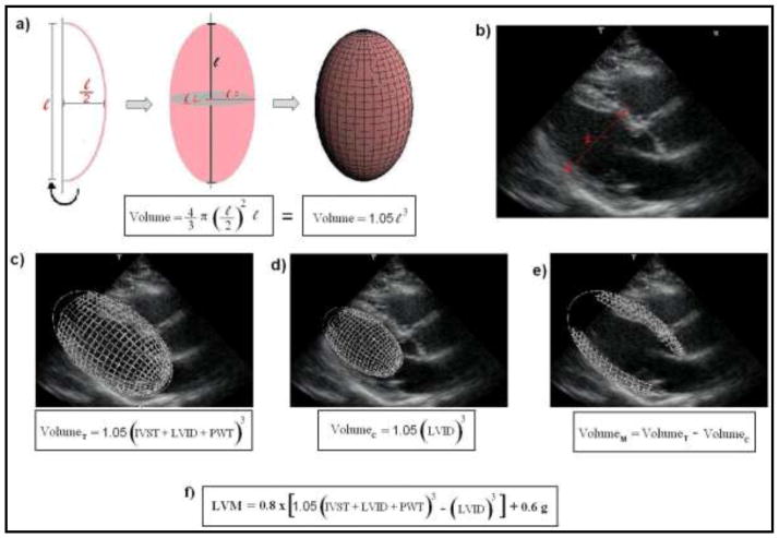
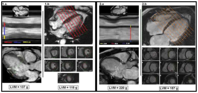
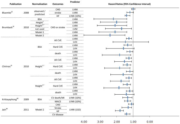
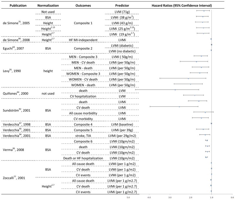
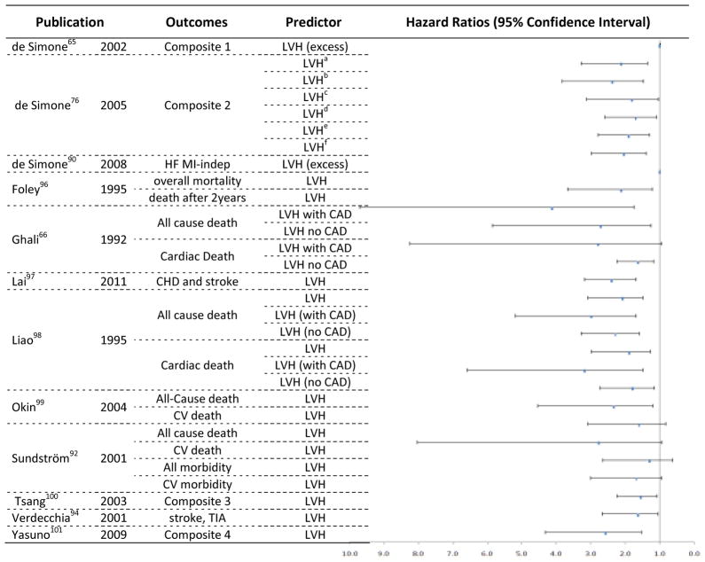
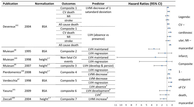
The authors investigated 3 important areas related to the clinical use of left ventricular mass (LVM): accuracy of assessments by echocardiography and cardiac magnetic resonance (CMR), the ability to predict cardiovascular outcomes, and the comparative value of different indexing methods. The recommended formula for echocardiographic estimation of LVM uses linear measurements and is based on the assumption of the left ventricle (LV) as a prolate ellipsoid of revolution. CMR permits a modeling of the LV free of cardiac geometric assumptions or acoustic window dependency, showing better accuracy and reproducibility. However, echocardiography has lower cost, easier availability, and better tolerability. From the MEDLINE database, 26 longitudinal echocardiographic studies and 5 CMR studies investigating LVM or LV hypertrophy as predictors of death or major cardiovascular outcomes were identified. LVM and LV hypertrophy were reliable cardiovascular risk predictors using both modalities. However, no study directly compared the methods for the ability to predict events, agreement in hypertrophy classification, or performance in cardiovascular risk reclassification. Indexing LVM to body surface area was the earliest normalization process used, but it seems to underestimate the prevalence of hypertrophy in obese and overweight subjects. Dividing LVM by height to the allometric power of 1.7 or 2.7 is the most promising normalization method in terms of practicality and usefulness from a clinical and scientific standpoint for scaling myocardial mass to body size. The measurement of LVM, calculation of LVM index, and classification for LV hypertrophy should be standardized by scientific societies across measurement techniques and adopted by clinicians in risk stratification and therapeutic decision making.


Conflict of interest statement

Potential Conflict of Interest

The authors have no competing interests in this study.

Figures

Figure 1
Figure 1. Principles for the assessment of LVM by echocardiography, as recommended by the American Society of Cardiology (ASE). Images from ‘c)’ to ‘e)’ refer to a schematic representation of the steps for the estimation of left ventricular myocardial volume, as initially proposed by Devereux et al. LVM is then calculated multiplying the myocardial volume by the specific gravity of myocardium (1.05 g)
a) A prolate ellipsoid of revolution, or prolate spheroid, is a three-dimensional figure formed by revolving an ellipse about its major axes. The ASE-recommended formula assumes that the left ventricle has a prolate ellipsoid of revolution shape, with minor radii that are half the major radius. b) Schematic representation of the linear measurements for the assessment of LVM by echocardiography (para-esternal view), according to the ASE recommendations. 1-interventricular septum thickness (IVST); 2- Left ventricular internal dimension (LVID); 3- posterior wall thickness (PWT). c) Calculation for the total left ventricular volume (VolumeT). d) Calculation for the left ventricular internal cavity volume (VolumeC). e) Calculation for the left ventricular myocardial volume (VolumeM). f) The current ASE-recommended formula for the assessment of LVM. It is based in the initial concepts, but includes correction factors derived from regression analysis.,
Figure 2
Figure 2. Images from cardiac magnetic resonance (CMR) of two patients with Chagas cardiomyopathy. Case 1 has preserved cardiac geometry, but case 2 shows left ventricular remodeling. The usual assessment of LVM (LVM) by CMR does not require cardiac geometry assumptions, in opposition to linear measurements used in echocardiography. (Courtesy of Dr. Gustavo Volpe)
For 1.a and 2.a - CMR-derived images representing usual echocardiography views for linear measurements assessing LVM. ASW corresponds to interventricular septal thickness; EDD corresponds to left ventricular internal dimension; and PLW corresponds to posterior wall thickness. At the bottom, the ASE-recommended formula was used to calculate LVM (see figure 1 for full description). For 1.b and 2.b - Usual CMR assessment for LVM, using contiguous short-axis slices covering the entire left ventricle from the atrio-ventricular ring to the apex (1 to 9). The estimated LVM is displayed at the bottom.
Figure 3
Figure 3
Hazards Ratios and 95% Confidence Interval for longitudinal studies using CMR to estimate LVM as predictor of clinical outcomes. Model 1 - LV mass divided by 42.5 x (height in meters)1.88 for women, and LV mass divided by 51.4 x (height in meters)1.88 for men; Model 2 - 6.82 x (height in meters)0.561 x (weight in kilograms)0.608 for women, and 8.17 x (height in meters)0.561 x (weight in kilograms)0.608 for men; MACE: cardiac death or nonfatal myocardial infarction or hospitalization due to heart failure or unstable angina, or life-threatening ventricular arrhythmia; CHD – coronary artery disease; LVMi – LVM index; HF – heart failure; BSA – body surface area; CVE – cardiovascular events; LVH – LVH; CV – cardiovascular; MI – myocardial infarct; HF – heart failure.
Figure 4
Figure 4. Hazards Ratios and 95% Confidence Interval for longitudinal studies using Echocardiography to estimate LVM or LVMi as predictor of clinical outcomes
composite 1 - fatal and nonfatal cardiovascular events, including sudden death and other cardiovascular deaths, myocardial infarction, stroke, heart failure requiring hospitalization, renal failure requiring dialysis, documented angina, transient ischemic attack, or peripheral occlusive arterial disease verified by angiography; Composite 2 - CV death, Ischemic heart disease, CHF, End-stage renal disease, peripheral arterial disease, and stroke; Composite 2 - coronary heart disease, congestive heart failure, stroke or transient ischemic attack and intermittent claudication; Composite 4 - new-onset coronary artery disease, stroke, transient cerebral ischemic attack, symptomatic aortoiliac occlusive disease verified at angiography, thrombotic occlusion of a retinal artery documented at fluoroangiography, progressive heart failure requiring hospitalization, and renal failure requiring dialysis; composite 5 - fatal and nonfatal MI, sudden cardiac death, fatal and nonfatal stroke, other cardiovascular deaths, all-cause death, severe heart failure requiring hospitalization and severe renal failure requiring dialysis; TIA - Transient ischemic attack; Composite 6 - end point of death from cardiovascular causes, reinfarction, heart failure, stroke, or resuscitation after cardiac arrest
Figure 5
Figure 5
Hazards Ratios and 95% Confidence Interval for longitudinal studies using Echocardiography to estimate LVH as predictor of clinical outcomes. Composite 1 - fatal or nonfatal myocardial infarction, sudden death, fatal or nonfatal stroke, other cardiovascular deaths, severe heart failure requiring hospitalization, severe renal failure requiring dialysis, documented angina, transient ischemic attack, and peripheral occlusive arterial disease verified by angiography; HF – heart failure; MI-indep – myocardial infarct independent; CAD – coronary artery disease; CHD - coronary heart disease; Composite 2: Fatal/nonfatal coronary heart disease, stroke, congestive heart failure, other cardiovascular disease. Composite 3 - myocardial infarction (MI), coronary revascularization, atrial fibrillation (AF), congestive heart failure (CHF), transient ischemic attack (TIA), stroke, or cardiovascular death; TIA - Transient ischemic attack; Composite 4 - sudden death, which is unexpected death that occurs within 24 h without external causes; cerebrovascular events including stroke or transient ischemic attack; cardiac events including heart failure, angina pectoris, or acute myocardial infarction; renal events including sCr more than or equal to 4.0 mg/dl, doubling of the sCr (however, sCr 2.0 mg/dl is not regarded as an event), or end-stage renal disease; and vascular events including dissecting aortic aneurysm or arteriosclerotic occlusion of a peripheral artery. LVHa – hypertrophy defined by non indexed LVM; LVHb– hypertrophy defined by LVM indexed by BSA; LVHc– hypertrophy defined by LVM indexed by BSA1.5; LVHd – hypertrophy defined by LVM indexed by height; LVHe– hypertrophy defined by LVM indexed by height2.7; LVHf– hypertrophy defined by LVM indexed by height2.13. LVHa to LVHf refer to the whole population included in the study.
Figure 6
Figure 6
Hazards Ratios and 95% Confidence Interval for longitudinal studies using Echocardiography to estimate LVM and LVH status serial changes as predictor of clinical outcomes. infarction, and stroke; composite 2 - stroke, MI, angina, HF; composite 3 - sudden death, fatal and nonfatal stroke, transient cerebral ischemic attack, fatal and nonfatal myocardial infarction, new-onset angina requiring hospitalization, progressive heart failure requiring hospitalization, coronary artery bypass or angioplasty, carotid endarterectomy, and renal failure requiring dialysis; composite 4 - fatal and non-fatal myocardial infarction, coronary revascularization, heart failure requiring hospitalization, and fatal and non-fatal stroke; composite 5 - new-onset coronary artery disease, stroke, transient cerebral ischemic attack, symptomatic aortoiliac occlusive disease verified at angiography, thrombotic occlusion of a retinal artery documented at fluoroangiography, progressive heart failure requiring hospitalization, and renal failure requiring dialysis; composite 6 - of sudden death, cerebrovascular events, heart failure, angina pectoris, acute myocardial infarction, renal events or end-stage renal disease, and vascular events; composite 7 - electrocardiographic-documented anginal episodes and myocardial infarction, heart failure, electrocardiographic-documented arrhythmia, transient ischemic attacks, stroke, and other thrombotic events) and death after the second echocardiogram. decrease of 6.6g/m2.7; ‡ reference: persistence of LVH after 1 Year; ¥ after 1 year; § 1 g/m2.7/month increase in an average of 18 months after baseline.

References

    1. Gidding SS. Controversies in the assessment of left ventricular mass. Hypertension. 2010;56:26–8. - PubMed
    1. Lorber R, Gidding SS, Daviglus ML, Colangelo LA, Liu K, Gardin JM. Influence of systolic blood pressure and body mass index on left ventricular structure in healthy African-American and white young adults: the CARDIA study. J Am Coll Cardiol. 2003;41:955–60. - PubMed
    1. Truong QA, Toepker M, Mahabadi AA, et al. Relation of left ventricular mass and concentric remodeling to extent of coronary artery disease by computed tomography in patients without left ventricular hypertrophy: ROMICAT study. J Hypertens. 2009;27:2472–82. - PMC - PubMed
    1. Dannenberg AL, Levy D, Garrison RJ. Impact of age on echocardiographic left ventricular mass in a healthy population (the Framingham Study) Am J Cardiol. 1989;64:1066–8. - PubMed
    1. Gardin JM, Siscovick D, Anton-Culver H, et al. Sex, age, and disease affect echocardiographic left ventricular mass and systolic function in the free-living elderly. The Cardiovascular Health Study. Circulation. 1995;91:1739–48. - PubMed

Publication types

MeSH terms