

Diffusion-weighted imaging in uterine artery embolisation: do findings correlate with contrast enhancement and volume reduction?
- PMID: 22898156
- PMCID: PMC3500802
- DOI: 10.1259/bjr/91540172
Diffusion-weighted imaging in uterine artery embolisation: do findings correlate with contrast enhancement and volume reduction?
Abstract
Objectives: The objective of this study was to determine the role of diffusion-weighted imaging (DWI) in uterine artery embolisation (UAE), and to assess the apparent diffusion coefficient (ADC) of the dominant fibroid and its relationship to contrast enhancement and fibroid volume reduction.
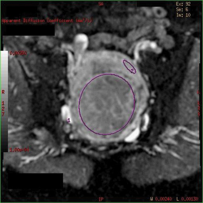
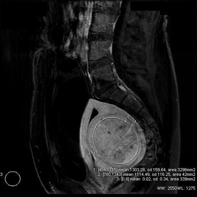
Methods: We carried out a retrospective study of 15 patients who underwent UAE. Calculations were performed at baseline and 6 months post-embolisation. Fibroid ADC (expressed in 10(-3) mm(2) s(-1)) was calculated using b=0 and b=1000 DWI values. Fibroid enhancement was compared with background myometrium by measuring signal-difference-to-noise ratio (SDNR). Fibroid volume was calculated using a prolate ellipse formula.
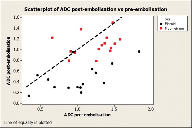
Results: There was a significant reduction (p<0.001) in fibroid ADC at 6 months (0.48; standard deviation, SD=0.26) as compared with baseline (1.01; SD=0.39). No significant change (p=0.07) was identified in 6-month myometrial ADC (1.09; SD=0.28) as compared with baseline (1.24; SD=0.20). Moderately strong and significant positive correlation was identified between baseline ADC and 6-month percentage volume reduction of the fibroid (correlation=0.66, p=0.007). No correlation was identified between SDNR and ADC at baseline or 6 months (r=0.01, p=0.97 and r=-0.13, p=0.64, respectively) or SDNR and percentage volume reduction at 6 months (correlation r=0.18, p=0.51).
Conclusion: Baseline ADC of dominant fibroids shows a moderately strong correlation with subsequent volume reduction at 6 months following UAE. No correlation was identified between ADC values and contrast enhancement on the baseline or 6-month scans. Further prospective evaluation is needed before DWI can be utilised in clinical practice. Advances in knowledge DWI imaging may provide additional information about UAE and possibly help to predict uterine volume reduction.
Figures
References
-
- Okolo S. Incidence, aetiology and epidemiology of uterine fibroids. Best Pract Res Obstet Gynaecol 2008;22:571–88 - PubMed
-
- Walker WJ, Pelage JP, Sutton C. Fibroid embolization. Clin Radiol 2002;57:325–31 - PubMed
-
- Kroncke TJ. Imaging before and after uterine artery embolization. Radiologe 2008;48:639–48 - PubMed
-
- Stejskal EO, Tanner JE. Spin diffusion measurements: spin-echo in the presence of a time dependent field gradient. J Chem Phys 1965;42:288–92
MeSH terms
LinkOut - more resources
Full Text Sources
