. 2012 Aug 16:9:200.
doi: 10.1186/1742-2094-9-200.

Neuroexcitatory effects of morphine-3-glucuronide are dependent on Toll-like receptor 4 signaling

Affiliations

Neuroexcitatory effects of morphine-3-glucuronide are dependent on Toll-like receptor 4 signaling

Michael R Due et al. J Neuroinflammation. .

Abstract

Background: Multiple adverse events are associated with the use of morphine for the treatment of chronic non-cancer pain, including opioid-induced hyperalgesia (OIH). Mechanisms of OIH are independent of opioid tolerance and may involve the morphine metabolite morphine-3-glucuronide (M3G). M3G exhibits limited affinity for opioid receptors and no analgesic effect. Previous reports suggest that M3G can act via the Toll-like receptor 4 (TLR4)/myeloid differentiation protein-2 (MD-2) heterodimer in the central nervous system to elicit pain.

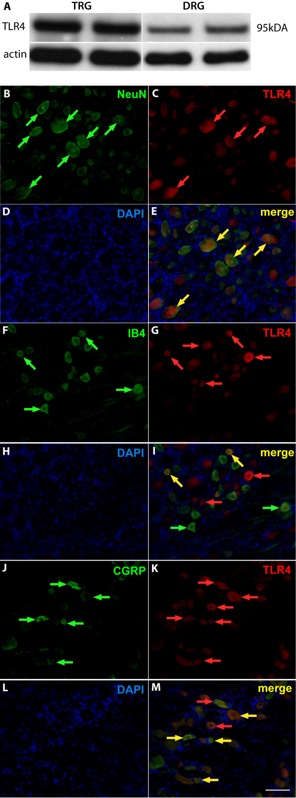
Methods: Immunoblot and immunocytochemistry methods were used to characterize the protein expression of TLR4 present in lumbar dorsal root ganglion (DRG). Using in vitro intracellular calcium and current clamp techniques, we determined whether TLR4 activation as elicited by the prototypical agonists of TLR4, lipopolysaccharide (LPS) and M3G, contributed to changes in intracellular calcium and increased excitation. Rodents were also injected with M3G to determine the degree to which M3G-induced tactile hyperalgesia could be diminished using either a small molecule inhibitor of the MD-2/TLR4 complex in rats or TLR4 knockout mice. Whole cell voltage-clamp recordings were made from small- and medium-diameter DRG neurons (25 μm < DRG diameter <45 μm) for both control and M3G-treated neurons to determine the potential influence on voltage-gated sodium channels (NaVs).

Results: We observed that TLR4 immunoreactivity was present in peptidergic and non-peptidergic sensory neurons in the DRG. Non-neuronal cells in the DRG lacked evidence of TLR4 expression. Approximately 15% of assayed small- and medium-diameter sensory neurons exhibited a change in intracellular calcium following LPS administration. Both nociceptive and non-nociceptive neurons were observed to respond, and approximately 40% of these cells were capsaicin-insensitive. Increased excitability observed in sensory neurons following LPS or M3G could be eliminated using Compound 15, a small molecule inhibitor of the TLR4/MD-2 complex. Likewise, systemic injection of M3G induced rapid tactile, but not thermal, nociceptive behavioral changes in the rat, which were prevented by pre-treating animals with Compound 15. Unlike TLR4 wild-type mice, TLR4 knockout mice did not exhibit M3G-induced hyperalgesia. As abnormal pain sensitivity is often associated with NaVs, we predicted that M3G acting via the MD-2/TLR4 complex may affect the density and gating of NaVs in sensory neurons. We show that M3G increases tetrodotoxin-sensitive and tetrodotoxin-resistant (NaV1.9) current densities.

Conclusions: These outcomes provide evidence that M3G may play a role in OIH via the TLR4/MD-2 heterodimer complex and biophysical properties of tetrodotoxin-sensitive and tetrodotoxin-resistant NaV currents.


Figures

Figure 1
Figure 1
Toll-like receptor 4 expression in sensory neurons. (A) Western blot analysis of Toll-like receptor 4 (TLR4) isolated from trigeminal root ganglion (TRG) and lumbar L3-L6 dorsal root ganglion (DRG) lysates. (B-M) Immunofluorescent images of TLR4 in peptidergic and non-peptidergic DRG populations from sectioned naïve L4 or L5 DRGs. NeuN is a neuronal marker (B, green arrowheads), which co-labeled with TLR4 (C, red arrowheads). Nuclei are stained with DAPI (D, H, L, blue). Merged images demonstrate co-labeling of NeuN containing neurons with TLR4 (E, yellow arrowheads). Calcitonin gene-related peptide (CGRP) is a marker for peptidergic-containing sensory neurons (F, green arrowheads), which co-labeled with TLR4 (G, red arrowheads). Merged images demonstrate co-labeling of CGRP-containing neurons with TLR4 (I, yellow arrowheads). Isolectin B4 (IB4) is a marker for non-peptidergic sensory neurons (J, green arrowheads), which co-labeled with TLR4 (K, red arrowheads). Merged images show co-labeling of IB4 non-peptidergic sensory neurons with TLR4 (M, yellow arrowheads). Scale bar is 60 μm (B-M).
Figure 2
Figure 2
Calcium imaging of functional Toll-like receptor 4 signaling in cultured dorsal root ganglion neurons. (A) Toll-like receptor 4 (TLR4)-IR (green) is largely restricted to neurons and not non-neuronal cell types (blue, DAPI) in dissociated dorsal root ganglion (DRG) cultures. (B) Representative recording of a transient intracellular calcium increase in a dissociated DRG sensory neuron via lipopolysaccharide (LPS) administration (1 μg/mL).
Figure 3
Figure 3
Lipopolysaccharide and morphine-3-glucuronide increase the excitability of nociceptive dorsal root ganglion neurons. Current clamp recordings were performed on small (≥30 μm) to medium (≥40 μm) dorsal root ganglion (DRG) neurons (L1-6) from naïve rats. Firing of two to four action potentials (APs) was elicited by a 1 second depolarizing current injection (ranging from 0.1 to 0.6 nA depending on the cell) every 30 seconds. (A) Representative recordings demonstrating that application of 2 μg/mL lipopolysaccharide (LPS) increases the number of elicited APs and Compound 15 can reverse this effect. (B) Group data demonstrating that LPS caused a significant increase in DRG AP firing that is reversed with Compound 15. (C) Representative recordings demonstrating that application of 3 μM morphine-3-glucuronide (M3G) increases the number of elicited APs and Compound 15 can reverse this effect. (D) Group data showing that M3G caused a significant increase in DRG AP firing that is reversed by Compound 15.
Figure 4
Figure 4
Systemic morphine-3-glucuronide administration fails to elicit tactile hyperalgesia in rats pretreated with the small molecule inhibitor of Toll-like receptor 4/myeloid differentiation protein-2 complex, Compound 15, or in Toll-like receptor 4 knockout mice. (A) Systemic morphine-3-glucuronide (M3G) (10 mg/kg, intraperitoneally) produced robust tactile hyperalgesia (n = 6, ANOVA, Bonferroni multiple comparison test, *P < 0.05), which could be prevented by pretreatment with Compound 15 (10 mg/kg, intraperitoneally, n = 6). (B) Shown is the force necessary to elicit paw withdrawal in Toll-like receptor 4 (Tlr4) wild-type and Tlr4 knockout mice. Systemic M3G (25 mg/kg, intraperitoneally) produced robust tactile hyperalgesia in Tlr4 wild-type mice (n = 6, *P < 0.05). Tlr4 knockout mice failed to display a decrease in paw withdrawal force following systemic M3G (n = 6).
Figure 5
Figure 5
Morphine-3-glucuronide-induced sensory neuron excitation is likely due to effects on voltage-gated sodium channels. (AB) Representative current traces from acutely dissociated control dorsal root ganglion (DRG) neurons evoked by 200-ms steps in 5-mV increments applied from a holding potential of −100 mV. (CE) Peak current densities (pA/pF) of DRGs exposed to extracellular recording solution (control) or 3 μM morphine-3-glucuronide (M3G) for 5 minutes. Tetrodotoxin-sensitive (TTX-S) current densities were estimated using a pre-pulse inactivation protocol (500 ms pre-pulses) with a 0 mV test pulse as well as using post hoc kinetic subtraction [18]. Tetrodotoxin-resistant (TTX-R) current densities were made in the presence of 500 nM TTX to pharmacologically isolate the properties of voltage-gated sodium channel (NaV)1.8 and NaV1.9 currents. TTX-R NaV1.8 currents were estimated from the current elicited from a 150 ms pulse to 0 mV from a holding potential of −100 mV whereas TTX-R NaV1.9 currents were estimated as the current remaining during the last 15 ms of a 150 msec test pulse to −60 mV from a holding potential of −100 mV(*P < 0.05, versus control). Error bars indicate mean ± SE from at least 10 cells per condition. Small- and medium-diameter DRG neurons were used for these experiments.

Similar articles

Cited by

References

    1. Coughtrie MW, Ask B, Rane A, Burchell B, Hume R. The enantioselective glucuronidation of morphine in rats and humans. Evidence for the involvement of more than one UDP-glucuronosyltransferase isoenzyme. Biochem Pharmacol. 1989;38:3273–3280. doi: 10.1016/0006-2952(89)90625-4. - DOI - PubMed
    1. Zelcer N, van de Wetering K, Hillebrand M, Sarton E, Kuil A, Wielinga PR, Tephly T, Dahan A, Beijnen JH, Borst P. Mice lacking multidrug resistance protein 3 show altered morphine pharmacokinetics and morphine-6-glucuronide antinociception. Proc Natl Acad Sci U S A. 2005;102:7274–7279. doi: 10.1073/pnas.0502530102. - DOI - PMC - PubMed
    1. Osborne R, Joel S, Trew D, Slevin M. Morphine and metabolite behavior after different routes of morphine administration: demonstration of the importance of the active metabolite morphine-6-glucuronide. Clin Pharmacol Ther. 1990;47:12–19. doi: 10.1038/clpt.1990.2. - DOI - PubMed
    1. Hasselstrom J, Sawe J. Morphine pharmacokinetics and metabolism in humans. Enterohepatic cycling and relative contribution of metabolites to active opioid concentrations. Clin Pharmacokinet. 1993;24:344–354. doi: 10.2165/00003088-199324040-00007. - DOI - PubMed
    1. van Dorp EL, Romberg R, Sarton E, Bovill JG, Dahan A. Morphine-6-glucuronide: morphine’s successor for postoperative pain relief? Anesth Analg. 2006;102:1789–1797. doi: 10.1213/01.ane.0000217197.96784.c3. - DOI - PubMed

Publication types

MeSH terms