. 2012 Sep 27;120(13):2704-7.
doi: 10.1182/blood-2012-05-431791. Epub 2012 Aug 16.

JAK2V617F homozygosity arises commonly and recurrently in PV and ET, but PV is characterized by expansion of a dominant homozygous subclone

Affiliations

JAK2V617F homozygosity arises commonly and recurrently in PV and ET, but PV is characterized by expansion of a dominant homozygous subclone

Anna L Godfrey et al. Blood. .

Abstract

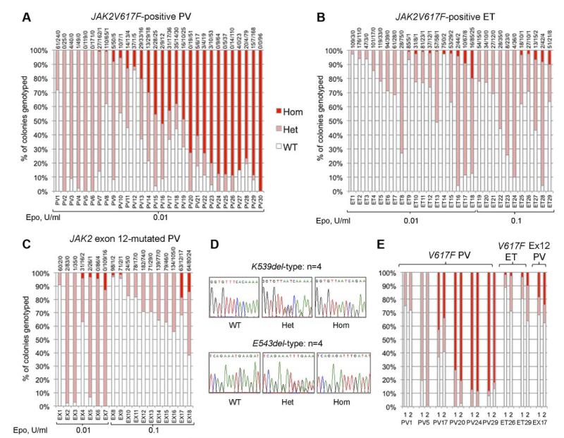
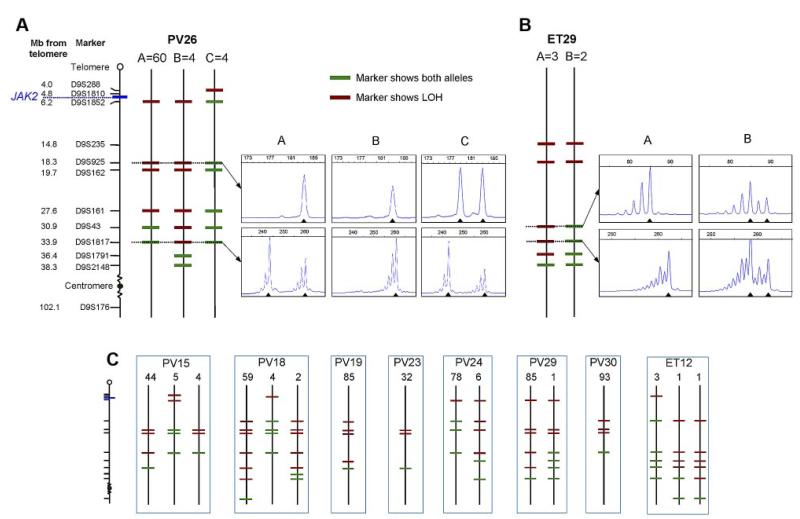
Subclones homozygous for JAK2V617F are more common in polycythemia vera (PV) than essential thrombocythemia (ET), but their prevalence and significance remain unclear. The JAK2 mutation status of 6495 BFU-E, grown in low erythropoietin conditions, was determined in 77 patients with PV or ET. Homozygous-mutant colonies were common in patients with JAK2V617F-positive PV and were surprisingly prevalent in JAK2V617F-positive ET and JAK2 exon 12-mutated PV. Using microsatellite PCR to map loss-of-heterozygosity breakpoints within individual colonies, we demonstrate that recurrent acquisition of JAK2V617F homozygosity occurs frequently in both PV and ET. PV was distinguished from ET by expansion of a dominant homozygous subclone, the selective advantage of which is likely to reflect additional genetic or epigenetic lesions. Our results suggest a model in which development of a dominant JAK2V617F-homzygous subclone drives erythrocytosis in many PV patients, with alternative mechanisms operating in those with small or undetectable homozygous-mutant clones.


Figures

Figure 1
Figure 1. Proportions of JAK2 genotypes in BFU-Es from patients with JAK2-mutated PV and ET
Each vertical bar represents 1 patient, divided according to the proportion of wild-type, heterozygous, and homozygous-mutant colonies obtained, with the absolute colony numbers shown above (WT/Het/Hom). BFU-E colonies were grown under low erythropoietin conditions as indicated. (A) Colony genotypes for 30 patients with JAK2V617F-positive PV (total 2287 colonies; mean 76 colonies per patient). (B) Colony genotypes for 29 patients with JAK2V617F-positive ET (total 2277 colonies; mean 79 per patient). (C) Colony genotypes for 18 patients with JAK2 exon 12-mutated PV (total 1931 colonies; mean 107 per patient). (D) Example sequence traces for patients with patients with homozygous JAK2 exon 12 mutations in colonies. (E) Examples of patients grown on 2 occasions show reproducibility of genotype proportions in JAK2V617F-positive PV, JAK2V617F-positive ET, and JAK2 exon 12-mutated PV (1 and 2 represent independent experiments). In total, 16 patients (5 “heterozygous-only” JAK2V617F-positive PV patients, 4 JAK2V617F-positive PV patients with homozygous and heterozygous clones, 3 JAK2V617F-positive ET patients with small homozygous clones, and 4 JAK2 exon 12-mutated PV patients with homozygous clones) were assessed in this way (mean time between experiments, 13 months; range, 2-32 months) and showed reproducibility of proportions of heterozygous and homozygous-mutant colonies.
Figure 2
Figure 2. Microsatellite mapping of 9p LOH in JAK2V617F-homozygous colonies
(A) Microsatellite mapping of 68 JAK2V617F-homozygous colonies from patient PV26 (patient codes are the same as in Figure 1). The panel of microsatellite markers on chromosome 9 is shown on the left (distances are to scale between the telomere and D9S2148), from which informative markers were selected for each patient. Three patterns of microsatellite markers were observed in colonies from this patient, indicating 3 distinct LOH breakpoints. Each subclone is represented by a vertical line denoted A (60 colonies), B (4 colonies), or C (4 colonies), with markers showing heterozygosity in green and those showing LOH in red. Example traces from fluorescence microsatellite PCR are shown for 2 markers (D9S925 and D9S1817) for the 3 subclones (right), with the position of alleles shown by arrowheads. (B) Microsatellite mapping of 5 JAK2V617F-homozygous colonies from patient ET29. Two patterns of microsatellite markers were observed; example traces are shown for 2 markers (D9S43 and D9S1817) on the right. (C) Microsatellite mapping of JAK2V617F-homozygous colonies in 8 other patients. The panel of markers is the same as in panel A. Numbers of colonies corresponding to each pattern are shown above.

References

    1. Baxter EJ, Scott LM, Campbell PJ, et al. Acquired mutation of the tyrosine kinase JAK2 in human myeloproliferative disorders. Lancet. 2005;365(9464):1054–1061. - PubMed
    1. Levine RL, Wadleigh M, Cools J, et al. Activating mutation in the tyrosine kinase JAK2 in polycythemia vera, essential thrombocythemia, and myeloid metaplasia with myelofibrosis. Cancer Cell. 2005;7(4):387–397. - PubMed
    1. James C, Ugo V, Le Couedic JP, et al. A unique clonal JAK2 mutation leading to constitutive signalling causes polycythaemia vera. Nature. 2005;434(7037):1144–1148. - PubMed
    1. Kralovics R, Passamonti F, Buser AS, et al. A gain-of-function mutation of JAK2 in myeloproliferative disorders. N Engl J Med. 2005;352(17):1779–1790. - PubMed
    1. Vannucchi AM, Antonioli E, Guglielmelli P, et al. Clinical profile of homozygous JAK2 617V>F mutation in patients with polycythemia vera or essential thrombocythemia. Blood. 2007;110(3):840–846. - PubMed

Publication types