. 2012 Nov;50(13):3004-14.
doi: 10.1016/j.neuropsychologia.2012.07.038. Epub 2012 Aug 7.

Neurophysiological evidence for a recollection impairment in amnesia patients that leaves familiarity intact

Affiliations

Neurophysiological evidence for a recollection impairment in amnesia patients that leaves familiarity intact

Richard James Addante et al. Neuropsychologia. 2012 Nov.

Abstract

In several previous behavioral studies, we have identified a group of amnestic patients that, behaviorally, appear to exhibit severe deficits in recollection with relative preservation of familiarity-based recognition. However, these studies have relied exclusively on behavioral measures, rather than direct measures of physiology. Event-related potentials (ERPs) have been used to identify putative neural correlates of familiarity- and recollection-based recognition memory, but little work has been done to determine the extent to which these ERP correlates are spared in patients with relatively specific memory disorders. ERP studies of recognition in healthy subjects have indicated that recollection and familiarity are related to a parietal old-new effect characterized as a late positive component (LPC) and an earlier mid-frontal old-new effect referred to as an 'FN400', respectively. Here, we sought to determine the extent to which the putative ERP correlates of recollection and familiarity are intact or impaired in these patients. We recorded ERPs in three amnestic patients and six age matched controls while they made item recognition and source recognition judgments. The current patients were able to discriminate between old and new items fairly well, but showed nearly chance-level performance at source recognition. Moreover, whereas control subjects exhibited ERP correlates of memory that have been linked to recollection and familiarity, the patients only exhibited the mid-frontal FN400 ERP effect related to familiarity-based recognition. The results show that recollection can be severely impaired in amnesia even when familiarity-related processing is relatively spared, and they also provide further evidence that ERPs can be used to distinguish between neural correlates of familiarity and recollection.


Figures

Figure 1
Figure 1. Coronal MRI sections from patient 03 (right) and from one of the age matched controls (left)
The hippocampus (arrows) was markedly reduced in volume in the patient, whereas other regions of the medial temporal lobe regions such as the perirhinal cortex, entorhinal cortex, and parahippocampal cortex showed no sign of atrophy(see Table 2 for quantification).
Figure 2
Figure 2. Experimental Design
Subjects made recognition memory judgments to a mixture of studied words and words that were new to the experiment. For each test item, subjects first made an item memory confidence judgment (i.e., is the item old or new to the experiment?), followed by a source memory confidence judgment (i.e., was the item encoded during the ‘animacy’ or ‘manmade’ conditions in the earlier study phase?). ERPs were averaged relative to the onset of the test word, and binned according to the item and source memory responses.
Figure 3
Figure 3. Behavioral Performance on Tests of Item Recognition and Source Memory
A) Recognition accuracy is plotted on the y-axis as the proportion of hits minus false alarms. B) Source memory accuracy is plotted on the y-axis as the percentage of source memory hits minus source memory false alarms.
Figure 4
Figure 4. ERPs of Item Recognition Confidence for Patients (N=3) and Controls (N=6)
(Top panel) FN400 effects at mid-frontal electrode (Fc1). The time window used to analyze the FN400 (400–600 ms) is highlighted in dashed blue box. (Bottom panel) Parietal effects at left parietal electrode (P3), LPC latency of 600–900 ms is shown in dashed blue box. ERP amplitudes (in microvolts) are plotted on the y-axis, and time relative to onset of the test item is plotted on the x-axis (−200 to 1500 ms). High confidence hits (“5” items) are plotted in black and lower confidence hits (“4” items) are plotted in red.
Figure 5
Figure 5. Topographic Distribution and Quantification of FN400 and LPC effects
A) Scalp topographies are plotted for the average amplitude of ERP differences between high vs. low confidence hits during the 400–600 ms (FN400) and 600–900ms (LPC) time windows for patients and controls. Note that FN400 effects are similar in magnitude and scalp topography for both patients and controls, whereas LPC effects are attenuated for patients. B) Mean amplitudes of ERP differences between high- and low-confidence item hits for patients (open bars) and controls (filled bars). At left, FN400 amplitudes are plotted for mid-frontal electrode Fc1 during the 400–600ms latency, and at right the LPC effect is plotted for left parietal electrode P3 during the 600–800ms latency. Error bars depict the standard error of the mean, ‘*’ indicates statistically significant differences, ‘ns’ indicates non-significant values.
Figure 6
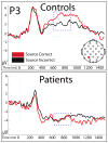
Figure 6
ERP correlates of source memory accuracy for patients and controls. ERPs are plotted for the left parietal electrode site (P3). The LPC time window (600–800ms) is indicated by the blue dashed box.
Figure 7
Figure 7
Late negative ERP shift related to source memory accuracy in patients. (Top Panel) ERPs for patients (N=3) reveal a prolonged negative shift for ERPs associated with correct source memory responses. ERPs are shown for left frontal (F7) and right parietal (P4) sites. (Bottom panel) This effect was attenuated in ERPs for Controls (N=6) at the same sites.

References

    1. Addante RJ, Ranganath C, Yonelinas AP. Examining ERP correlates of recognition memory: Evidence of accurate source recognition without recollection. Neuroimage 2012 - PMC - PubMed
    1. Aggleton JP, Brown MW. Episodic memory, amnesia, and the hippocampal-anterior thalamic axis. Behavioral Brain Science. 1999;223:425–444. discussion 444–489. - PubMed
    1. Aly M, Knight RT, Yonelinas AP. Faces are special but not too special: spared face recognition in amnesia is based on familiarity. Neuropsychologia. 2010;48(13):3941–3948. - PMC - PubMed
    1. Baddeley A, Vargha-Khadem F, Mishkin M. Preserved recognition in a case of developmental amnesia: implications for the acquisition of semantic memory? Journal of Cognitive Neuroscience. 2001;13(3):357–369. - PubMed
    1. Bell AJ, Sejnowski TJ. An information-maximization approach to blind separation and blind deconvolution. Neural Computation. 1995;7(6):1129–1159. - PubMed

Publication types