. 2012 Nov;30(9):1279-90.
doi: 10.1016/j.mri.2012.06.003. Epub 2012 Aug 13.

3-T MR-guided brachytherapy for gynecologic malignancies

Affiliations

3-T MR-guided brachytherapy for gynecologic malignancies

Tina Kapur et al. Magn Reson Imaging. 2012 Nov.

Abstract

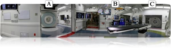
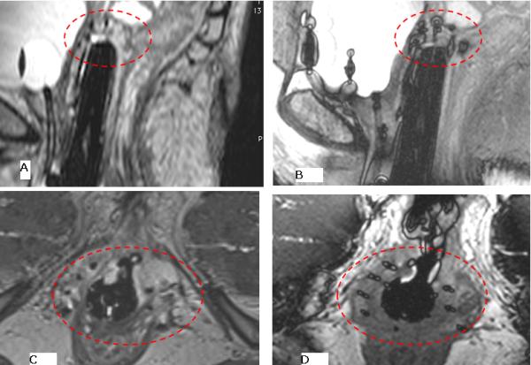
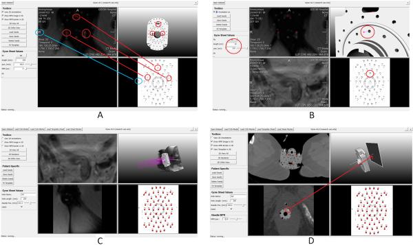
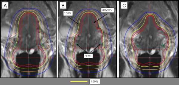
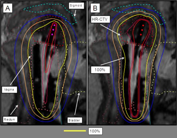
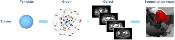
Gynecologic malignancies are a leading cause of death in women worldwide. Standard treatment for many primary and recurrent gynecologic cancer cases includes external-beam radiation followed by brachytherapy. Magnetic resonance (MR) imaging is beneficial in diagnostic evaluation, in mapping the tumor location to tailor radiation dose and in monitoring the tumor response to treatment. Initial studies of MR guidance in gynecologic brachytherapy demonstrate the ability to optimize tumor coverage and reduce radiation dose to normal tissues, resulting in improved outcomes for patients. In this article, we describe a methodology to aid applicator placement and treatment planning for 3 Tesla (3-T) MR-guided brachytherapy that was developed specifically for gynecologic cancers. This methodology has been used in 18 cases from September 2011 to May 2012 in the Advanced Multimodality Image Guided Operating (AMIGO) suite at Brigham and Women's Hospital. AMIGO comprises state-of-the-art tools for MR imaging, image analysis and treatment planning. An MR sequence using three-dimensional (3D)-balanced steady-state free precession in a 3-T MR scanner was identified as the best sequence for catheter identification with ballooning artifact at the tip. 3D treatment planning was performed using MR images. Items in development include software designed to support virtual needle trajectory planning that uses probabilistic bias correction, graph-based segmentation and image registration algorithms. The results demonstrate that 3-T MR image guidance has a role in gynecologic brachytherapy. These novel developments have the potential to improve targeted treatment to the tumor while sparing the normal tissues.


Figures

Figure 1
Figure 1
AMIGO Suite with A) MR room, B) OR, and C) PET/CT Scanner.
Figure 2
Figure 2
Lay-out of the AMIGO floor plan with the MRI room and the MRI ceiling track (left), the OR and the angio ceiling (middle) and the PET/CT Scanner room (right) corresponding to Figure 1.
Figure 3
Figure 3
Applicators used in gynecologic brachytherapy include A) Interstitial catheters with central tungsten alloy obturators placed through a central vaginal obturator (white) and through a disposable template; B) tandem and ovoids; and C) tandem and ring; B and C may be used with or without interstitial catheters.
Figure 4
Figure 4
Ballooning created at catheter tip using the 3D-bSSFP sequence. A) a sagittal image using using fat suppressed 3D-FSE 1.2 mm slice width, taken over approximately 5 minutes shows the difficulty in identifying catheter tips, whereas the B) 3D fat suppressed balanced SSFP, 1.6 mm slice width, over approximately 1.2 minutes allows rapid identification of the catheter tip. This determines the deepest point of insertion, in order to avoid bowel insertion, and covers the length of the tumor. All subsequent needles are inserted to a similar depth based on tumor location. Similar results are seen on axial images C) 3D-FSE and D) 3D balanced SSFP, where instead of the balloon configuration seen on the sagittal image, a cross centered on each catheter can be visualized.
Figure 5
Figure 5
Workflow of the treatment planning process for AMIGO procedures.
Figure 6
Figure 6
Tandem and ring (T&R) with interstitial needles. A) The preliminary plan consists of a standard loading to the T&R, resulting to a pear-shaped distribution with prescription dose (100% isodose line in yellow) to the A points. B) Interstitial needles are uniformly loaded, with dwell times amounting to 15% of the total dwell loading time. C) 3D optimization of T&R and interstitial needles results in increased HR-CTV coverage and sparing the OARs.
Figure 7
Figure 7
Interstitial treatment planning with the 100% isodose line in yellow. A) The preliminary plan consists of a uniform loading of all dwell positions. B) 3D optimization of the dwell loading results in increased coverage to the HR-CTV and sparing the OARs.
Figure 8
Figure 8
Principle for fitting a gynecological template for brachytherapy with the initial MRI image. The three red circles indicate corresponding needle holes in the template and the MRI image. The fitting is realized via a rigid transformation between these corresponding point sets. The blue circles are used to ensure that the left and right sides of the patient and the template are matched correctly (A). Virtual fitted gynecological brachytherapy template and selection of a specific interstitial needle (Ba, red circles in the screenshot). As shown on the left side of the prototype interface, individual needle insertion can be planned by defining parameters such as the needle length and depth (B). Virtual placement of several interstitial needles (purple) with different lengths and depths as shown in the settings in the menu in the left column. This allows the radiation oncologist to plan the placement of needles (C).Needle (white line in the upper right window) that has been selected for visualization of multiplanar reconstructions (MPR) along the needle path (lower left window). The MPR at the position of the arrow (tip of red arrow in the upper right image) is displayed in the lower left window as a 2D slice. In the MPR of the lower left window the needle cross section (white) is surrounded by a red circle (D).
Figure 9
Figure 9
Segmentation principle of the bladder with the Nugget-Cut approach: a spherical template (left image) is used to construct a directed graph (second image from the left). The graph is fused with the CT dataset of a patient (third image from the left) and the s-t-cut provides the segmented bladder (red, rightmost image).

References

    1. Cancer Facts & Figures 2012. [ http://www.cancer.org/Research/CancerFactsFigures/CancerFactsFigures/can...]
    1. Viswanathan AN, Thomadsen B. American Brachytherapy Society consensus guidelines for locally advanced carcinoma of the cervix. Part I: General principles. Brachytherapy. 2012;11(1):33–46. - PubMed
    1. Viswanathan AN, Kirisits C, Erickson B, Pötter R. Gynecologic Radiation Therapy: Novel Approaches to Image-Guidance and Management. Springer; Berlin; London: 2011.
    1. Viswanathan AN, Moughan J, Small W, Jr., Levenback C, Iyer R, Hymes S, Dicker AP, Miller B, Erickson B, Gaffney DK. The Quality of Cervical Cancer Brachytherapy Implantation and the Impact on Local Recurrence and Disease-Free Survival in Radiation Therapy Oncology Group Prospective Trials 0116 and 0128. Int J Gynecol Cancer. 2012;22(1):123–131. - PMC - PubMed
    1. Hricak H, Gatsonis C, Coakley FV, Snyder B, Reinhold C, Schwartz LH, Woodward PJ, Pannu HK, Amendola M, Mitchell DG. Early invasive cervical cancer: CT and MR imaging in preoperative evaluation - ACRIN/GOG comparative study of diagnostic performance and interobserver variability. Radiology. 2007;245(2):491–498. - PubMed

Publication types