. 2012 Sep;11(9):814-26.
doi: 10.1016/S1474-4422(12)70188-6.

Impaired consciousness in epilepsy

Affiliations

Impaired consciousness in epilepsy

Hal Blumenfeld. Lancet Neurol. 2012 Sep.

Abstract

Consciousness is essential to normal human life. In epileptic seizures consciousness is often transiently lost, which makes it impossible for the individual to experience or respond. These effects have huge consequences for safety, productivity, emotional health, and quality of life. To prevent impaired consciousness in epilepsy, it is necessary to understand the mechanisms that lead to brain dysfunction during seizures. Normally the consciousness system-a specialised set of cortical-subcortical structures-maintains alertness, attention, and awareness. Advances in neuroimaging, electrophysiology, and prospective behavioural testing have shed light on how epileptic seizures disrupt the consciousness system. Diverse seizure types, including absence, generalised tonic-clonic, and complex partial seizures, converge on the same set of anatomical structures through different mechanisms to disrupt consciousness. Understanding of these mechanisms could lead to improved treatment strategies to prevent impairment of consciousness and improve the quality of life of people with epilepsy.


Figures

Figure 1
Figure 1. The consciousness system
Anatomical structures known to regulate the level of consciousness. A. Medial view. B. Lateral view. Cortical components of the consciousness system (shown in blue) include the medial and lateral fronto-parietal association cortex, anterior and posterior cingulate, precuneus and retrosplenial cortex. Subcortical components (shown in red) include the basal forebrain, hypothalamus, thalamus and upper brainstem activating systems. Note that other circuits such as the basal ganglia and cerebellum may also participate in attention and other aspects of consciousness. (Reproduced from Blumenfeld H. Neuroanatomy through clinical cases. 2nd edition. Sunderland (MA): Sinauer Associates; 2010 with permission).
Figure 2
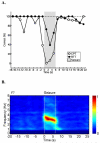
Figure 2. Absence seizures: transient behavioral and EEG changes
A. Behavioral impairment during seizures. Percent correct responses are shown over time (2s time bins) before, during and after seizures (shaded region). Performance on the more difficult continuous performance task (CPT) declined rapidly for letters presented just before seizure onset and recovered quickly after seizures end. Impaired performance on the simpler repetitive tapping task (RTT) task was more transient than on CPT, did not begin until after seizure onset, and was less severely impaired during seizures than the CPT task (F = 15.3, P = 0.017; ANOVA). Results are based on a total of 53 seizures in 8 patients. B. EEG signal power changes abruptly at beginning and end of seizures. Average time-frequency dynamics of spike-wave discharges are shown for EEG channel F7. A total of 54 seizures (9 patients) were analyzed. (Reproduced with permission from Bai X etal, 2010, Journal of Neuroscience 30:5884-5893.)
Figure 3
Figure 3. Absence seizures: early and late fMRI changes in cortical-subcortical networks
fMRI percent change increases (warm colors) and decreases (cool colors) are shown, with a display threshold of 0.5% The ictal time period of seizures was scaled to 6.6s (mean seizure duration), and the preictal, ictal, and postictal time periods temporally aligned across all seizures. Early fMRI signal increases were seen well before seizure onset (0s) in medial orbital frontal (OF), frontal polar (FP), cingulate (CG), lateral parietal (LP), precuneus (PC), and lateral occipital (LO) cortex. After seizure onset, fMRI increases progressed to also involve lateral frontal (LF) and temporal (LT) cortex. Following the end of seizures, fMRI increases were seen in the medial occipital (MO) cortex, and lastly in the thalamus (Th). fMRI signal decreases occurred later and continued well after seizure end, showing initial strong involvement of fronto-parietal association cortex. Data are from group analysis of 51 seizures in 8 patients. (Reproduced with permission from Bai X etal, 2010, Journal of Neuroscience 30:5884-5893.)
Figure 4
Figure 4. Generalized tonic-clonic seizures: network changes in cerebellum, thalamus and cortex
Positive (red) and negative (green) correlations are shown between cerebellum and other brain regions. A. Surface rendering. B. Coronal sections. Significant positive correlations with cerebellar blood flow changes were found in the upper brainstem tegmentum and thalamus. Negative correlations were found with the bilateral fronto-parietal association cortex, anterior and posterior cingulate and precuneus. Statistical parametric mapping (SPM) analysis was across patients (n=59) with extent threshold, k = 125 voxels (voxel size = 2×2×2 mm), and height threshold, p = 0.01. (Reproduced with permission from Blumenfeld H et al, 2009, Brain 132:999-1012.)
Figure 5
Figure 5. Network inhibition hypothesis for impaired consciousness in temporal lobe complex partial seizures
A. Under normal conditions, the upper brainstem-diencephalic activating systems interact with the cerebral cortex to maintain normal consciousness. B. A focal seizure involving the mesial temporal lobe. If the seizure remains confined, then a simple-partial seizure will occur without impairment of consciousness. Intracranial EEG recordings (inset) show fast polyspike activity in the temporal lobe. C. Spread of seizure activity from the temporal lobe to midline subcortical structures. Propagation often occurs to the contralateral mesial temporal as well (not shown). D. Inhibition of subcortical activating systems leads to depressed activity in bilateral fronto-parietal association cortex, and to loss of consciousness. Intracranial EEG recordings from fronto-parietal association cortex (inset) show slow wave activity resembling deep sleep. (A-D modified with permission from Blumenfeld and Taylor, 2003, The Neuroscientist, 9:301 – 310; B, D insets modified from Englot et al., 2010, Brain 133(12): 3764 – 3777).
Figure 6
Figure 6. Complex partial temporal lobe seizures
Complex partial seizures arising from the temporal lobe are associated with significant cerebral blood flow increases and decreases in widespread brain regions. Statistical parametric maps depict SPECT increases in red and decreases in green. Changes ipsilateral to seizure onset are shown on the left side of the brain, and contralateral changes on the right side of the brain (combining patients with left and right onset seizures, n=10). Data are from >90s after seizure onset, when consciousness was markedly impaired. Note that at earlier times there were SPECT increases in the ipsilateral mesial temporal lobe (not shown). A-D. Horizontal sections progressing from inferior to superior, and E, F. coronal sections progressing from anterior to posterior showing blood flow increases in the bilateral midbrain, hypothalamus, medial thalamus, and midbrain. Decreases are seen in the bilateral association cortex. G. 3-dimensional surface renderings show increases mainly in the bilateral medial diencephalon, upper brainstem and medial cerebellum, while decreases occur in the ipsilateral > contralateral frontal and parietal association cortex (same data as A-F). Extent threshold, k = 125 voxels (voxel size = 2 × 2×2 mm). Height threshold, P = 0.01. (Reproduced with permission from Blumenfeld et al, 2004, Cerebral Cortex 14:892-902.)

References

    1. Sperling MR. The consequences of uncontrolled epilepsy. CNS spectrums. 2004;9(2):98–101. - PubMed
    1. Vickrey BG, Berg AT, Sperling MR, Shinnar S, Langfitt JT, Bazil CW, et al. Relationships between seizure severity and health-related quality of life in refractory localization-related epilepsy. Epilepsia. 2000;41(6):760–4. - PubMed
    1. Laureys S, Schiff ND. Disorders of Consciousness. Annals of the New York Academy of Sciences, Wiley-Blackwell; 2009. - PubMed
    1. Laureys S, Tononi G. The Neurology of Consciousness: Cognitive Neuroscience and Neuropathology. Academic Press; 2008.
    1. Nagel T. What is it Like to be a Bat? Philosophical Review. 1974;82:435–56.

Publication types