. 2012 Oct 19;287(43):36147-57.
doi: 10.1074/jbc.M112.389676. Epub 2012 Aug 16.

Cryptococcus neoformans activates RhoGTPase proteins followed by protein kinase C, focal adhesion kinase, and ezrin to promote traversal across the blood-brain barrier

Affiliations

Cryptococcus neoformans activates RhoGTPase proteins followed by protein kinase C, focal adhesion kinase, and ezrin to promote traversal across the blood-brain barrier

Jong-Chul Kim et al. J Biol Chem. .

Abstract

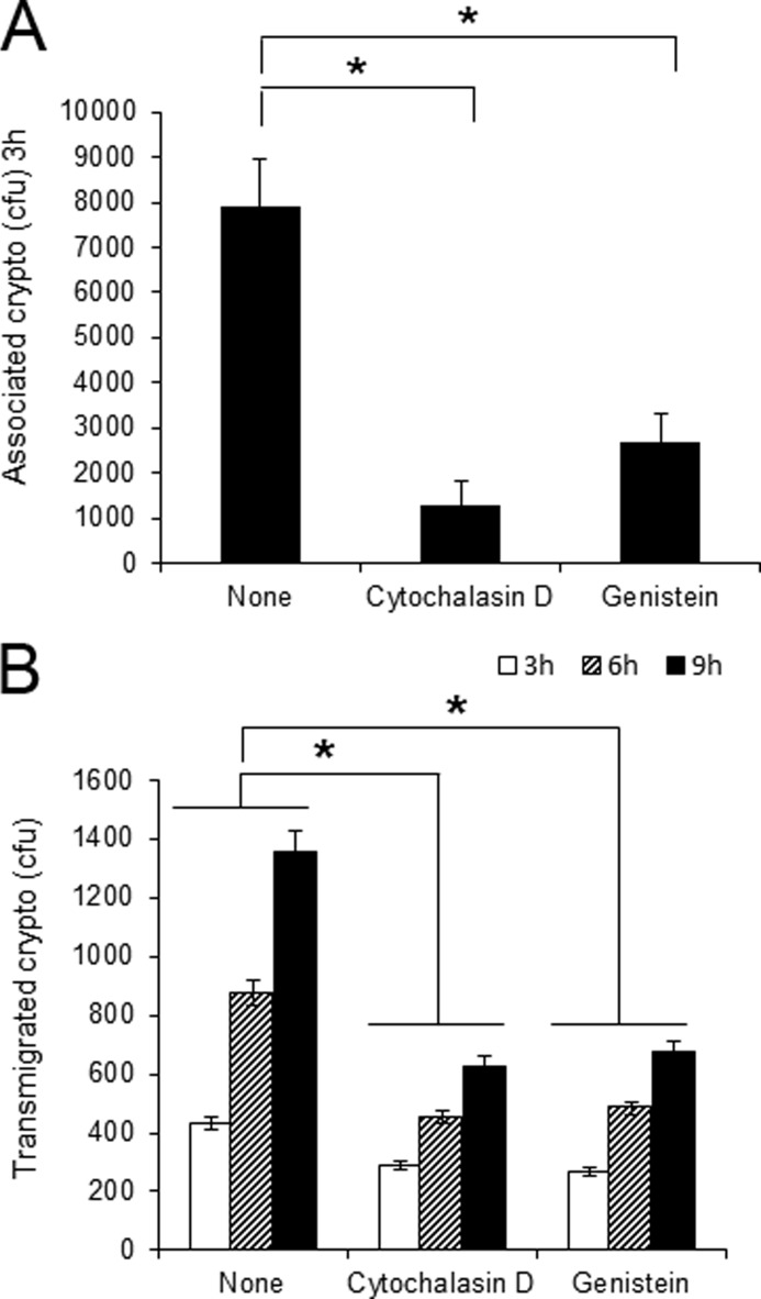
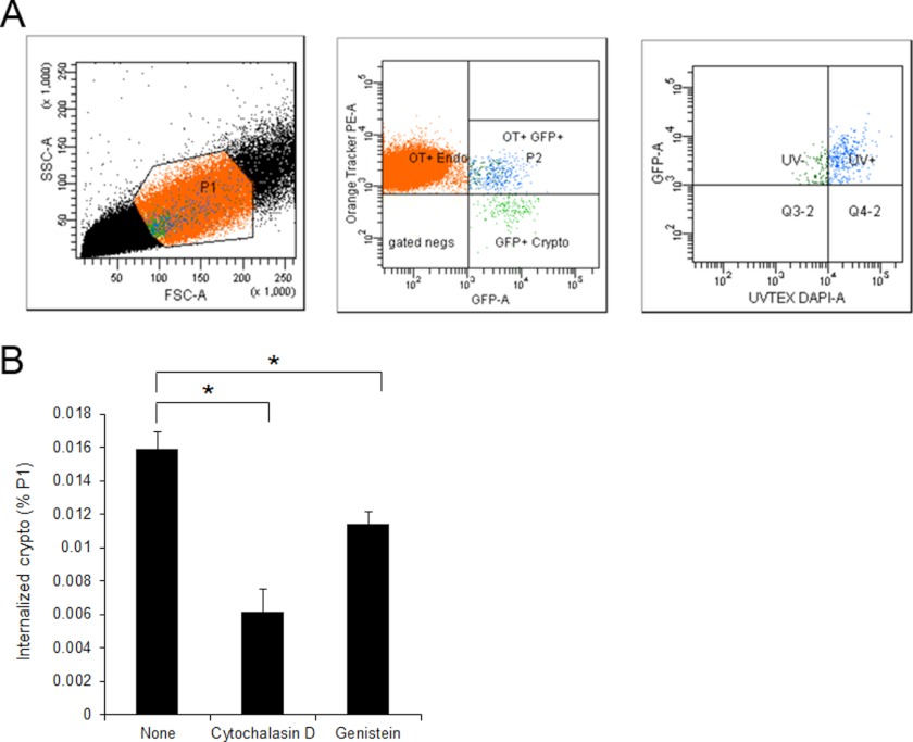
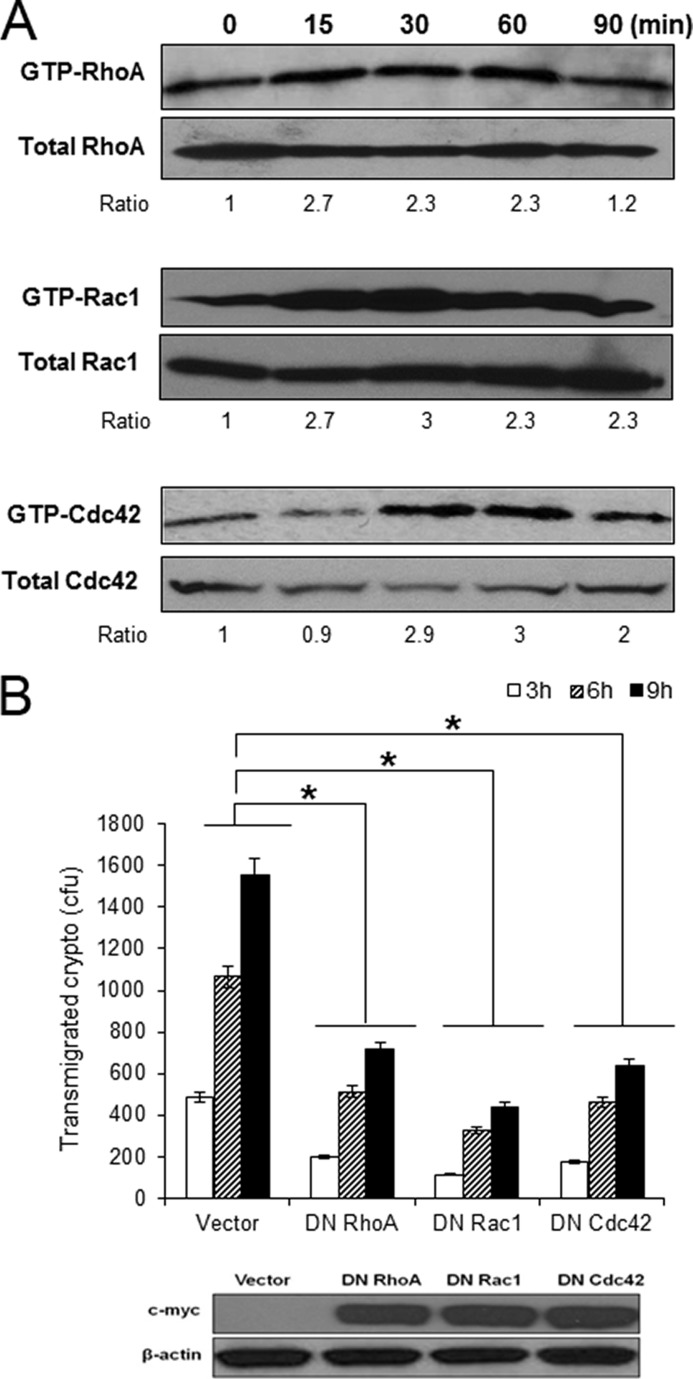
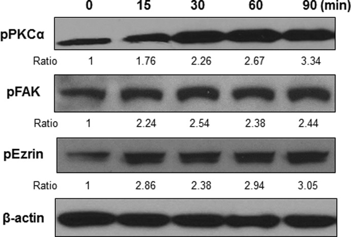
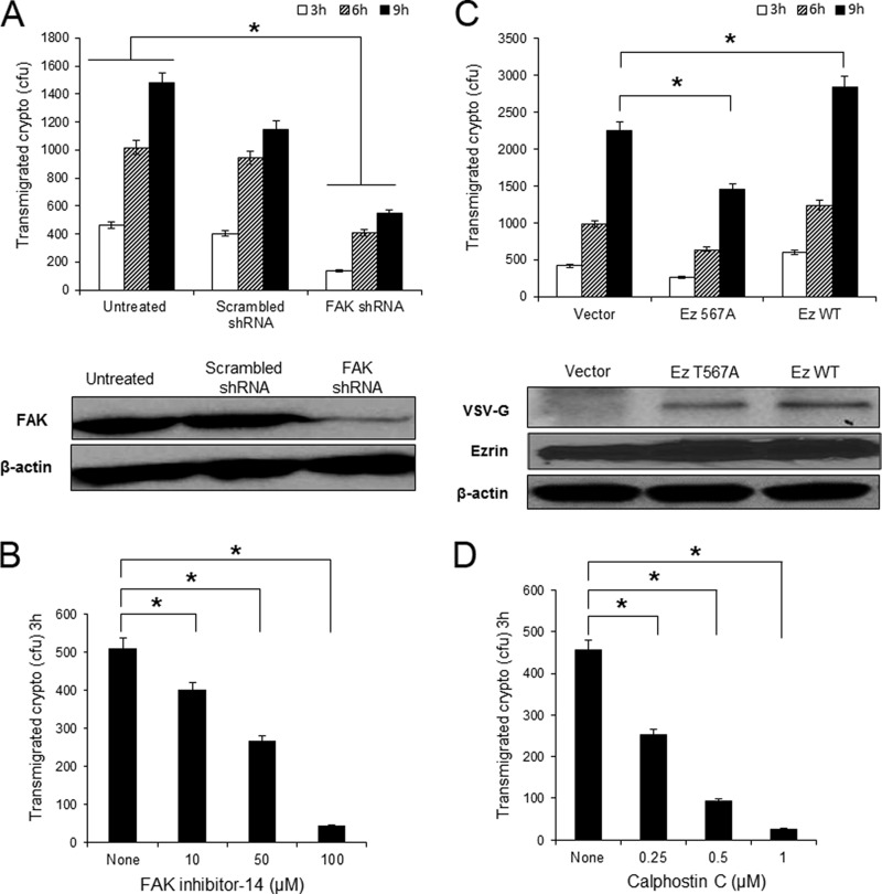
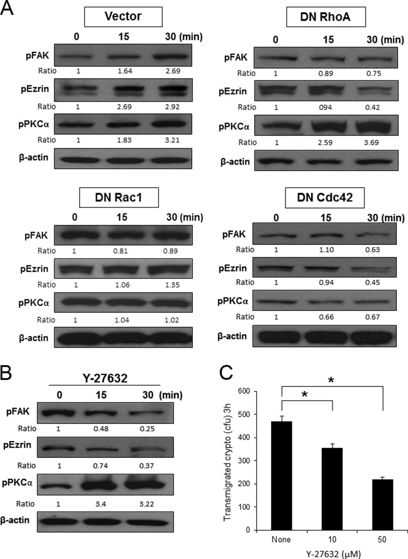
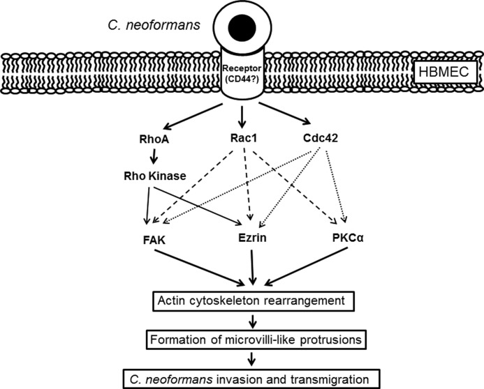
Cryptococcus neoformans is an opportunistic fungal pathogen that causes meningoencephalitis. Previous studies have demonstrated that Cryptococcus binding and invasion of human brain microvascular endothelial cells (HBMEC) is a prerequisite for transmigration across the blood-brain barrier. However, the molecular mechanism involved in the cryptococcal blood-brain barrier traversal is poorly understood. In this study we examined the signaling events in HBMEC during interaction with C. neoformans. Analysis with inhibitors revealed that cryptococcal association, invasion, and transmigration require host actin cytoskeleton rearrangement. Rho pulldown assays revealed that Cryptococcus induces activation of three members of RhoGTPases, e.g. RhoA, Rac1, and Cdc42, and their activations are required for cryptococcal transmigration across the HBMEC monolayer. Western blot analysis showed that Cryptococcus also induces phosphorylation of focal adhesion kinase (FAK), ezrin, and protein kinase C α (PKCα), all of which are involved in the rearrangement of host actin cytoskeleton. Down-regulation of FAK, ezrin, or PKCα by shRNA knockdown, dominant-negative transfection, or inhibitors significantly reduces cryptococcal ability to traverse the HBMEC monolayer, indicating their positive role in cryptococcal transmigration. In addition, activation of RhoGTPases is the upstream event for phosphorylation of FAK, ezrin, and PKCα during C. neoformans-HBMEC interaction. Taken together, our findings demonstrate that C. neoformans activates RhoGTPases and subsequently FAK, ezrin, and PKCα to promote their traversal across the HBMEC monolayer, which is the critical step for cryptococcal brain infection and development of meningitis.


Figures

FIGURE 1.
FIGURE 1.
Association and transmigration of C. neoformans require actin cytoskeleton rearrangement in HBMEC. Cryptococcal association (A) and transmigration assay (B) were performed in the presence of either cytochalasin D or genistein. HBMEC were grown in 24-well plates or Transwells for association or transmigration assays, respectively. Confluent culture of HBMEC was preincubated with reagents for 1 h before the addition of C. neoformans. The numbers (colony forming units (cfu)) of associated or transmigrated Cryptococcus were determined after 3 h (Association assay) or 3, 6, and 9 h of incubation (Transmigration assay). Values are the means ± S.D. of three independent experiments done in triplicate. *, p < 0.001.
FIGURE 2.
FIGURE 2.
C. neoformans internalization into HBMEC involves host actin cytoskeleton rearrangement. Cryptococcal internalization was determined using flow cytometry with GFP-expressing Cryptococcus cells. HBMEC monolayers were labeled with CellTracker CMTMR Orange (red) and incubated with GFP-expressing cryptococci (crypto, green) for 3 h. The HBMEC were washed with PBS to remove unbound fungi followed by incubation with trypsin/EDTA to dissociate bound fungi from HBMEC as well as to collect HBMEC. The cell mixture was subsequently stained with Uvitex-2B (blue). A, the mixed population of HBMEC and fungal cells was analyzed by flow cytometry using triple filter sets. P1 region (left) of total population was analyzed with red (Orange Tracker) and green (GFP) filters, and then the population showing double-positive (P2 region, middle) was further analyzed with blue (DAPI) filter. The population exhibiting Green+/Red+/Blue− was determined as the HBMEC with internalized fungal cells (right). B, a cryptococcal internalization assay was repeated in the presence of inhibitors. The percentages of HBMEC with internalized fungal cells among the P1 region were plotted. These experiments were repeated twice independently. *, p < 0.001.
FIGURE 3.
FIGURE 3.
C. neoformans induces activation of RhoGTPases in HBMEC required for their transmigration. A, confluent HBMEC monolayers were incubated with C. neoformans (1 × 10⁶) for the indicated times, and Rho pulldown assays were performed to determine activation of RhoA, Rac1, and Cdc42. The level of activation was determined by densitometry analysis (measuring the ratios of GTP-Rho/total Rho). B, HBMEC was transduced with adenoviral constructs of DN RhoGTPases mutants (N19RhoA, N17Rac1 or N17Cdc42). Expression of DN RhoGTPases in the transduced HBMEC was verified by Western blotting with anti-myc antibody (bottom). Transmigration assay with HBMEC expressing DN RhoGTPases were carried out. These experiments were repeated three times independently. *, p < 0.001.
FIGURE 4.
FIGURE 4.
C. neoformans induces phosphorylation of PKCα, FAK, and ezrin in HBMEC. Confluent cultures of HBMEC monolayer were incubated with C. neoformans for the indicated times, and the lysates were analyzed by Western blotting with phospho-specific antibodies of PKCα, FAK, and ezrin. β-Actin was detected as an internal loading control. The amount of phosphorylated proteins was determined by densitometric analysis by measuring the relative ratios of amount of each band against that of the untreated control. The representative image of three independent experiments was shown.
FIGURE 5.
FIGURE 5.
FAK, ezrin, and PKCα are required for cryptococcal transmigration of HBMEC monolayer. A, HBMEC was transduced with FAK shRNA to knock down FAK expression. Western blotting with anti-FAK antibody showed the reduction of FAK in the cells with FAK shRNA but not in the scrambled shRNA cells. The transmigration assay was performed with the FAK knockdown cells. B, transmigration assay was done with normal HBMEC in the presence of FAK inhibitor-14. The number of transmigrated Cryptococcus cells was determined by plating the medium collected from the bottom compartment after 3 h of incubation. The result was normalized to show the relative transmigration rate compared with the untreated control. C, HBMEC was transfected with the wild type or DN T567A ezrin mutant construct. Transfection was verified by Western blotting with anti-VSVG antibody (bottom). The transmigration assay was performed with the ezrin transfected cells. D, a transmigration assay carried out with normal HBMEC in the presence of calphostin C, an inhibitor for PKC. *, p < 0.001.
FIGURE 6.
FIGURE 6.
RhoGTPases activation contributes to phosphorylation of PKCα, FAK, and ezrin in HBMEC. A, C. neoformans cells (1 × 10⁶) were incubated with the HBMEC-expressing DN mutant of RhoGTPases (N19RhoA, N17Rac1, and N17Cdc42) for 15 and 30 min. B, normal HBMEC was incubated with C. neoformans cells (1 × 10⁶) in the presence of Y27632, an inhibitor of Rho kinase, for 15 and 30 min. The cell lysates was analyzed by Western blotting with phospho-specific antibodies of PKCα, FAK, and ezrin. β-Actin was detected as an internal loading control. The amounts of phosphorylated proteins were determined by densitometry analysis. C, cryptococcal transmigration assay was done in the presence of Y-27632. *, p < 0.001.
FIGURE 7.
FIGURE 7.
Current model of the host signaling events involved in cryptococcal traversal of the BBB. Cryptococcus binding to the host receptor(s) on HBMEC triggers activation of RhoGTPases such as RhoA, Rac1, and Cdc42. Subsequently, active RhoGTPases induces phosphorylation of other host signaling proteins regulating actin cytoskeleton. Both Rac1 and Cdc42 are involved in phosphorylation of FAK, ezrin, and PKCα, whereas RhoA leads to phosphorylation of FAK and ezrin but not PKCα. Activation of those host proteins results in the host actin cytoskeleton rearrangement and formation of microvilli-like membrane protrusions associated with invading Cryptococcus cells followed by the cryptococcal invasion and transmigration across the HBMEC monolayer. CD44 has shown to play a role as a host cellular receptor for hyaluronic acid of Cryptococcus. However, it is not fully understood how Cryptococcus-CD44 interaction triggers activation of host proteins yet.

References

    1. Alspaugh J. A., Perfect J. R., Heitman J. (1997) Cryptococcus neoformans mating and virulence are regulated by the G-protein α subunit GPA1 and cAMP. Genes Dev. 11, 3206–3217 - PMC - PubMed
    1. Casadevall A., Perfect J. R. (1998) Cryptococcus neoformans, American Society for Microbiology, Washington, D. C.
    1. Chayakulkeeree M., Perfect J. R. (2006) Cryptococcosis. Infect. Dis. Clin. North Am. 20, 507–544 - PubMed
    1. Park B. J., Wannemuehler K. A., Marston B. J., Govender N., Pappas P. G., Chiller T. M. (2009) Estimation of the current global burden of cryptococcal meningitis among persons living with HIV/AIDS. AIDS 23, 525–530 - PubMed
    1. Pukkila-Worley R., Mylonakis E. (2008) Epidemiology and management of cryptococcal meningitis. Developments and challenges. Expert Opin. Pharmacother. 9, 551–560 - PubMed

Publication types

MeSH terms