. 2012 Aug 16;3(8):e374.
doi: 10.1038/cddis.2012.115.

Slow development of ALS-like spinal cord pathology in mutant valosin-containing protein gene knock-in mice

Affiliations

Slow development of ALS-like spinal cord pathology in mutant valosin-containing protein gene knock-in mice

H Z Yin et al. Cell Death Dis. .

Abstract

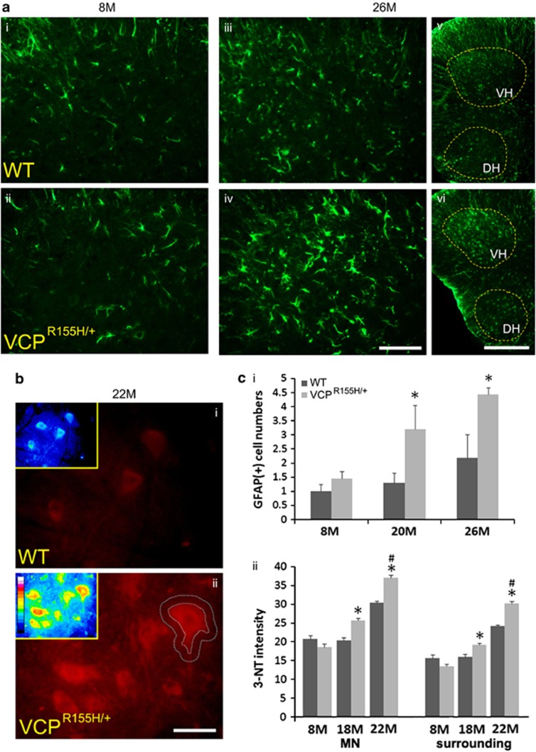
Pathological features of amyotrophic lateral sclerosis (ALS) include, in addition to selective motor neuron (MN) degeneration, the occurrence of protein aggregates, mitochondrial dysfunction and astrogliosis. SOD1 mutations cause rare familial forms of ALS and have provided the most widely studied animal models. Relatively recent studies implicating another protein, TDP-43, in familial and sporadic forms of ALS have led to the development of new animal models. More recently, mutations in the valosin-containing protein (VCP) gene linked to the human genetic disease, Inclusion Body Myopathy associated with Paget's disease of bone and frontotemporal dementia (IBMPFD), were found also to be associated with ALS in some patients. A heterozygous knock-in VCP mouse model of IBMPFD (VCP(R155H/+)) exhibited muscle, bone and brain pathology characteristic of the human disease. We have undertaken studies of spinal cord pathology in VCP(R155H/+) mice and find age-dependent degeneration of ventral horn MNs, TDP-43-positive cytosolic inclusions, mitochondrial aggregation and progressive astrogliosis. Aged animals (~24-27 months) show electromyography evidence of denervation consistent with the observed MN loss. Although these animals do not develop rapidly progressive fatal ALS-like disease during their lifespans, they recapitulate key pathological features of both human disease and other animal models of ALS, and may provide a valuable new model for studying events preceding onset of catastrophic disease.


Figures

Figure 1
Figure 1
VCP expression levels, muscle pathology, mild weakness and modestly decreased lifespan in Neo cassette free VCPR155H/+ mice. (a) Western blots showing VCP expression in spinal cord of WT and VCPR155H/+ knock-in mice at 7 and 18 months of age. Note that WT and VCPR155H/+ mice show very similar levels of VCP at both 7 and 18 months of age (two animals of each genotype at each age). Alpha tubulin was used as a loading control. (b) Muscle pathology in VCPR155H/+ mice. Hematoxylin and eosin staining was carried out on quadriceps muscle from 20-month-old WT (left), and VCPR155H/+ knock-in (right) mice. Pathological features noted include variation in muscle fiber size, basophilic rimmed cracks (black arrows) and centrally located nucleii (white arrow). Bar=50 μm. (c) Mild weight, strength and survival deficits in VCPR155H/+ mice. (i) Mildly decreased weight in VCPR155H/+ mice at 24–28 months, but not at 18 months of age (n=18 each WT and VCPR155H/+ mice at 18 months, n=4 each strain at 24–28 months; *P<0.05 by two-tailed t-test). (ii) Mild progressive weakness in VCPR155H/+ mice. Grip strength was assessed using a Grip Strength Meter apparatus (TSE Systems Gmbh); for each measurement on each animal, the maximum force achieved among five repetitions was used. Note the mild but significant decrease in grip strength in VCPR155H/+ animals starting at 9 months of age. (14–16 WT animals and 12 VCPR155H/+ animals were examined at each age. * indicates significant difference by paired t-test at P<0.01; **P<0.005). (iii) Survival curve comparing the natural history of WT and VCPR155H/+ heterozygous mice. Mice were allowed to age until they died naturally. Note that although the maximal lifespan was close to 30 months in VCPR155H/+ as well as WT mice, some VCPR155H/+ mice died prematurely. n=22 VCPR155H/+ and 22 WT mice; P<0.02 by the log rank (Mantel–Cox) method
Figure 2
Figure 2
EMG evidence of muscle denervation in VCPR155H/+ mice. EMG studies were carried out on four VCPR155H/+ animals (average age 24.98±0.41 months), in comparison with four WT littermates. (a) Traces show representative pathological changes, which were seen only in the VCPR155H/+ animals. (i) Fasciculation potential; (ii) fibrillation potentials; (iii) reduced motor unit recruitment. This is a neurogenic recruitment pattern. Firing frequency in these animals was increased at 50 Hz. (iv) Co-existing neurogenic and myopathic potentials. Motor units exhibit mixed morphology with small amplitude, polyphasic potentials, suggestive of a myogenic disturbance. However, recruitment frequencies are decreased, suggesting a neurogenic pattern. (b) Tabular chart showing frequency of various abnormalities seen on EMG from the sampling of seven muscles in each animal. Note frequent abnormalities in VCPR155H/+ animals and absence of abnormalities in WT mice (MUP, motor unit potential)
Figure 3
Figure 3
Progressive age-dependent degenerative changes and loss of ventral horn MNs from heterozygous VCP knock-in (VCPR155H/+) mice. (a) Age-dependent degenerative changes in ventral horn MNs VCPR155H/+ mice. Lumbar sections were stained with toluidine blue. Images show representative fields (400 × ) from ventral horn of WT and sibling VCPR155H/+ mice at the indicated ages (±2 months). In the WTs, virtually all MNs appeared healthy, with mild atrophic changes noted in a small number of the older MNs. In contrast, in VCPR155H/+ animals, clear age-dependent degenerative changes are seen (panels vii and viii) with eccentric nuclei, fragmentation of dendrites, and some MNs showing cellular and nuclear constriction (stars), and others showing prominent swelling and vacuolar changes in cytoplasm (arrowheads). In some cases, degenerated MNs were replaced by glial nodules (arrows). Bottom panels (iv and ix) show low power views from 26–month-old spinal cords, with regions from ventral horn (VH) and dorsal horn (DH) highlighted. Arrows indicate high power views of regions indicated. Note the relative preservation of neuronal morphology in the DH region, despite extensive degenerative changes in the VH region of the 26 month VCPR155H/+ mouse (panels viii, ix, and x). Comparative examination of VH and DH regions from >40 slices from 26±2-month-old WT and VCPR155H/+ mice revealed similar condition of VH and DH neurons in all WT's but distinctly greater degenerative changes in VH MNs compared to DH regions in all slices from VCPR155H/+ mice. Bar=500 μm (low power panels); 50 μm (all other panels). (b) Age-dependent loss of ventral horn MNs in VCPR155H/+ mice. Lumbar sections were immunostained for the non-phosphoryylated neurofilament marker, SMI-32. Images show representative fields (400 × ) from ventral horn of WT and sibling VCPR155H/+ mice at the indicated ages (±2 months). Note the age-dependent atrophy and loss of MNs in the VCPR155H/+ animals. Bar=50 μm. (c) Quantification of MN cell loss. Surviving MNs were counted in SMI-32 immunostained slices from each condition. Values represent mean number of surviving ventral horn MNs per section (containing two ventral horns), as assessed under direct microscopic examination. Each data point reflects counts from 3–7 animals (for SMI-32); all ventral horn MNs were counted in each of 10–15 slices for each animal. Note the modest age-dependent MN loss in WTs in contrast to the marked loss in the VCPR155H/+ mice. * indicates difference from WT animals at the same age by two-tailed t-test (P<0.025)
Figure 4
Figure 4
Age-dependent increases in total spinal cord TDP-43 and its appearance in cytosolic inclusions in ventral horn MNs of VCPR155H/+ mice. (a) Western blots showing TDP-43 expression in spinal cord of WT and VCPR155H/+ knock-in mice at 7 and 18 months of age. Note that WT and VCPR155H/+ mice show very similar levels of TDP-43 at 7 months of age, whereas TDP-43 levels were markedly increased in the VCPR155H/+ spinal cord at 18 months. Alpha tubulin was used as a loading control. (b) Immunocytochemical assessment of cytosolic TDP-43 aggregation as a function of age-representative photomicrographs. Lumbar sections were immunostained for TDP-43 and photographed (400 × ). Representative fields are shown from ventral horn of WT (i, ii, and iii) and sibling VCPR155H/+ mice (iv, v, and vi) at the indicated ages (±2 months). In WT animals, faint and generally cytosolic or nuclear labeling was generally seen at young ages, with more prominent but generally still diffuse staining seen by 20 months. In the VCPR155H/+ animals, cytosolic TDP-43 labeling was both stronger, and far more prone to appear as discrete inclusions or aggregates with increasing age (arrowheads). By way of comparison, we also show stains from young (0.5 months) homozygous VCPR155H/R155H animals (vii). Note the markedly accelerated appearance of strong cytosolic TDP-43 aggregation in MN from these young mice (arrowheads). Bar=50 or 25 μm (detail). (c) Quantification of cytosolic TDP-43 aggregation. Each MN in examined sections was rated for cytosolic TDP-43 aggregation according to a five point scale as described (see Materials and methods). Each data point represents all MNs (>160) present in 5–10 ventral horn micrographs from each of 2–5 animals. * indicates difference from WT animals at the same age by two-tailed t-test (P<0.001)
Figure 5
Figure 5
Colocalization of mitochondrial aggregates with TDP-43, and occurrence of ubiquitin and SQSTM1/ p62 aggregates in aging VCPR155H/+ MNs. (a) Representative images. Slices were double fluorescence immunostained for the mitochondrial marker, COX-IV (top) and TDP-43 (bottom) (400 × ); inserts show overlay. Note in the 18-month-old WT animals, the COX-IV labeling is speckled and distributed throughout the cytoplasm. TDP-43 labeling shows mild early aggregation in a small number of MNs, that co-localizes with some early mitochondrial aggregation (arrowheads). In contrast, in age-matched VCPR155H/+ animals, the COX-IV labeling is highly aggregated and colocalizes with TDP-43 (arrows). Bar=25 μm. (b) Quantification of COX-IV and TDP-43 co-aggregation. Graph shows the % of examined MNs containing COX-IV and TDP-43 co-aggregation at each age (each point based on examination of >40 MNs). Note the paucity of co-aggregation at 8 months in all animals, and the sharp increase of co-aggregation in the VCPR155H/+ mice by 18 months. (c and d): Ubiquitin (c) and SQSTM1/ p62 (d) labeling (24-month-old animals) revealed similar patterns of age-dependent appearance of cytosolic aggregates as seen for TDP-43. Arrows denote aggregates in VCPR155H/+ MNs. Bar=25 μm
Figure 6
Figure 6
Progressive astrogliosis and increased 3-NT labeling in ventral horn of VCPR155H/+ mice. (a) Representative images of GFAP labeled slices. Lumbar sections were immunostained for GFAP and the ventral horns photographed under fluorescence (200 × ). Note the marked increase in reactive astrocytes in VCPR155H/+ mice at 26 months (iv). Low magnification images of the hemi-spinal cord show the greater degree of astrogliosis in the ventral horn (VH) compared with the dorsal horn (DH) of the VCPR155H/+ mice (vi). Bar=50 μm (high power); 400 μm (low power). (b) Representative images of 3-NT labeled slices. Lumber slices from 22-month-old WT and VCPR155H/+ mice were immunostained for 3-NT, and photographed under immunofluorescence. Inserts show pseudocolor (8 bit) representation of fluorescence, in order to highlight the gradient in labeling intensity. Note the moderate labeling of WT MNs, but the markedly increased intensity of labeling not only in MNs of the VCPR155H/+ mice, but also in the neuropil surrounding and between MNs. (Dotted lines show representative regions marked for one MN, as was used for fluorescence quantification, below). Note that the region of increased 3-NT labeling between ventral horn MNs in the VCPR155H/+ mice may correspond generally to the region of increased ventral horn astrogliosis (see a, panels iv and vi above) in these animals. Bar=50 μm. (c): Quantification of GFAP and 3-NT labeling. (i) GFAP immunoreactive astrocytes were counted from lumbar ventral horns of WT and VCPR155H/+ mice of the indicated ages. Each data point reflects counts from 3–5 animals; for each animal, all GFAP labeled astrocytes were counted from 200 × ventral horn microscope fields from five sections. Note the marked age-dependent increase in numbers of reactive astrocytes in the VCPR155H/+ animals. * indicates difference from WT animals at the same age by two-tailed t-test (P<0.025). (ii) 3-NT labeling intensity was assessed in MNs, and in 10 μm wide zones surrounding MNs, in WT and VCPR155H/+ mice as indicated. Each data point represents all MNs (>75) present in 5–10 ventral horn micrographs from each of 2–5 animals. Note the age-dependent increase in 3-NT labeling in WT mice, and the greater increase in VCPR155H/+ mice, both within the MNs (MN) and in the neuropil surrounding them (surrounding). * indicates difference from WT animals at the same age by two-tailed t-test (P<0.001); #indicates difference from 18-month-old VCPR155H/+ mice (P<0.001)

References

    1. Rothstein JD. Current hypotheses for the underlying biology of amyotrophic lateral sclerosis. Ann Neurol. 2009;65 (Suppl 1:S3–S9. - PubMed
    1. Manfredi G, Xu Z. Mitochondrial dysfunction and its role in motor neuron degeneration in ALS. Mitochondrion. 2005;5:77–87. - PubMed
    1. Neumann M, Sampathu DM, Kwong LK, Truax AC, Micsenyi MC, Chou TT, et al. Ubiquitinated TDP-43 in frontotemporal lobar degeneration and amyotrophic lateral sclerosis. Science. 2006;314:130–133. - PubMed
    1. Sreedharan J, Blair IP, Tripathi VB, Hu X, Vance C, Rogelj B, et al. TDP-43 mutations in familial and sporadic amyotrophic lateral sclerosis. Science. 2008;319:1668–1672. - PMC - PubMed
    1. Chen-Plotkin AS, Lee VM, Trojanowski JQ. TAR DNA-binding protein 43 in neurodegenerative disease. Nat Rev Neurol. 2010;6:211–220. - PMC - PubMed

Publication types

MeSH terms