. 2012 Sep 1;26(17):1911-25.
doi: 10.1101/gad.193169.112. Epub 2012 Aug 16.

C16orf57, a gene mutated in poikiloderma with neutropenia, encodes a putative phosphodiesterase responsible for the U6 snRNA 3' end modification

Affiliations

C16orf57, a gene mutated in poikiloderma with neutropenia, encodes a putative phosphodiesterase responsible for the U6 snRNA 3' end modification

Seweryn Mroczek et al. Genes Dev. .

Abstract

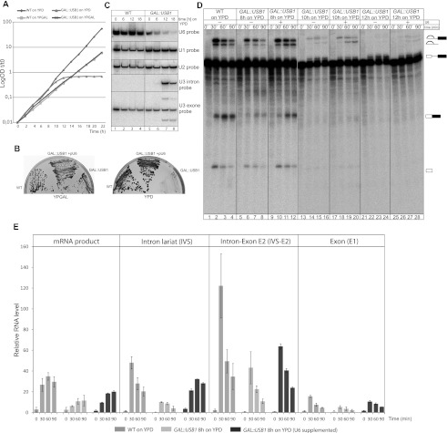
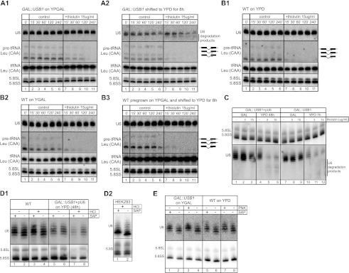
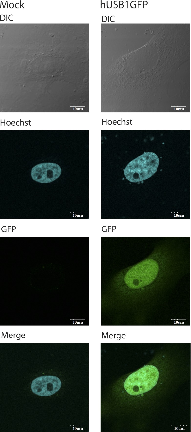
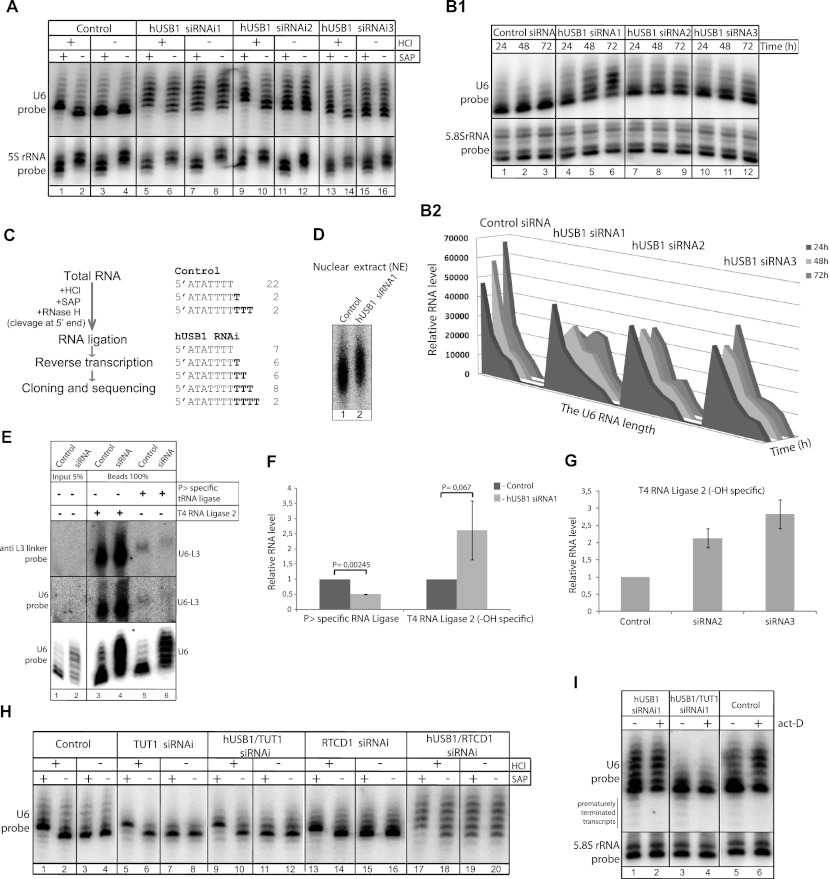
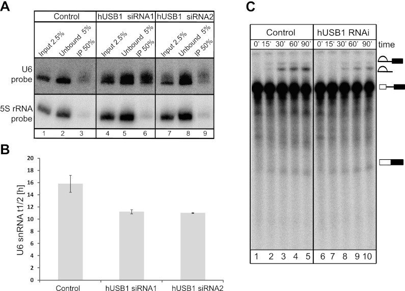
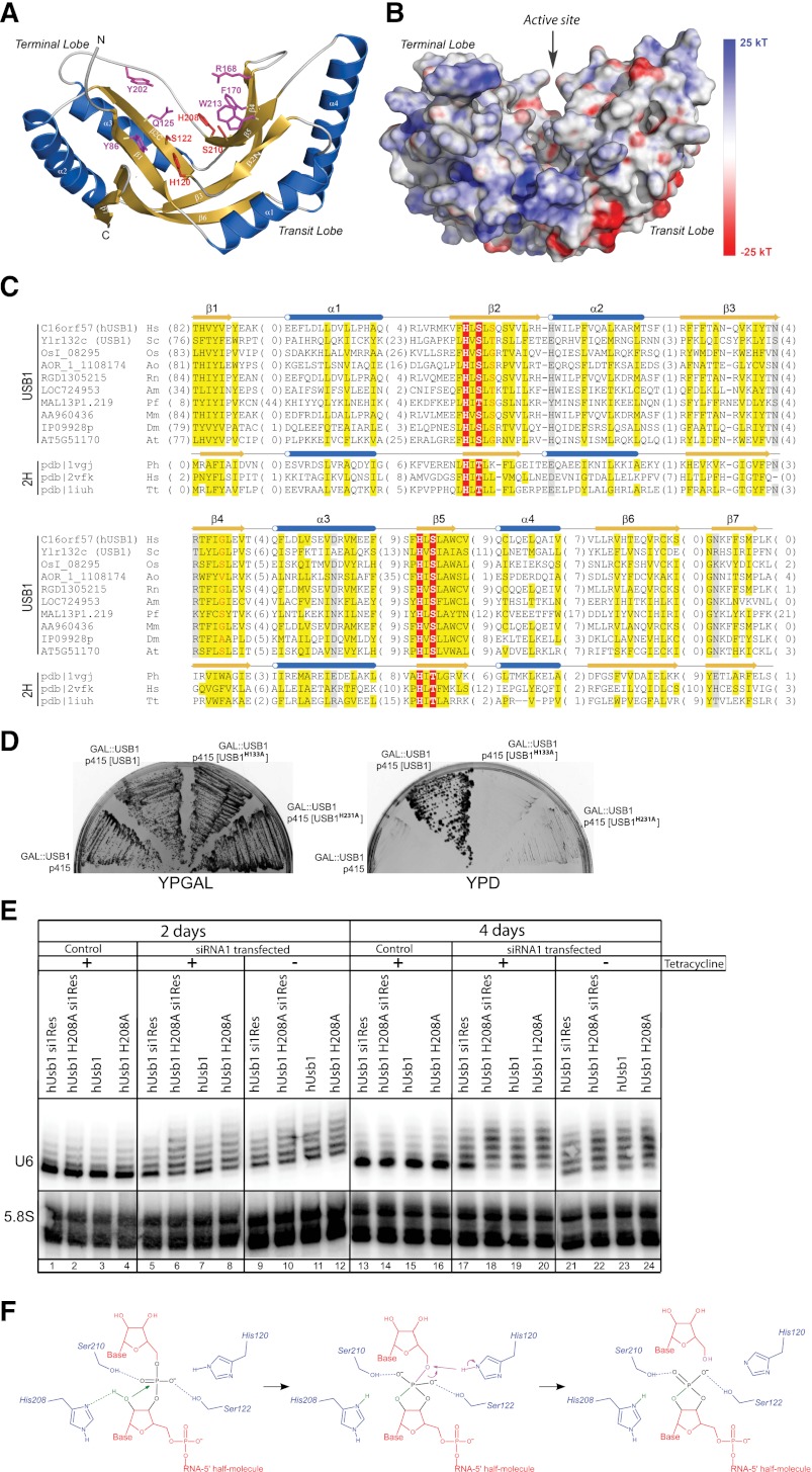
C16orf57 encodes a human protein of unknown function, and mutations in the gene occur in poikiloderma with neutropenia (PN), which is a rare, autosomal recessive disease. Interestingly, mutations in C16orf57 were also observed among patients diagnosed with Rothmund-Thomson syndrome (RTS) and dyskeratosis congenita (DC), which are caused by mutations in genes involved in DNA repair and telomere maintenance. A genetic screen in Saccharomyces cerevisiae revealed that the yeast ortholog of C16orf57, USB1 (YLR132C), is essential for U6 small nuclear RNA (snRNA) biogenesis and cell viability. Usb1 depletion destabilized U6 snRNA, leading to splicing defects and cell growth defects, which was suppressed by the presence of multiple copies of the U6 snRNA gene SNR6. Moreover, Usb1 is essential for the generation of a unique feature of U6 snRNA; namely, the 3'-terminal phosphate. RNAi experiments in human cells followed by biochemical and functional analyses confirmed that, similar to yeast, C16orf57 encodes a protein involved in the 2',3'-cyclic phosphate formation at the 3' end of U6 snRNA. Advanced bioinformatics predicted that C16orf57 encodes a phosphodiesterase whose putative catalytic activity is essential for its function in vivo. Our results predict an unexpected molecular basis for PN, DC, and RTS and provide insight into U6 snRNA 3' end formation.


Figures

Figure 1.
Figure 1.
Usb1 depletion leads to splicing defects caused by a reduction in U6 snRNA levels. (A) Usb1 depletion causes growth arrest in yeast. Growth curves of wild type (WT) and the GAL#x2237;USB1 strain (with the USB1 gene under the control of a galactose-inducible promoter) cultured on permissive YPGAL and repressive YPD medium. The presented values were corrected for dilution and are shown as log OD600, where t is the time in hours. (B) U6 snRNA overexpression suppresses the growth arrest caused by Usb1 depletion. The expression of additional copies of U6 snRNA or USB1 fully restores growth of the GAL#x2237;USB1 strain on glucose-rich repressive medium (YPD). (C) Depletion of Usb1 leads to in vivo splicing defects and the accumulation of precursor RNAs, which is correlated with reduced levels of the U6 snRNA. Northern blot analysis for the U1, U2, and U6 snRNAs, and precursor and mature U3 snRNA is shown. Total RNA was isolated from wild-type and GAL#x2237;USB1 strains grown in repressive YPD medium for up to 18 h. All hybridizations were performed with the same blot. (D) Extracts lacking Usb1 have reduced splicing activity in vitro, a defect that is partially restored by the addition of U6 snRNA isolated from a TAP-purified Prp24 fraction to the splicing reaction. Splicing reactions were performed using extracts prepared from wild-type (lanes 1–4) or GAL#x2237;USB1 cells after 8 h (lanes 5–12), 10 h (lanes 13–20), and 12 h (lanes 21–28) of Usb1 depletion. Reactions were carried out for 0, 30, 60, and 90 min. Reaction products were fractionated in 15% polyacrylamide gels and detected by autoradiography. The cell extracts were supplemented with the U6 snRNA in order to resume splicing activity. (E) Quantifications of the in vitro splicing reaction product and its intermediates. The graph shows relative levels of mature RP51a mRNA and its splicing intermediates, intron–lariat (IVS), intron–exon2 (IVS-E2), and exon1 (E1), in splicing reactions performed using extracts from wild type and the GAL#x2237;USB1 strain after 8 h of Usb1 depletion. All values are averages from three independent splicing reactions. Autoradiograms were quantified using MultiGauge software.
Figure 2.
Figure 2.
Depletion of Usb1 reduces U6 snRNA half-life and leads to accumulation of decay intermediates. (A,B) The GAL#x2237;USB1 strain was grown in permissive YPGAL medium (A1) or repressive YPD medium (A2) for 8 h. The wild-type (WT) strain was grown on YPD (B1) or YPGAL (B2) or pregrown on YPGAL medium and shifted to YPD for 8 h (B3). Cells were treated with the RNA polymerase inhibitor thiolutin, then total RNA was extracted at the indicated times for Northern blot hybridizations against U6 snRNA, 5.8S rRNA as the loading control, and tRNALeu (CAA) precursor as the thiolutin treatment indicator. Depletion of Usb1 led to U6 snRNA instability and decreased half-life from 10 h to 2.5 h (cf. A2, lanes 7–11 and A1, lanes 7–11). For additional quantification analyses, see Supplemental Fig. S6A,B. (C) High-resolution Northern blot analysis of RNA from GAL#x2237;USB1 + pU6 and GAL#x2237;USB1 growing in permissive YPGAL (lanes 1–3,7–9, respectively) or repressive YPD (lanes 4–6,10–12, respectively) medium and treated with thiolutin for 4 h. (Lanes 5,6) The degradation products of U6 snRNA were detectable in the samples after 48 h of depletion. (Lanes 1,4) Long periods of Usb1 depletion caused changes in the migration of U6 snRNA. (D1) Analysis of U6 snRNA 3′ termini after Usb1 depletion. Total RNA from wild type (lanes 1–4) and GAL#x2237;USB1 + pU6 (lanes 5–8) growing in repressive YPD medium for 48 h was treated with HCl and/or shrimp alkaline phosphatase (SAP) and analyzed by Northern blotting. Only the U6 snRNA isolated from wild-type cells changed its migration toward the longer species upon phosphate removal by SAP, whereas HCl treatment, which can open the cyclic phosphate ring, had no effect on migration. (D2) Total RNA from HEK293 cells was treated with SAP and HCl similar to that for yeast, and U6 snRNA was detected by high-resolution Northern blot. (E) The U6 snRNA is terminated with 3′ phosphate at the 3′ end. Total RNA from GAL#x2237;USB1 grown on YPGAL (lanes 1–4) and wild type cultured on YPD (lanes 5–8) were treated with T4 PNK (lanes 3,7) or SAP (lanes 1,5) as control.
Figure 3.
Figure 3.
hUSB1 is a nuclear protein. HeLa cells were transiently transfected with a plasmid bearing a hUSB1-GFP fusion gene. Twenty-four hours after transfection, cells were examined by confocal microscopy. The fluorescence images for GFP and Hoechst nucleic acid stain, a differential interference contrast (DIC) image, and a merged image are presented for hUSB1GFP and the negative control.
Figure 4.
Figure 4.
hUSB1 is essential for the presence of the cyclic phosphate at the U6 snRNA 3′ end. (A) U6 snRNA molecules are more heterogeneous and elongated after hUSB1 depletion by RNAi. High-resolution Northern blot analysis of RNA from control (lanes 1–4) and siRNA-treated (lanes 5–16) HeLa cells is shown. In order to examine the phosphorylation status, RNA was treated with HCl and/or SAP phosphatase, which in control cells led to a shift in U6 snRNA on the gel caused by cyclic phosphate removal. (B) Kinetics of the effect of three siRNAs against hUsb1 on the U6 snRNA. The HEK293 cells were treated with three siRNAs against hUSB1 and then harvested at the indicated time points. (B1) Total RNA was analyzed with high-resolution Northern blots. (B2) The U6 snRNA molecules were quantified with MultiGauge software and are shown as a 3D plot. (C) U6 snRNA molecules are more heterogeneous at the 3′ end upon hUSB1 depletion. CR-RT–PCR analysis of U6 snRNA from control and siRNA-treated HeLa cells is shown. Before ligation, the RNA was treated with HCl and SAP in order to remove the phosphate moiety from the 3′ end. RNase H cleavage was performed in order to remove the γ-monomethylguanosine triphosphate (meGTP) structure from the U6 5′ end. After reverse transcription and PCR, the reaction products were cloned and sequenced. This confirmed increased heterogeneity at the 3′ ends of U6 snRNA molecules after siRNA treatment. The numbers indicate the amount of independent clones that were sequenced. (D) RNA labeling in nuclear extracts confirms the changes in U6 snRNA mobility in high-resolution gels. Native U6 snRNA was labeled by incubating nuclear extracts from control (lane 1) or siRNA-treated (lane 2) HeLa cells with α-32PUTP. Isolated RNA was separated on 6% sequencing polyacrylamide gels and detected by autoradiography. (E–G) The formation of the U6 snRNA cyclic phosphate moiety is hUSB1-dependent. (E) U6 snRNA from control (lanes 1,3,5) and siRNA-treated (lanes 2,4,6) cells was captured with streptavidin magnetic beads with biotinylated antisense oligonucleotide complementary to its 5′ end. Subsequently, RNA was ligated with the preadenylated L3 linker using T4 RNA ligase 2 or cyclic phosphate-specific tRNA ligase from Arabidopsis thaliana. Reaction products were separated on 6% sequencing polyacrylamide gels and analyzed by Northern blotting using probes against the L3 linker and U6 snRNA. Larger blots with unligated U6 snRNA are presented in Supplemental Figure S8. (F,G) After hUSB depletion, the relative number of U6 snRNA molecules terminated with 2′,3′-cyclic phosphate decreases, and those terminated with OH groups increases, as quantified by MultiGauge software. All values are averages from three independent experiments, and the respective P-values calculated with an unpaired two-tailed Student's t-test are presented. Please note that the low signal intensity for the tRNA ligase reaction products compared with the T4 reaction was due to a lower reaction efficiency. (H) TUTase or RTCD1 activity is not required for the formation of the U6 snRNA 3′ end cyclic phosphate. RNA from cells treated with siRNAs against TUTase (lanes 5–8) or RTCD1 (lanes 13–16) and from control HeLa cells (lanes 1–4) was analyzed using high-resolution Northern blots. The RNA phosphorylation status was determined as described in A. (I) Depletion of hUSB1 or TUTase does not change the stability of mature U6 snRNA. High-resolution Northern blot analysis of RNA from cells treated with siRNAs against hUSB1 (lanes 1,2) and both hUSB1 and TUTase (lanes 3,4) and from control cells (lanes 5,6) after 2 h of actinomycin-D treatment is shown. (Lanes 2,6) The oligouridylated transcripts are more stable compared with mature forms and hence accumulate. (Lanes 2,4,6) Note that shorter precursor transcripts are eliminated after actinomycin-D treatment.
Figure 5.
Figure 5.
hUSB1 knockdown does not significantly change the half-life of human U6 snRNA or its in vitro splicing activity. (A) Northern blot analysis of RNA transcripts after thiouridine labeling of HeLa cells in vivo. Total RNA from control cells (lanes 1–3) and cells treated with siRNA1 (lanes 4–6) and siRNA2 (lanes 7–9) was biotinylated and captured with streptavidin beads. Captured RNA was separated on 6% polyacrylamide gels, blotted, and hybridized against the U6 snRNA and 5S rRNA. Quantitative analyses were performed with MultiGauge software, as described in the Materials and Methods. (B) The graph shows measurements of the U6 snRNA half-life in control and siRNA-treated HeLa cells. The values are averages from three independent experiments. (C) hUSB1 depletion slightly affects the in vitro splicing activity. Nuclear extracts from HeLa cells after hUsb1 depletion (lanes 6–10) exhibited a slightly decreased splicing activity compared with extracts from control cells (lanes 1–5). The reactions were carried out for 90 min, after which RNA was purified, fractionated on 15% polyacrylamide gels, and detected by autoradiography.
Figure 6.
Figure 6.
USB1 encodes a putative phosphodiesterase whose potential catalytic activity is essential for its function. (A) A 3D model of the hUSB1 protein. Invariant catalytic histidines and serines from HXS/T motifs are shown in red, whereas other conserved residues potentially responsible for substrate binding are shown in magenta. (B) The electrostatic potential surface for hUSB1 suggests that the terminal lobe has the ability to bind nucleic acids. Negatively charged regions are colored in red, and positively charged regions are colored in blue (ranging from −25 kT up to 25 kT). The protein is in the same orientation as in A. Please see Supplemental Figure S10 for more details. (C) Usb1 belongs to the 2H phosphodiesterase superfamily. Multiple sequence alignments of USB1 family representatives and selected distantly related 2H phosphodiesterase structures. Numbers of the residues that are not shown are specified in parentheses. Residue conservation is denoted by the following scheme: uncharged are highlighted in yellow; charged or polar are highlighted in gray; small are letters in red; and invariant catalytic residues of HXT/S motifs are highlighted in red. Locations of predicted (hUSB1) and observed (Protein Data Bank [PDB], 1VGJ) secondary structure elements are marked above the corresponding sequences. (Hs) Homo sapiens; (Sc) Saccharomyces cerevisiae; (Os) Oryza sativa; (Ao) Aspergillus oryzae; (Rn) Rattus norvegicus; (Am) Apis mellifera; (Pf) Plasmodium falciparum; (Mm) Mus musculus; (Dm) Drosophila melanogaster; (At) Arabidopsis thaliana; (Ph) Pyrococcus horikoshii; (Tt) Thermus thermophilus. (D) A complementation assay reveals that the Usb1 catalytic mutant cannot restore cell growth. Plasmids containing either wild-type (WT) or H133A, H231A mutant versions of the USB1 gene were transformed into the GAL#x2237;USB1 strain. The resulting strains were plated on permissive (YPGAL) and restrictive (YPD) media. (E) Expression of siRNA-insensitive wild-type hUSB1 mRNA, but not H208A hUSB1 mutant mRNA, leads to partial rescue of the molecular phenotype caused by hUSB1 depletion. HEK293 stable cell lines expressing constructs under the control of a tetracycline-regulated promoter, including hUsb1 mRNA resistant to siRNA (si1Res hUsb1) or its corresponding H208A mutant mRNA (H208A si1Res) as well as hUsb1 mRNA sensitive to siRNA (hUsb1) or its corresponding H208A mutant mRNA (hUsb1 H208A), were transfected with siRNA against hUSB1 and then induced with tetracycline. (Lines 5–8,17–20) Cells were collected 2 and 4 d after transfection, and U6 snRNA was detected by high-resolution Northern blots. Untransfected cells (lines 1–4,13–16) and uninduced cells (lines 9–12,21–24) were used as controls. (F) Proposed reaction mechanism catalyzed by hUSB1. The nucleophilic attack on the phosphate group results in the formation of a cyclic phosphate moiety. Groups involved in the first and second steps of the reaction are shown in green and magenta, respectively, whereas the catalytic residues are denoted in blue.

Similar articles

Cited by

References

    1. Achsel T, Brahms H, Kastner B, Bachi A, Wilm M, Luhrmann R 1999. A doughnut-shaped heteromer of human Sm-like proteins binds to the 3′-end of U6 snRNA, thereby facilitating U4/U6 duplex formation in vitro. EMBO J 18: 5789–5802 - PMC - PubMed
    1. Alexander RD, Barrass JD, Dichtl B, Kos M, Obtulowicz T, Robert MC, Koper M, Karkusiewicz I, Mariconti L, Tollervey D, et al. 2010. RiboSys, a high-resolution, quantitative approach to measure the in vivo kinetics of pre-mRNA splicing and 3′-end processing in Saccharomyces cerevisiae. RNA 16: 2570–2580 - PMC - PubMed
    1. Altschul SF, Madden TL, Schaffer AA, Zhang J, Zhang Z, Miller W, Lipman DJ 1997. Gapped BLAST and PSI-BLAST: A new generation of protein database search programs. Nucleic Acids Res 25: 3389–3402 - PMC - PubMed
    1. Aravind L, Koonin EV 2001. A natural classification of ribonucleases. Methods Enzymol 341: 3–28 - PubMed
    1. Armanios M 2009. Syndromes of telomere shortening. Annu Rev Genomics Hum Genet 10: 45–61 - PMC - PubMed

Publication types

MeSH terms