. 2012 Nov 1;125(Pt 21):5188-95.
doi: 10.1242/jcs.109884. Epub 2012 Aug 16.

Syndecan-1 controls cell migration by activating Rap1 to regulate focal adhesion disassembly

Affiliations

Syndecan-1 controls cell migration by activating Rap1 to regulate focal adhesion disassembly

William A Altemeier et al. J Cell Sci. .

Abstract

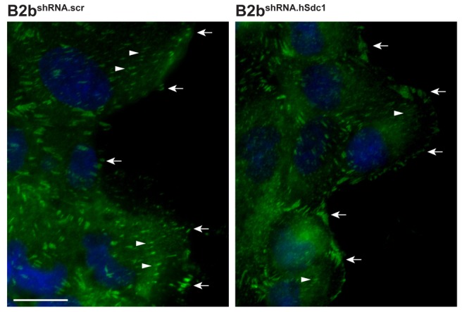
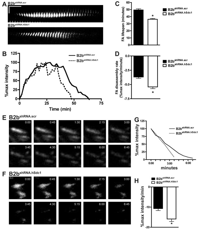
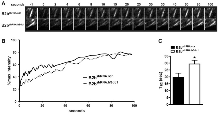
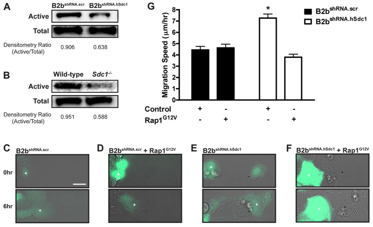
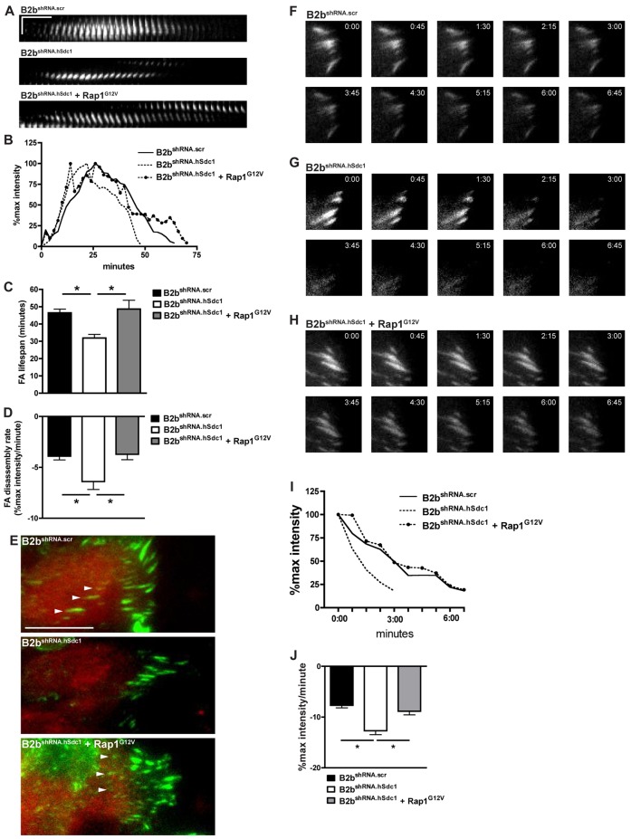
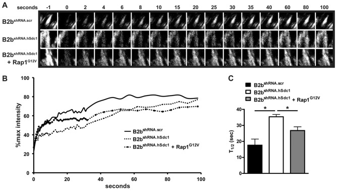
After injury, residual epithelial cells coordinate contextual clues from cell-cell and cell-matrix interactions to polarize and migrate over the wound bed. Protrusion formation, cell body translocation and rear retraction is a repetitive process that allows the cell to move across the substratum. Fundamental to this process is the assembly and disassembly of focal adhesions that facilitate cell adhesion and protrusion formation. Here, we identified syndecan-1 as a regulator of focal adhesion disassembly in migrating lung epithelial cells. Syndecan-1 altered the dynamic exchange of adhesion complex proteins, which in turn regulates migration speed. Moreover, we provide evidence that syndecan-1 controls this entire process through Rap1. Thus, syndecan-1 restrains migration in lung epithelium by activating Rap1 to slow focal adhesion disassembly.


Figures

Fig. 1.
Fig. 1.
Syndecan-1 increases FAs. B2bshRNA.scr and B2bshRNA.hSdc1 cells were allowed to migrate for 2 h and immunostained for paxillin (green) and DAPI (blue). Nascent adhesions were seen at the cell front (arrows). FAs (arrowheads) are seen throughout migrating B2bshRNA.scr cells. In contrast, B2bshRNA.hSdc1 cells had fewer and less prominent FAs in migrating cells. These are representative images from reproducible experiments. Scale bar: 20 µm.
Fig. 2.
Fig. 2.
Syndecan-1 slows FA disassembly in lung epithelial cells. (A–D) Migrating B2bshRNA.scr and B2bshRNA.hSdc1 cells stably expressing paxillin–eGFP were observed by time-lapse TIRF microscopy (supplementary material Movie 1). (A) Kymographs revealed a longer FA lifespan in B2bshRNA.scr compared to B2bshRNA.hSdc1 cells. Horizontal line: 10 min; vertical line: 5 µm. (B) Normalized fluorescent intensity (% of max intensity) of the FAs in panel A over time. (C) The total FA lifespan and (D) the rate of the FA disassembly were determined for migrating lung epithelial cells. *P<0.0001 by Student's t-test; n = 4 independent experiments. (E–H) B2bshRNA.scr and B2bshRNA.hSdc1 stably expressing paxillin–eGFP were treated with nocodazole (10 µM; 4 h). After washing the cells with fresh medium, FA disassembly was observed with time-lapse TIRF microscopy in (E) B2bshRNA.scr and (F) B2bshRNA.hSdc1 cells. (G) The intensity curves of the FAs in panels E and F. (H) The FA disassembly rate was determined in B2bshRNA.scr and B2bshRNA.hSdc1 cells. *P<0.05 by Student's t-test; n = 3 independent experiments.
Fig. 3.
Fig. 3.
Syndecan-1 facilitates the recovery of fluorescence after photobleaching in migrating lung epithelial cells. A scratch wound was created on monolayers of B2bshRNA.scr and B2bshRNA.hSdc1 cells stably expressing paxillin–eGFP, and FAs at the wound front were identified for photobleaching. (A) Representative FA intensities prior to (−1 second) and after photobleaching. (B) The normalized intensity of the representative FA in A was plotted over time. (C) The FRAP recovery half-time was determined for B2bshRNA.scr and B2bshRNA.hSdc1 cells. More than 15 FAs were analyzed across three independent experiments. *P<0.05 by Student's t-test.
Fig. 4.
Fig. 4.
Syndecan-1 restrains lung epithelial cell migration by activating Rap1. Western blot for active and total Rap1 in (A) B2bshRNA.scr and B2bshRNA.hSdc1 cells and (B) wild-type and Sdc1−/− primary ALI cultures. (C–F) Migration of (C) B2bshRNA.scr transduced with eGFP, (D) B2bshRNA.scr co-transduced with eGFP and Rap1G12V, (E) B2bshRNA.hSdc1 cells transduced with eGFP, or (F) B2bshRNA.hSdc1 cells co-transduced with eGFP and Rap1G12V (supplementary material Movie 2). The white asterisk identifies the same cell at 0 h and 6 h after migration. (G) Cell migration speed was measured for all conditions. *P<0.001 by one-way ANOVA and post-test Bonferroni analysis; n = 3 independent experiments.
Fig. 5.
Fig. 5.
Rap1 slows FA disassembly in lung epithelial cells lacking syndecan-1 expression. (A–E) A scratch wound was created on monolayers of B2bshRNA.scr and B2bshRNA.hSdc1 cells stably expressing paxillin–eGFP, and FA assembly and disassembly was observed by time-lapse TIRF microscopy. All cells were identified by expression of mCherry. In control conditions (B2bshRNA.scr and B2bshRNA.hSdc1), cells were transduced with mCherry only, whereas B2bshRNA.hSdc1 + Rap1G12V cells were transduced with both mCherry and Rap1G12V. (A) Kymographs (horizontal line: 10 min; vertical line: 5 µm) and (B) intensity curves of FAs of the conditions indicated. (C) The total FA lifespan and (D) the rate of the FA disassembly were determined. *P<0.05 by one-way ANOVA and post-test Bonferroni analysis; n = 3 independent experiments. (E) FAs (arrowheads) are present in the center of B2bshRNA.scr cells and B2bshRNA.hSdc1 cells + Rap1G12V, and lacking in B2bshRNA.hSdc1 cells. Green, paxillin; Red, mCherry; Scale bar: 20 µm. (F–J) Cells stably expressing paxillin–eGFP were treated with nocodazole (10 µM; 4 h). In control conditions (B2bshRNA.scr and B2bshRNA.hSdc1), cells were transduced with mCherry only, whereas B2bshRNA.hSdc1 + Rap1G12V cells were transduced with both mCherry and Rap1G12V. After washing with fresh medium, FA disassembly was observed by time-lapse TIRF microscopy in (F) B2bshRNA.scr, (G) B2bshRNA.hSdc1 and (H) B2bshRNA.hSdc1 + Rap1G12V cells. (I) The intensity curves of the FAs in panels F–H. (J) The FA disassembly rate was determined in all conditions. *P<0.001 by one-way ANOVA and post-test Bonferroni analysis; n = 3 independent experiments.
Fig. 6.
Fig. 6.
Rap1 accelerates the recovery of fluorescent after photobleaching in migrating lung epithelial cells lacking syndecan-1 expression. A scratch wound was created on monolayers of B2bshRNA.scr and B2bshRNA.hSdc1 cells stably expressing paxillin–eGFP, and FAs at the wound front were identified for photobleaching. In control conditions (B2bshRNA.scr and B2bshRNA.hSdc1), cells were transduced with mCherry only whereas B2bshRNA.hSdc1 + Rap1G12V cells were transduced with both mCherry and Rap1G12V. (A) Representative FA intensities prior to (−1 sec) and after photobleaching. (B) The normalized intensity of the representative FA in A was plotted over time. (C) The FRAP recovery half-time was determined for B2bshRNA.scr and B2bshRNA.hSdc1 cells. More than 15 FAs were analyzed across three independent experiments. *P<0.01 by one-way ANOVA and post-test Bonferroni analysis.

References

    1. Alexandrova A. Y., Arnold K., Schaub S., Vasiliev J. M., Meister J-J., Bershadsky A. D., Verkhovsky A. B. (2008). Comparative dynamics of retrograde actin flow and focal adhesions: formation of nascent adhesions triggers transition from fast to slow flow. PLoS ONE 3, e3234 10.1371/journal.pone.0003234 - DOI - PMC - PubMed
    1. Bass M. D., Morgan M. R., Humphries M. J. (2009). Syndecans shed their reputation as inert molecules. Sci. Signal. 2, pe18 10.1126/scisignal.264pe18 - DOI - PMC - PubMed
    1. Beauvais D. M., Rapraeger A. C. (2003). Syndecan-1-mediated cell spreading requires signaling by alphavbeta3 integrins in human breast carcinoma cells. Exp. Cell Res. 286, 219–232 10.1016/S0014-4827(03)00126-5 - DOI - PubMed
    1. Beauvais D. M., Rapraeger A. C. (2010). Syndecan-1 couples the insulin-like growth factor-1 receptor to inside-out integrin activation. J. Cell Sci. 123, 3796–3807 10.1242/jcs.067645 - DOI - PMC - PubMed
    1. Beauvais D. M., Burbach B. J., Rapraeger A. C. (2004). The syndecan-1 ectodomain regulates alphavbeta3 integrin activity in human mammary carcinoma cells. J. Cell Biol. 167, 171–181 10.1083/jcb.200404171 - DOI - PMC - PubMed

Publication types