. 2013 Jan;6(1):95-105.
doi: 10.1242/dmm.010116. Epub 2012 Aug 16.

A zebrafish model of congenital disorders of glycosylation with phosphomannose isomerase deficiency reveals an early opportunity for corrective mannose supplementation

Affiliations

A zebrafish model of congenital disorders of glycosylation with phosphomannose isomerase deficiency reveals an early opportunity for corrective mannose supplementation

Jaime Chu et al. Dis Model Mech. 2013 Jan.

Abstract

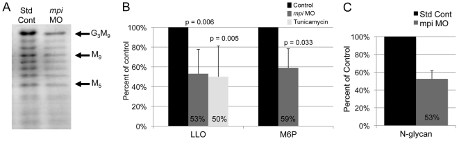
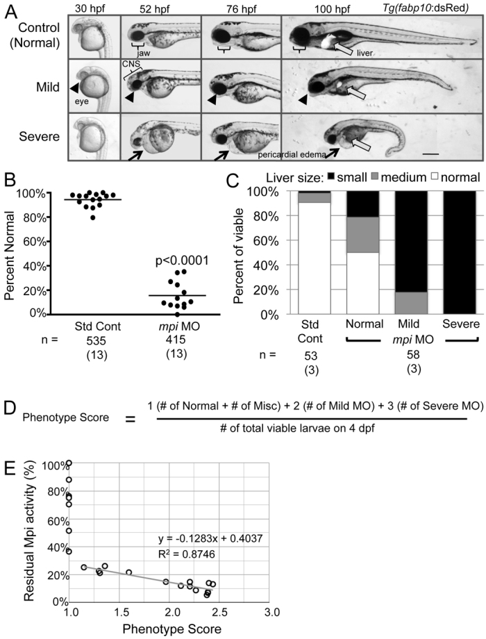
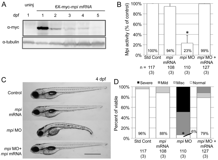
Individuals with congenital disorders of glycosylation (CDG) have recessive mutations in genes required for protein N-glycosylation, resulting in multi-systemic disease. Despite the well-characterized biochemical consequences in these individuals, the underlying cellular defects that contribute to CDG are not well understood. Synthesis of the lipid-linked oligosaccharide (LLO), which serves as the sugar donor for the N-glycosylation of secretory proteins, requires conversion of fructose-6-phosphate to mannose-6-phosphate via the phosphomannose isomerase (MPI) enzyme. Individuals who are deficient in MPI present with bleeding, diarrhea, edema, gastrointestinal bleeding and liver fibrosis. MPI-CDG patients can be treated with oral mannose supplements, which is converted to mannose-6-phosphate through a minor complementary metabolic pathway, restoring protein glycosylation and ameliorating most symptoms, although liver disease continues to progress. Because Mpi deletion in mice causes early embryonic lethality and thus is difficult to study, we used zebrafish to establish a model of MPI-CDG. We used a morpholino to block mpi mRNA translation and established a concentration that consistently yielded 13% residual Mpi enzyme activity at 4 days post-fertilization (dpf), which is within the range of MPI activity detected in fibroblasts from MPI-CDG patients. Fluorophore-assisted carbohydrate electrophoresis detected decreased LLO and N-glycans in mpi morphants. These deficiencies resulted in 50% embryonic lethality by 4 dpf. Multi-systemic abnormalities, including small eyes, dysmorphic jaws, pericardial edema, a small liver and curled tails, occurred in 82% of the surviving larvae. Importantly, these phenotypes could be rescued with mannose supplementation. Thus, parallel processes in fish and humans contribute to the phenotypes caused by Mpi depletion. Interestingly, mannose was only effective if provided prior to 24 hpf. These data provide insight into treatment efficacy and the broader molecular and developmental abnormalities that contribute to disorders associated with defective protein glycosylation.


Figures

Fig. 1.
Fig. 1.
Titration of mpi morpholino results in dose-dependent mortality and Mpi enzyme knockdown. (A) Abridged schematic of N-glycosylation focused on the MPI and PMM2 enzymatic steps. (B) Zebrafish embryos were injected with mpi ATG blocking morpholino (MO) and collected at 4 days post-fertilization (dpf). Cumulative mortality on 4 dpf for the increasing amounts of mpi MO injected are shown. * and **, P<0.0001 by Fisher’s exact test. Numbers in parentheses under n values indicate the number of experiments. (C) Mpi activity was measured in 4 dpf lysates (normalized to total protein) using the standard coupled assay for this enzyme. Mpi activity expressed as the percent of control activity is shown. Gray bar indicates the range of Mpi activity demonstrated in MPI-CDG individuals (3–18%). *P=0.04; **P=7.52E–06; ***P=1.38E–18 by unpaired t-test. (D) Mpi enzyme activity is 13% (as compared with standard control MO) when 6.7 ng of mpi MO is injected. *P<0.0001 by Fisher’s exact test.
Fig. 2.
Fig. 2.
Mpi knockdown decreases full-length LLO and mannose-6-phosphate levels in zebrafish. (A) Full-length LLO (G3M9) levels are decreased by Mpi knockdown, as seen on FACE analysis. (B) G3M9 LLO (normalized to number of fish) and mannose-6-phosphate (M6P) levels (normalized to 100 microgram protein) in lysates from 4 dpf zebrafish larvae. Values from larvae injected with the standard control morpholino were set as 100%. Average from three experiments. (C) N-glycans are decreased to 53% in mpi morphants on FACE analysis as compared with standard controls (n=2 experiments).
Fig. 3.
Fig. 3.
mpi morphants develop multi-systemic abnormalities. (A) Injection of 6.7 ng of mpi MO results in morphants that are characterized by a small head, microphthalmia, pericardial edema, jaw defects and reduced liver size as visualized in live fish expressing dsRed in hepatocytes [Tg(fabp10:dsRed)] from 30 to 100 hpf. Scale bar: 50 μm. (B) Embryo clutches injected with mpi MO have an average of 15% phenotypically normal embryos as compared with 93% of control embryos. Range from 0–35% normal. P<0.0001 by Fisher’s exact test. Numbers in parentheses under n values indicate the number of experiments. (C) Injection of 6.7 ng of mpi MO results in abnormal liver development. Liver size correlated with severity of phenotype. (D) ‘Phenotype score’ equation. (E) Linear regression analysis shows that phenotype score is inversely correlated with residual Mpi enzyme activity.
Fig. 4.
Fig. 4.
mpi morphants are rescued with mpi mRNA. Full-length zebrafish mpi mRNA was cloned using degenerate sequencing primers surrounding the ATG site so that no significant overlap with MO sequence would occur. The embryos were injected with either standard control MO, mpi MO, 6×-myc-mpi mRNA or mpi MO + 6×-myc-mpi mRNA. (A) Western blot showing Mpi expression in embryos from 1 to 5 dpf after mpi mRNA injection at t=0. (B) Restoration of Mpi enzyme activity in embryos injected with either mpi mRNA only or co-injected with mpi MO + mRNA (P=0.47 and 0.80 by paired t-test, respectively, as compared with standard control MO). *P=0.047 by paired t-test. Results from three experiments. (C) mpi morphants show phenotypic rescue when co-injected with 6×-myc-mpi mRNA. Scale bar: 50 μm. (D) At 4 dpf, 78% of morphants injected with 6×-myc-mpi mRNA were rescued to normal as compared with 6% normal in mpi MO embryos; * P<0.0001 by Fisher’s exact test. Numbers in parentheses under n values indicate the number of experiments.
Fig. 5.
Fig. 5.
Mannose supplementation rescues mpi morphants. (A) Phenotype rescue of mpi morphants treated with 50 mM mannose at 4 dpf. Liver visualized using Tg(fabp10:dsRed). Scale bar: 100 μm. (B) 77% of mpi morphants treated with 50 mM mannose from fertilization through 4 dpf appear completely normal, whereas no rescue is seen with glucose. *P<0.0001 by Fisher’s exact test as compared with mpi MO + water or mpi MO + glucose supplementation. (C) Liver size is improved in mpi morphants treated with mannose supplementation. *P<0.0001 by Fisher’s exact test. (D) Mpi activity is not significantly changed with mannose supplementation. (E) FACE analysis with quantification showing restoration of G3M9 LLO levels in mpi morphants treated with mannose as compared with mpi MO alone. Average of two experiments. (F) LLO and M6P levels are increased in mpi morphants treated with mannose to 105% and 68%, respectively. Average of two experiments. Numbers in parentheses under n values indicate the number of experiments.
Fig. 6.
Fig. 6.
Embryonic rescue is only effective with mannose supplementation early in development. (A) Time course of mannose removal. Mannose was added to control and mpi MO embryos at 1 hpf and then subsequently removed at 24, 48 or 72 hpf, or not removed at all until scored at 96 hpf. *P<0.0001, 0.0003, 0.0003 and 0.0001, respectively, by Fisher’s exact test compared with mpi MO alone. (B) Time course of mannose addition. Mannose was added to control and mpi MO embryos at 0, 24 or 48 hpf. Significant rescue only occurred in embryos in which mannose was added at 0 hpf (*P<0.0001 by Fisher’s exact test), whereas no rescue was seen in embryos where mannose was added at 24 hpf or at 48 hpf (P=0.2045 and 0.3624, respectively, by Fisher’s exact test). Numbers in parentheses under n values indicate the number of experiments.

References

    1. Babovic-Vuksanovic D., Patterson M. C., Schwenk W. F., O’Brien J. F., Vockley J., Freeze H. H., Mehta D. P., Michels V. V. (1999). Severe hypoglycemia as a presenting symptom of carbohydrate-deficient glycoprotein syndrome. J. Pediatr. 135, 775–781 - PubMed
    1. Blomme B., Van Steenkiste C., Callewaert N., Van Vlierberghe H. (2009). Alteration of protein glycosylation in liver diseases. J. Hepatol. 50, 592–603 - PubMed
    1. Bradford M. M. (1976). A rapid and sensitive method for the quantitation of microgram quantities of protein utilizing the principle of protein-dye binding. Anal. Biochem. 72, 248–254 - PubMed
    1. Chu J., Sadler K. C. (2009). New school in liver development: lessons from zebrafish. Hepatology 50, 1656–1663 - PMC - PubMed
    1. Cinaroglu A., Gao C., Imrie D., Sadler K. C. (2011). Activating transcription factor 6 plays protective and pathological roles in steatosis due to endoplasmic reticulum stress in zebrafish. Hepatology 54, 495–508 - PMC - PubMed

Publication types

MeSH terms