. 2012;7(8):e41273.
doi: 10.1371/journal.pone.0041273. Epub 2012 Aug 10.

O-antigen modulates infection-induced pain states

Affiliations

O-antigen modulates infection-induced pain states

Charles N Rudick et al. PLoS One. 2012.

Abstract

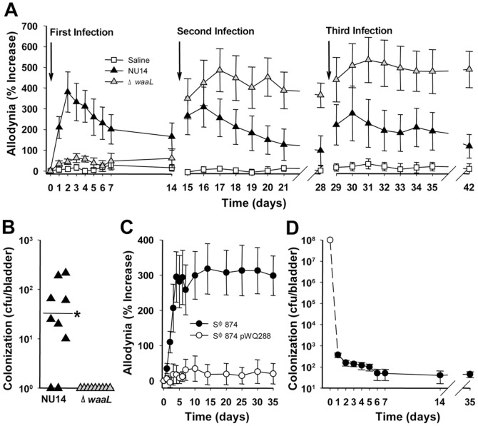
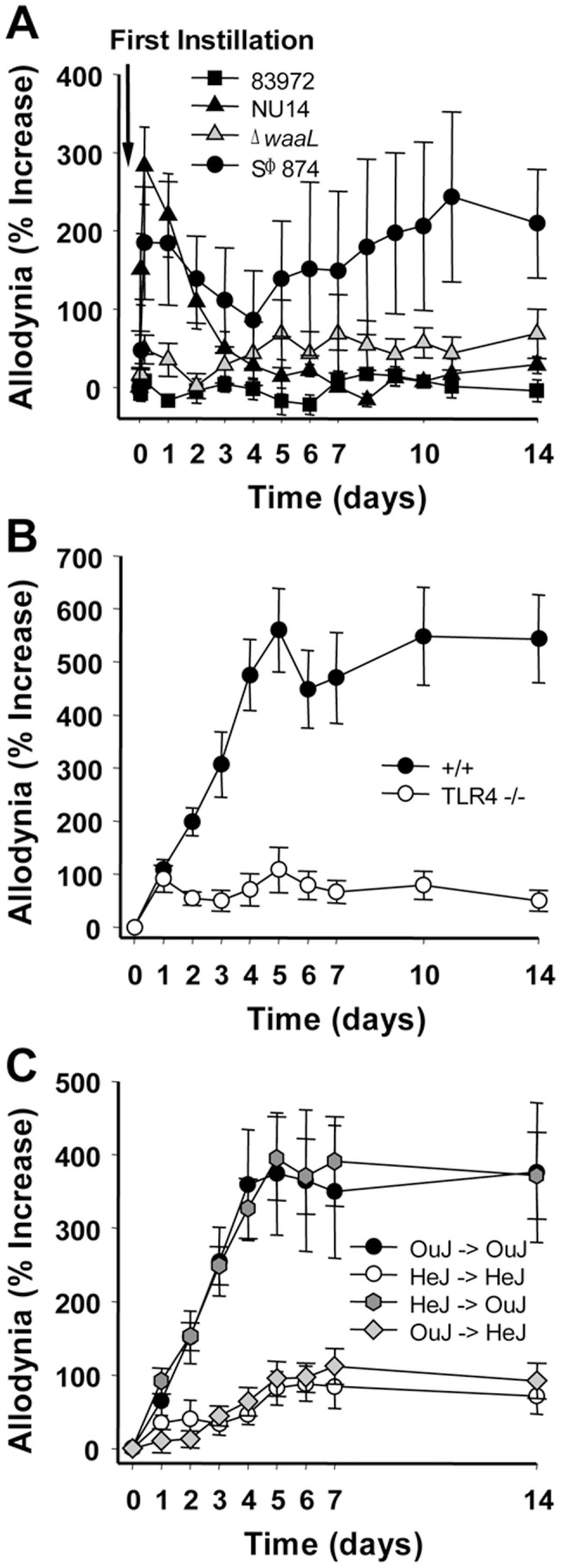
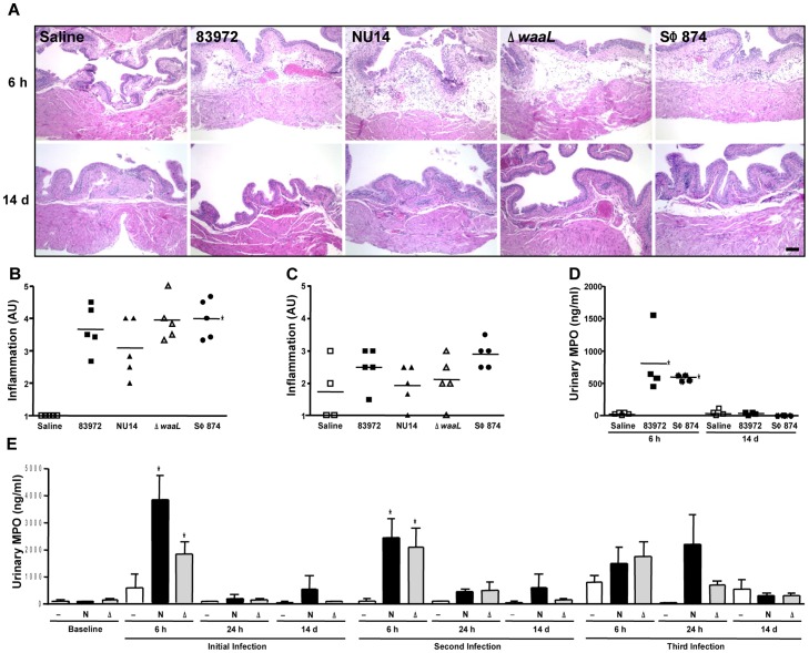
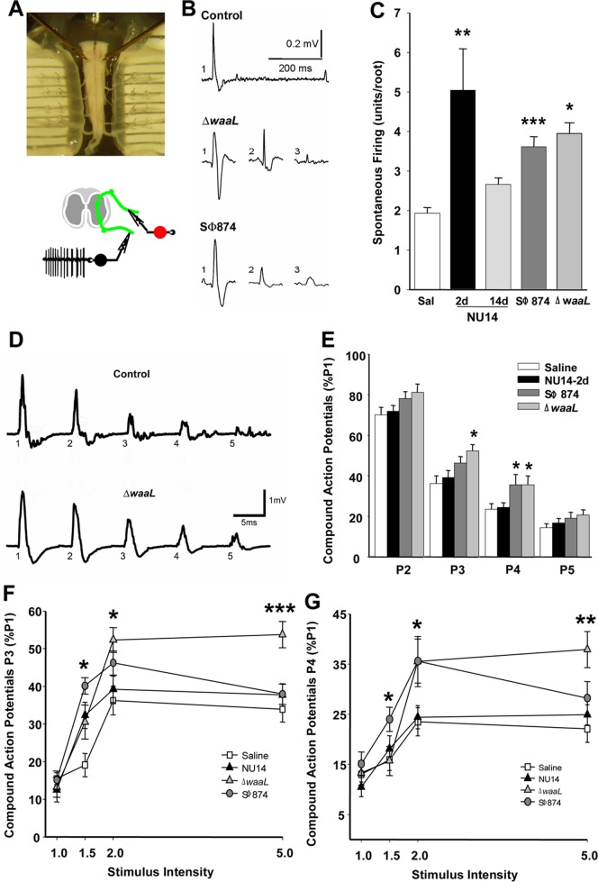
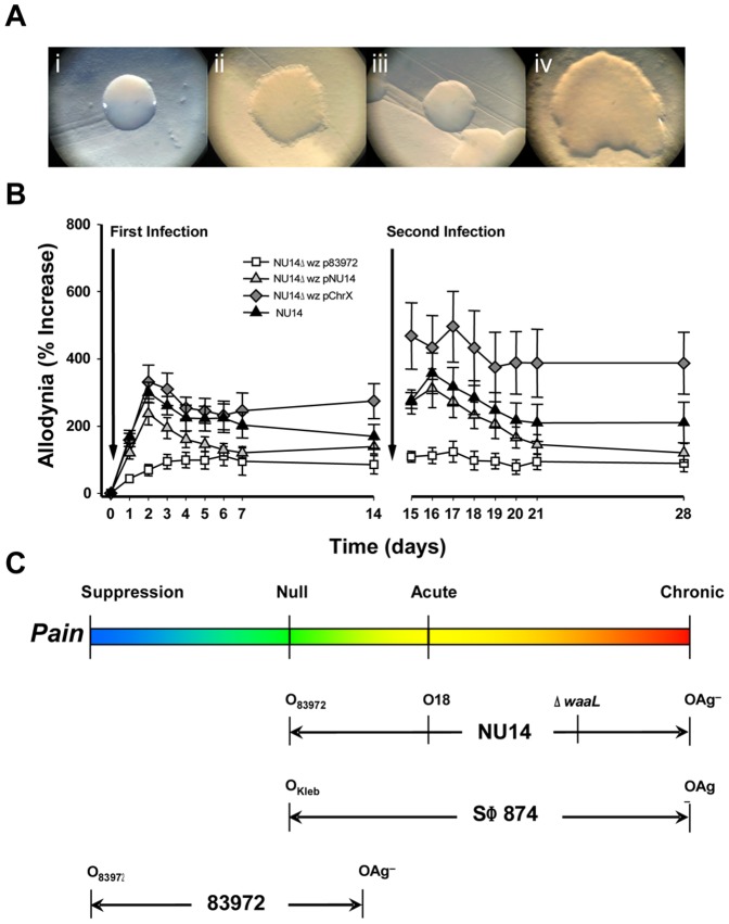
The molecular initiators of infection-associated pain are not understood. We recently found that uropathogenic E. coli (UPEC) elicited acute pelvic pain in murine urinary tract infection (UTI). UTI pain was due to E. coli lipopolysaccharide (LPS) and its receptor, TLR4, but pain was not correlated with inflammation. LPS is known to drive inflammation by interactions between the acylated lipid A component and TLR4, but the function of the O-antigen polysaccharide in host responses is unknown. Here, we examined the role of O-antigen in pain using cutaneous hypersensitivity (allodynia) to quantify pelvic pain behavior and using sacral spinal cord excitability to quantify central nervous system manifestations in murine UTI. A UPEC mutant defective for O-antigen biosynthesis induced chronic allodynia that persisted long after clearance of transient infections, but wild type UPEC evoked only acute pain. E. coli strains lacking O-antigen gene clusters had a chronic pain phenotype, and expressing cloned O-antigen gene clusters altered the pain phenotype in a predictable manner. Chronic allodynia was abrogated in TLR4-deficient mice, but inflammatory responses in wild type mice were similar among E. coli strains spanning a wide range of pain phenotypes, suggesting that O-antigen modulates pain independent of inflammation. Spinal cords of mice with chronic allodynia exhibited increased spontaneous firing and compromised short-term depression, consistent with centralized pain. Taken together, these findings suggest that O-antigen functions as a rheostat to modulate LPS-associated pain. These observations have implications for an infectious etiology of chronic pain and evolutionary modification of pathogens to alter host behaviors.


Conflict of interest statement

Competing Interests: The authors have declared that no competing interests exist.

Figures

Figure 1
Figure 1. TLR4 mediates chronic pain.
UTI was induced in female mice by instilling 108 E. coli into the bladder, and tactile allodynia and bladder colonization were quantified. (A) Mice were instilled repeatedly with saline, NU14, or ΔwaaL (n = 9). NU14 induced resolving acute pain (P<0.001 Days 2–5, P<0.01 Days 1 and 6, P<0.05 Days 7–10), but ΔwaaL induced chronic pain following the second infection. (B) Bladders from mice in (A) had detectable NU14 colonization but not ΔwaaL colonization (P<0.044). (C) SΦ874 pain (n = 10) was abrogated in mice infected with SΦ874 pWQ288 (n = 10). (D) SΦ874 is cleared rapidly from the bladder; open circle indicates inoculum (n = 5 Days 1–14, n = 10 Day 35).
Figure 2
Figure 2. Purified LPS mimics effects of intact E. coli, and chronic pain is TLR4-dependent.
(A) Mice (n = 8) were instilled 25 µl of 2 µg/ml of LPS purified from NU14, 83972, ΔwaaL, or SΦ874 and then evaluated for pelvic allodynia. (B) SΦ874-induced pain in +/+ mice is reduced in TLR4−/− mice (n = 5; P<0.05 Days 4–14). (C) +/+ mice (C3H/HeJOuJ, “OuJ") or TLR4-deficient mice (C3H/HeJ, “HeJ") were used as bone marrow donors for γ-irradiated recipients; legend arrow indicates donor bone marrow into recipient (n = 9, 9, 14 and 15 respectively). C3H/HeJOuJ recipients exhibited SΦ874-induced pain that was reduced in C3H/HeJ recipients (P<0.01 Days 3–14).
Figure 3
Figure 3. E. coli with differential pain phenotypes do not elicit differential inflammation.
(A) Hematoxylin-eosin stained sections of bladders from mice instilled with saline, 83972, NU14, ΔwaaL, and SΦ874 appeared similar at 6 hours and 14 days. Calibration mark is 100 µm. (B) Inflammation that was scored by a blinded reviewer and expressed as arbitrary units (AU) was significantly elevated for 83972-, NU14-, ΔwaaL-, and SΦ874-infected bladders harvested at 6 hours, relative to saline (P<0.001) but was not significantly different among E. coli. (C) Inflammation scores were not significantly different for 83972-, NU14-, ΔwaaL-, and SΦ874-infected bladders harvested at 14 days (P = 0.11). (D) Myeloperoxidase (MPO) was quantified in mouse urine by ELISA. Urines were collected at 6 h and 14 d following instillation of saline, 83972, or SΦ874. (E) MPO was quantified in mouse urines obtained at baseline or at 6 h, 24 h, and 14 d following serial instillation of saline (—, n = 4), NU14 (N, n = 4), or ΔwaaL (Δ, n = 7). *P<0.05. No significant differences were observed in urinary MPO of mice with treated with NU14 or ΔwaaL.
Figure 4
Figure 4. Pelvic pain behavior is associated with sacral spinal cord excitability.
Spontaneous action potentials and evoked potentials were quantified in sacral spinal cords ex vivo at ventral roots S1–S3. (A) A sacral spinal cord is mounted in a recording chamber (upper panel), and spontaneous activity is recorded from the ventral root (lower panel). (B) Representative action potentials from spontaneous firing of individual neurons identified by pCLAMP. (C) Firing activity in sacral spinal cords is higher in ΔwaaL at 14 d (n = 5, P = 0.0099), SΦ874 at 20 days (n = 9, P = 0.0001), and NU14 mice at 2 d (n = 6, P = 0.0298) than in saline controls (n = 7) or resolved NU14 (n = 5). (D) Evoked ventral root responses to dorsal root current at 2× current intensity for spinal cord of saline mouse at 2 d (upper trace) and ΔwaaL-infected mouse after serial infection. (E) Normalized responses at P2–P5 relative to P1 in ventral roots of mice instilled with saline (n = 23), NU14 (n = 26), SΦ874 (n = 21), or ΔwaaL (n = 12). *P<0.05 and **P<0.01 by Student t test relative to saline. (F and G) Responses across stimulus intensities at P3 and P4, respectively.
Figure 5
Figure 5. O-antigen modulates pain states.
Mice were infected with NU14Δwz* bearing a deletion of the O-antigen gene clusters and harboring like or heterologous complementation constructs. (A) NU14 smooth colony morphology (i) is rough in the NU14Δwz* mutant with a human X chromosome plasmid (ii) or a 83972 wz* plasmid (iv) but is rescued by an NU14wz* plasmid (iii). (B) Tactile allodynia of mice in response to sequential infection with NU14 or NU14Δwz* containing plasmids with the wz* cluster of 83972, NU14, or a fragment of the human X chromosome (n = 10). (C) Summary of O-antigen modulation of pain responses.

References

    1. Basbaum AI, Bautista DM, Scherrer G, Julius D (2009) Cellular and molecular mechanisms of pain. Cell 139: 267–284. - PMC - PubMed
    1. Scholz J, Woolf CJ (2007) The neuropathic pain triad: neurons, immune cells and glia. Nat Neurosci 10: 1361–1368. - PubMed
    1. Foxman B, Barlow R, D'Arcy H, Gillespie B, Sobel JD (2000) Urinary tract infection: self-reported incidence and associated costs. Ann Epidemiol 10: 509–515. - PubMed
    1. Schappert SM (1999) Ambulatory care visits to physician offices, hospital outpatient departments, and emergency departments: United States, 1997.PG - i–iv. Vital Health Stat 13. - PubMed
    1. Schaeffer AJ, Schaeffer EM (2007) Infections of the urinary tract. In: Wein A, Kavoussi L, Novick A, Partin A, Peters C, editors. Campbell-Walsh Urology. 8th ed. Philadelphia: Elsevier. pp. 223–303.

Publication types