. 2012;7(8):e42921.
doi: 10.1371/journal.pone.0042921. Epub 2012 Aug 10.

Sensitivity to TOP2 targeting chemotherapeutics is regulated by Oct1 and FILIP1L

Affiliations

Sensitivity to TOP2 targeting chemotherapeutics is regulated by Oct1 and FILIP1L

Huarui Lu et al. PLoS One. 2012.

Abstract

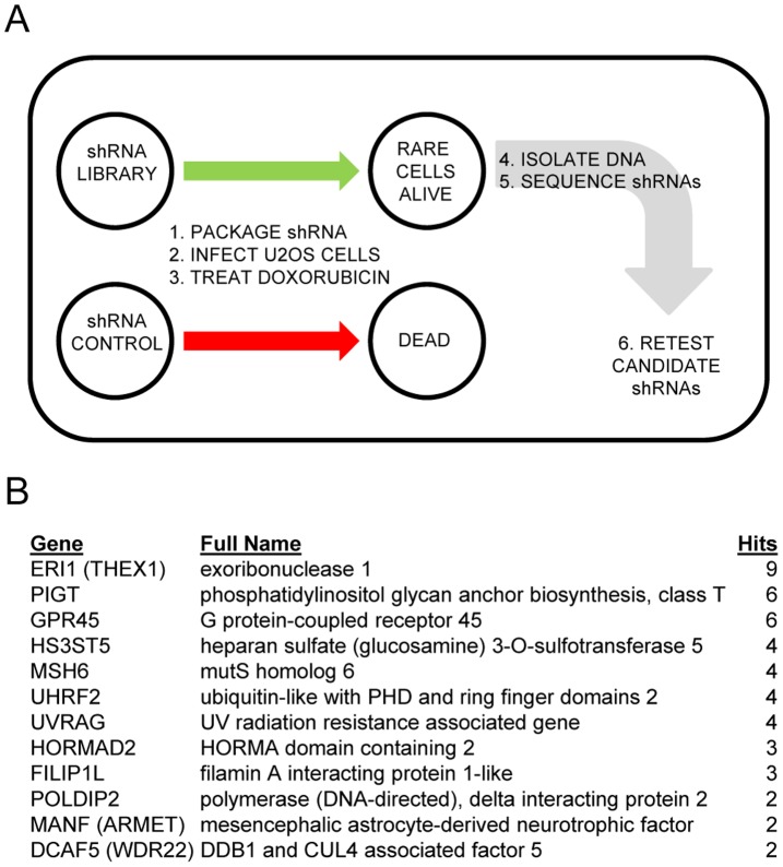
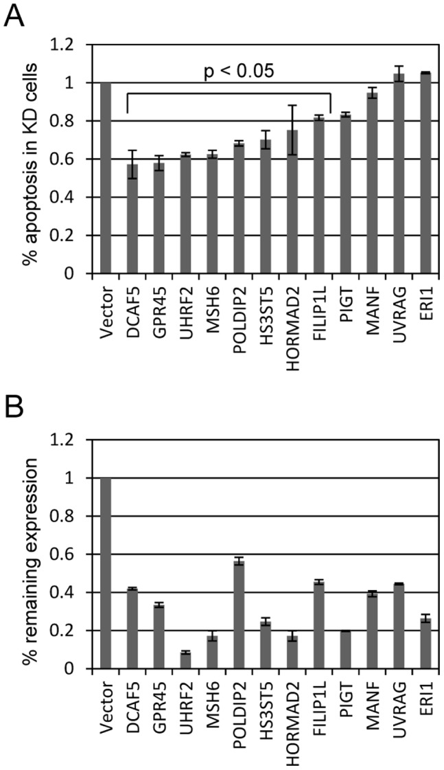
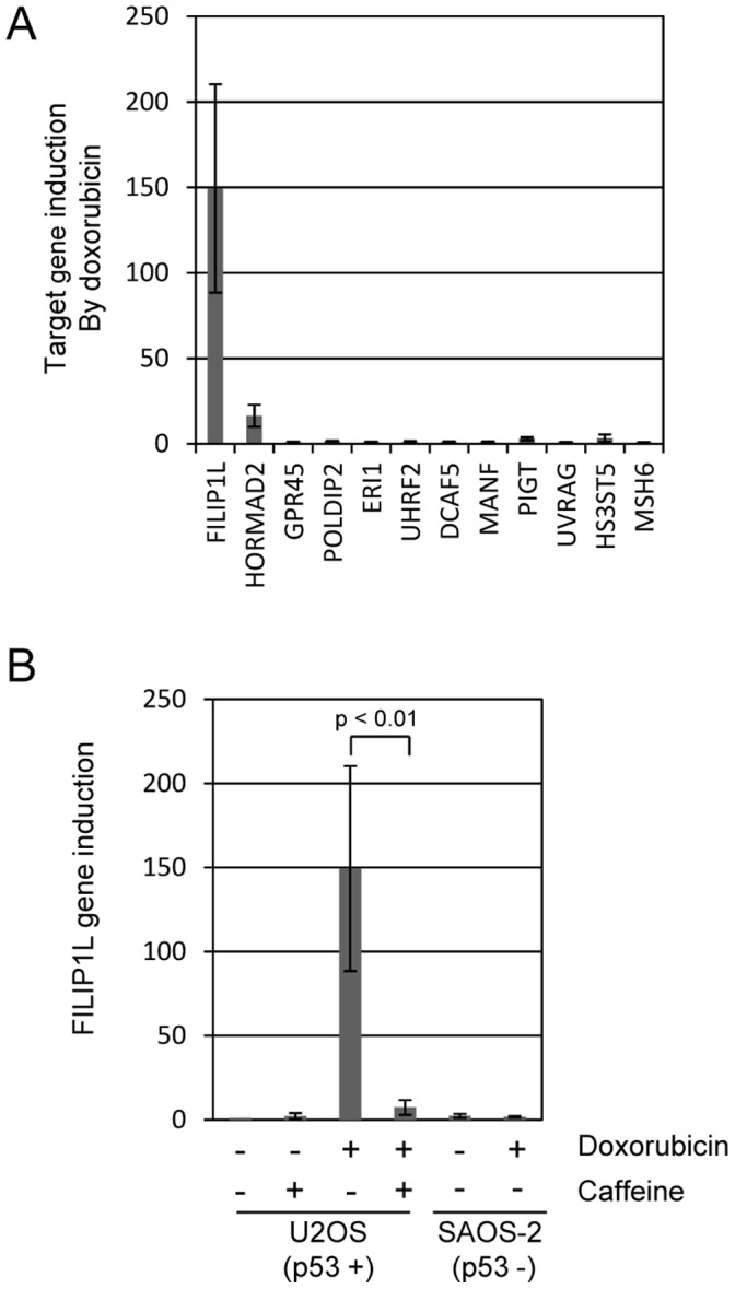
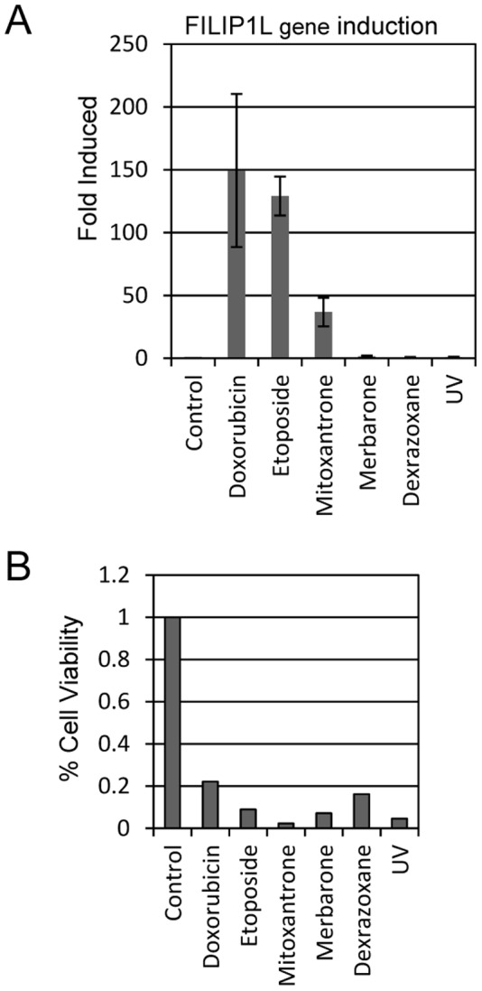
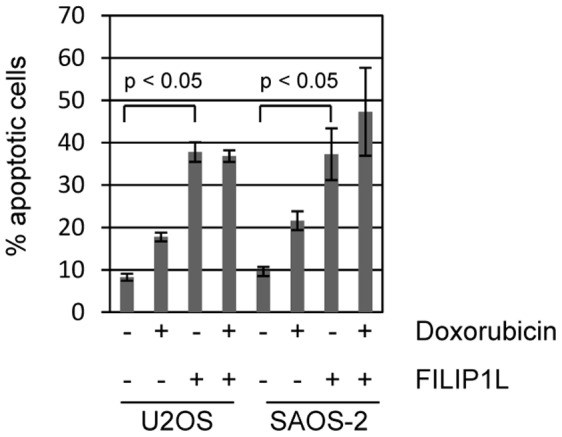
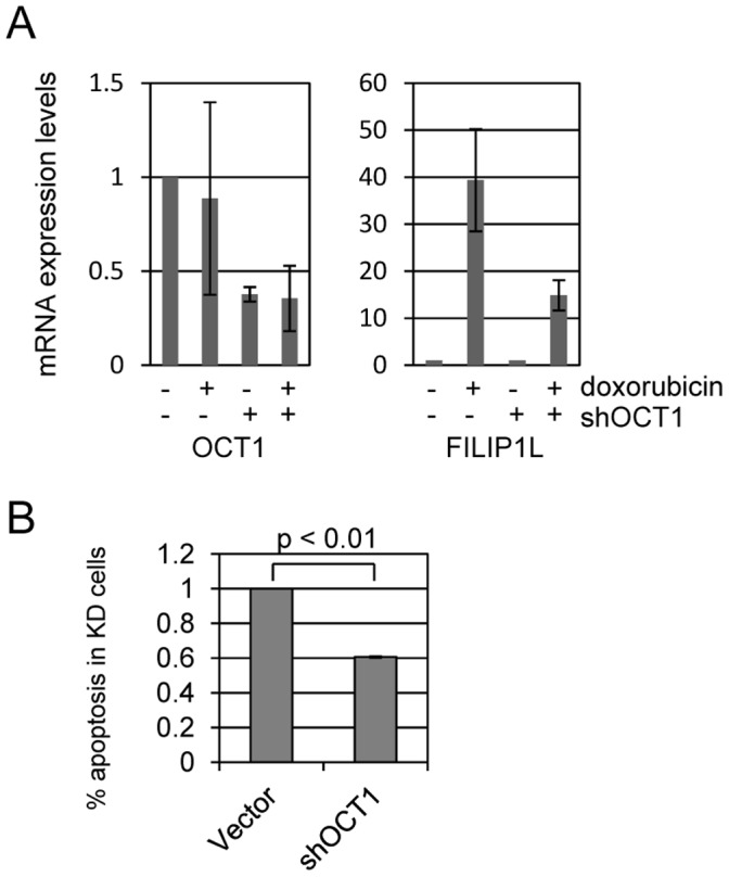
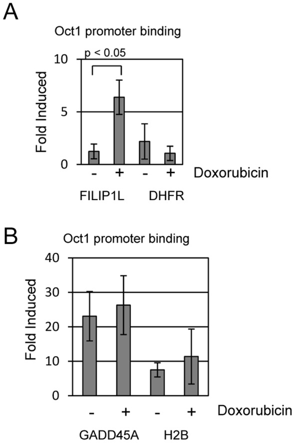
Topoisomerase II (TOP2) targeting drugs like doxorubicin and etoposide are frontline chemotherapeutics for a wide variety of solid and hematological malignancies, including breast and ovarian adenocarcinomas, lung cancers, soft tissue sarcomas, leukemias and lymphomas. These agents cause a block in DNA replication leading to a pronounced DNA damage response and initiation of apoptotic programs. Resistance to these agents is common, however, and elucidation of the mechanisms causing resistance to therapy could shed light on strategies to reduce the frequency of ineffective treatments. To explore these mechanisms, we utilized an unbiased shRNA screen to identify genes that regulate cell death in response to doxorubicin treatment. We identified the Filamin A interacting protein 1-like (FILIP1L) gene as a crucial mediator of apoptosis triggered by doxorubicin. FILIP1L shares significant similarity with bacterial SbcC, an ATPase involved in DNA repair. FILIP1L was originally described as DOC1, or "down-regulated in ovarian cancer" and has since been shown to be downregulated in a wide variety of human tumors. FILIP1L levels increase markedly through transcriptional mechanisms following treatment with doxorubicin and other TOP2 poisons, including etoposide and mitoxantrone, but not by the TOP2 catalytic inhibitors merbarone or dexrazoxane (ICRF187), or by UV irradiation. This induction requires the action of the OCT1 transcription factor, which relocalizes to the FILIP1L promoter and facilitates its expression following doxorubicin treatment. Our findings suggest that the FILIP1L expression status in tumors may influence the response to anti-TOP2 chemotherapeutics.


Conflict of interest statement

Competing Interests: The authors have declared that no competing interests exist.

Figures

Figure 1
Figure 1. A functional shRNA screen for regulators of doxorubicin induced apoptosis.
(A) Outline of the doxorubicin induced apoptosis bypass screen using U2OS cells. Pools of shRNA were transfected into retroviral packaging cell lines, and retrovirus transduced into U2OS cells followed by puromycin selection. Transduced U2OS cells were treated with 225 ng/ml Doxorubicin for 5 days, which led to apoptotic death of approximately 99.8% of the library infected cells. We harvested cells that survived treatment, isolated genomic DNA, PCR amplified the region containing shRNA sequences, shotgun cloned and sequenced. A total of approximately 1500 inserts were sequenced. (B) Twelve genes identified by this screen are listed. Full gene names and the number of times identified are also listed.
Figure 2
Figure 2. Identification of mediators of doxorubicin induced apoptosis.
To determine which genes identified by our screen were true or false positives, we targeted each for degradation by shRNA. (A) Individual genes listed in 1B were targeted for shRNA mediated degradation in U2OS cells. shRNA targeted and control cells were treated with 400 ng/ml doxorubicin and measured by propidium iodide (PI) assay 72 hours later. Levels of apoptosis are reported as % apoptosis in shRNA targeted cell compared with vector control cells. Five of the cell lines appeared to be false positives and did not display reduced doxorubicin induced apoptosis. The other lines were impaired by 20–40% for doxorubicin induced apoptosis. (B) Knockdown levels in these cell lines were determined by qPCR comparing with vector control cells and listed as % remaining expression in target cells in 2A. Genes are listed in the order presented in 2B.
Figure 3
Figure 3. Doxorubicin treatment induces FILIP1L expression.
(A) U2OS cells were treated with 200 ng/ml doxorubicin and mRNA isolated 24 hours later for qPCR analysis. The twelve genes identified in the shRNA screen were tested for induction by doxorubicin. Expression of most genes was unaffected by doxorubicin treatment. However, two genes, expression of FILIP1L and HORMAD2 were significantly induced by doxorubicin treatment, particularly FILIP1L which showed >200-fold induction. (B) FILIP1L induction by doxorubicin impaired following ATM/ATR inhibition in U2OS. Doxorubicin treatment induces DNA damage that activates the ATM and ATR kinases. Caffeine (4 mM) was used to inhibit ATM and ATR. FILIP1L induction by doxorubicin is reduced by over 90% by treatment with caffeine. SAOS-2 cells, which unlike U2OS do not contain wild-type p 53, fail to induce FILIP1L following doxorubicin treatment.
Figure 4
Figure 4. FILIP1L is induced by TOP2 poisons but not by catalytic inhibitors.
(A) U2OS cells were treated with DMSO (Control), the TOP2 poisons doxorubicin (200 ng/ml), etoposide and mitoxantrone, or the TOP2 catalytic inhibitors merbarone or dexrazoxane. After 24 hours of treatment, mRNA was harvested from cells and FILIP1L expression levels were measured by qPCR analysis. (B) To allay concern that chosen drug dosages of the TOP2 inhibitors was too low to affect FILIP1L expression, we measured % cell viability after drug treatment. Treated cells were harvested after 24 hours and cell viability was measured using an Invitrogen Countess automated cell counter.
Figure 5
Figure 5. FILIP1L expression induces cell death.
Ectopic expression of one of the identified genes, FILIP1L, caused significant induction of apoptosis on its own. U2OS and SAOS-2 cells were transfected with vector control (designated as “–” in the FILIP1L legend) or V5/His tagged FILIP1L expression plasmid. Cells were additionally treated with control or 200 ng/ml doxorubicin. Cells were harvested 24 hours after transfection and apoptotic cells were quantitated by measuring sub-G1 DNA content by propidium iodide staining. Apoptosis caused by FILIP1L expression in either cell type was not further augmented by treatment with doxorubicin.
Figure 6
Figure 6. The transcription factor OCT1 mediates doxorubicin induced FILIP1L expression and apoptosis.
OCT1, also called POU2F1 (POU domain, class 2, transcription factor 1), is a transcription factor that plays a role in the DNA damage response. We identified potential OCT1 binding sites in the FILIP1L promoter and tested if OCT1 is involved in Doxorubicin induced FILIP1L expression and apoptosis. (A) We targeted Oct1 for shRNA degradation in U2OS cells and used qPCR to verify 60% knockdown of target mRNA. Control and shOct1 cells were treated with 200 ng/ml doxorubicin and mRNA harvested 24 hours later for qPCR analysis. We determined that Oct1 mRNA levels are not affected by doxorubicin treatment. However, FILIP1L induction by doxorubicin is markedly reduced (∼65%) in shOct1 cells compared to control. (B) Control and shOct1 cells were treated with 400 ng/ml doxorubicin and measured for apoptosis at 24 hours. We found that Oct1 knockdown cells displayed 50% reduced doxorubicin induced apoptosis compared to control cells.
Figure 7
Figure 7. Doxorubicin induces OCT1 recruitment to the FILIP1L promoter.
(A) We performed chromatin immunoprecipitation assays to determine if OCT1 binds to the FILIP1L promoter and if binding is influenced by doxorubicin. U2OS cells were treated with 0 or 400 ng/ml doxorubicin for 4 hours and then harvested. Chromatin was isolated and immunoprecipitated with control IgG or anti-Oct1 antisera. Units in this figure are “fold-induced” binding compared to binding we detect with IgG control (arbitrarily set to 1) used for the ChIP assay. ChIP analysis indicated that Oct1 does not bind to the FILIP1L promoter in unstressed conditions (no doxorubicin). However, treatment with doxorubicin resulted in a 6-fold induction of OCT1 binding to the FILIP1L promoter. OCT1 does not bind to the negative control dihydrofolate reductase (DHFR) promoter in either doxorubicin treated or untreated cells. (B) GADD45A and H2B are also OCT1 transcriptional target genes. We used ChIP to ask if doxorubicin induces OCT1 binding to these promoters. We determined that OCT1 binds to both promoters in the absence of doxorubicin, and treatment did not further stimulate OCT1 binding to either promoter.
Figure 8
Figure 8. Model describing doxorubicin induction of FILIP1L and apoptosis.
Our data suggests that doxorubicin treatment significantly activates FILIP1L expression and requires both OCT1 and ATM/ATR/p 53 activity for this expression. Ectopic expression of FILIP1L is sufficient for significant apoptosis induction. It is unclear how and if ATM/ATR and p 53 interact with Oct1, or if they function in via a parallel pathway to modulate FILIP1L expression. These findings suggest that FILIP1L expression may mediate doxorubicin induced apoptosis during chemotherapy and pose the hypothesis that cancers with down-regulated FILIP1L expression may display elevated doxorubicin resistance.

References

    1. Gros P, Ben Neriah Y, Croop JM, Housman DE (1986) Isolation and expression of a complementary DNA that confers multidrug resistance. Nature 323: 728–731. - PubMed
    1. Cole SP, Bhardwaj G, Gerlach JH, Mackie JE, Grant CE, et al. (1992) Overexpression of a transporter gene in a multidrug-resistant human lung cancer cell line. Science 258: 1650–1654. - PubMed
    1. Gottesman MM, Pastan I (1993) Biochemistry of multidrug resistance mediated by the multidrug transporter. Annu Rev Biochem 62: 385–427. - PubMed
    1. Kobayashi S, Boggon TJ, Dayaram T, Jänne PA, Kocher O, et al. (2005) EGFR mutation and resistance of non-small-cell lung cancer to gefitinib. N Engl J Med 352: 786–792. - PubMed
    1. Pao W, Miller VA, Politi KA, Riely GJ, Somwar R, et al. (2005) Acquired resistance of lung adenocarcinomas to gefitinib or erlotinib is associated with a second mutation in the EGFR kinase domain. PLoS Med 2: e73. - PMC - PubMed

Publication types

MeSH terms