. 2012;7(8):e42990.
doi: 10.1371/journal.pone.0042990. Epub 2012 Aug 10.

A single protofilament is sufficient to support unidirectional walking of dynein and kinesin

Affiliations

A single protofilament is sufficient to support unidirectional walking of dynein and kinesin

Keitaro Shibata et al. PLoS One. 2012.

Abstract

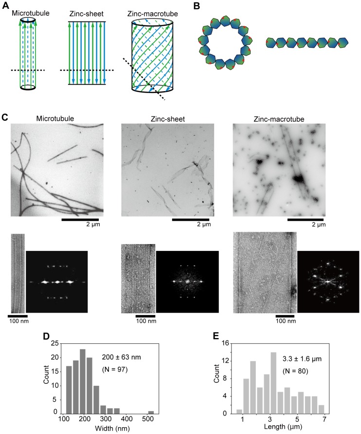
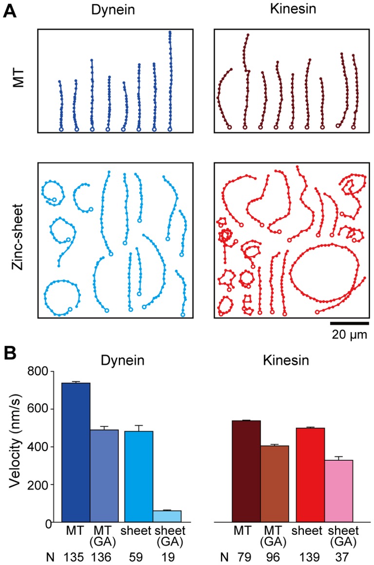
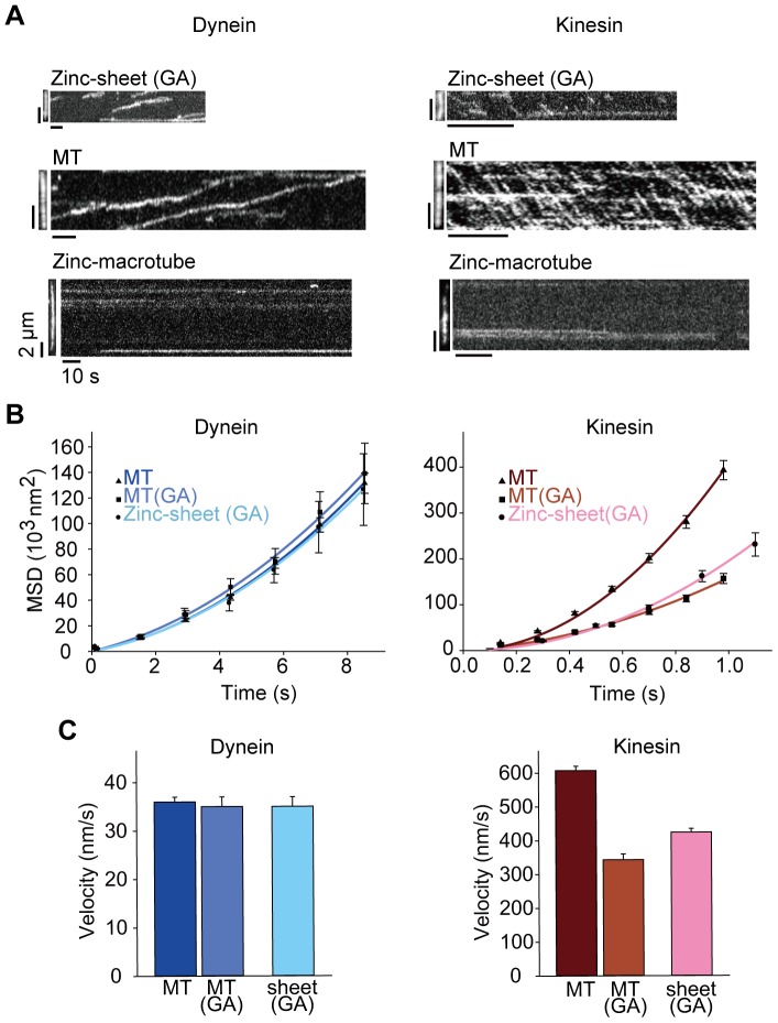
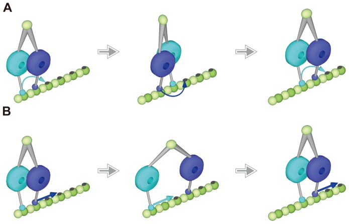
Cytoplasmic dynein and kinesin are two-headed microtubule motor proteins that move in opposite directions on microtubules. It is known that kinesin steps by a 'hand-over-hand' mechanism, but it is unclear by which mechanism dynein steps. Because dynein has a completely different structure from that of kinesin and its head is massive, it is suspected that dynein uses multiple protofilaments of microtubules for walking. One way to test this is to ask whether dynein can step along a single protofilament. Here, we examined dynein and kinesin motility on zinc-induced tubulin sheets (zinc-sheets) which have only one protofilament available as a track for motor proteins. Single molecules of both dynein and kinesin moved at similar velocities on zinc-sheets compared to microtubules, clearly demonstrating that dynein and kinesin can walk on a single protofilament and multiple rows of parallel protofilaments are not essential for their motility. Considering the size and the motile properties of dynein, we suggest that dynein may step by an inchworm mechanism rather than a hand-over-hand mechanism.


Conflict of interest statement

Competing Interests: The authors have declared that no competing interests exist.

Figures

Figure 1
Figure 1. Structure of MTs, zinc-sheets and zinc-macrotubes.
(A) The protofilament arrangements are modified from Wolf et al. (1993). Arrows represent the polarity of a protofilament, and color discriminates the outer side (green) from the inner side (blue) of a MT. Dashed lines indicate the cut plane of the cross-cut views in B. (B) The cross-cut views of a MT (left), zinc-sheet and zinc-macrotube (right). Red stars denote a helix 12 that is closely related to dynein and kinesin binding sites. (C) General views, close-up views and diffraction patterns of MTs (left), zinc-sheets (middle) and zinc-macrotubes (right). (D and E) The width and length distribution of zinc-sheets measured in EM images. The mean ± SD and number of counted zinc-sheets (N) are shown.
Figure 2
Figure 2. MT and zinc-sheet gliding movements on dynein- and kinesin-coated glass surfaces.
(A) Movement trajectories of tubulin polymers plotted by positions marked every 3 s from the starting points (open circles). MT movements on dynein (upper left) and RK430-Avi (upper right). Zinc-sheet movements on dynein (lower left) and RK430-Avi (lower right). (B) Velocities of the gliding movement. Bars represent the mean ± SEM, and (N) is the number of measured gliding tubulin polymers.
Figure 3
Figure 3. Single molecule motility of dynein and kinesin on tubulin polymers.
(A) Tubulin polymers (left panels) and kymographs of GFP-GST-Dyn1331kDa and RK430-GFP movements (right panels). (B) Mean-square displacement (MSD) plots of GFP-GST-Dyn1331kDa (left) and RK430-GFP (right) movements on MTs, MTs (GA) and zinc-sheets (GA) with fitted quadratic curves (solid line). Each plot represents the mean ± SEM. (C) Velocities of GFP-GST-Dyn1331kDa (left) and RK430-GFP (right) movements on MTs, MTs (GA) and zinc-sheets (GA) determined from MSD plots. Bars represent the mean ± SEM.
Figure 4
Figure 4. Schematic illustration of dynein walking on a single protofilament of a MT.
(A) Partially overlapping hand-over-hand model. The rear head dissociates from the protofilament to overtake the front head and the two motor domains partially overlap side by side. (B) Inchworm model. The front head is always leading and the rear head is always trailing. Each head can slide and step advance without complete dissociation from the protofilament by keeping a weak interaction with the protofilament.

References

    1. Vallee RB, Sheetz MP (1996) Targeting of motor proteins. Science 271: 1539–1544. - PubMed
    1. Hirokawa N (1998) Kinesin and Dynein Superfamily Proteins and the Mechanism of Organelle Transport. Science 279: 519–526. - PubMed
    1. Vale RD (2003) The molecular motor toolbox for intracellular transport. Cell 112: 467–480. - PubMed
    1. Chevalier-Larsen E, Holzbaur ELF (2006) Axonal transport and neurodegenerative disease. Biochim Biophys Acta 1762: 1094–1108. - PubMed
    1. Hirokawa N, Niwa S, Tanaka Y (2010) Molecular motors in neurons: transport mechanisms and roles in brain function, development, and disease. Neuron 68: 610–638. - PubMed

Publication types