Review
. 2013 Jul 20;19(3):269-84.
doi: 10.1089/ars.2012.4875. Epub 2012 Oct 5.

Mitochondrial fatty acid oxidation in obesity

Affiliations
Review

Mitochondrial fatty acid oxidation in obesity

Dolors Serra et al. Antioxid Redox Signal. .

Abstract

Significance: Current lifestyles with high-energy diets and little exercise are triggering an alarming growth in obesity. Excess of adiposity is leading to severe increases in associated pathologies, such as insulin resistance, type 2 diabetes, atherosclerosis, cancer, arthritis, asthma, and hypertension. This, together with the lack of efficient obesity drugs, is the driving force behind much research.

Recent advances: Traditional anti-obesity strategies focused on reducing food intake and increasing physical activity. However, recent results suggest that enhancing cellular energy expenditure may be an attractive alternative therapy.

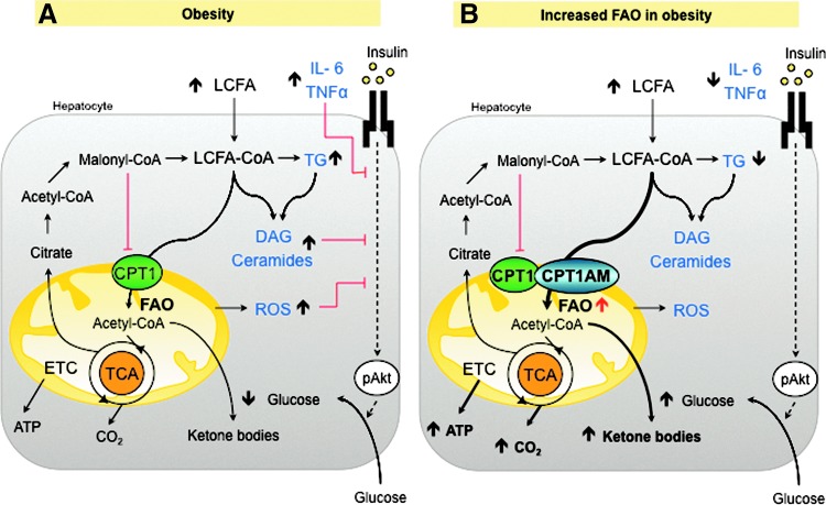
Critical issues: This review evaluates recent discoveries regarding mitochondrial fatty acid oxidation (FAO) and its potential as a therapy for obesity. We focus on the still controversial beneficial effects of increased FAO in liver and muscle, recent studies on how to potentiate adipose tissue energy expenditure, and the different hypotheses involving FAO and the reactive oxygen species production in the hypothalamic control of food intake.

Future directions: The present review aims to provide an overview of novel anti-obesity strategies that target mitochondrial FAO and that will definitively be of high interest in the future research to fight against obesity-related disorders.


Figures

FIG. 1.
FIG. 1.
Mitochondrial fatty acid oxidation (FAO). Long-chain fatty acid (LCFA) catalysis implies activation by acyl-CoA synthase (ACS) of LCFAs into LCFA-CoA, which is a substrate for the mitochondrial carnitine palmitoyltransferase 1 (CPT1) enzyme. The CPT system, which includes CPT1, acylcarnitine translocase (CACT), and CPT2, allows LCFA-CoA to enter the mitochondrial matrix, via transesterification reactions, to then be β-oxidized. CPT1 is the rate-limiting enzyme in FAO since its activity is tightly regulated by the glucose-derived malonyl-CoA, generated by acetyl-CoA carboxylase (ACC) during fatty acid (FA) de novo formation (in energetically abundant situations) or degraded by malonyl-CoA decarboxylase (MCD) in a process regulated by AMP-activated protein kinase (AMPK). Acetyl-CoA generated in FAO eventually enters the tricarboxylic acid (TCA) cycle to obtain reductive power for cellular respiration and produce ATP. To see this illustration in color, the reader is referred to the web version of this article at www.liebertpub.com/ars
FIG. 2.
FIG. 2.
Bioenergetics and mitochondrial metabolism. The mitochondrial fuels, glucose, and FAs, are converted to acetyl-CoA, which can be further metabolized to obtain energy. The TCA cycle generates protons (H+) and electrons that are carried by NADH and FADH to the electron transport chain (ETC), where the protons are transported to the mitochondrial intermembrane (MIM) space to generate energy as ATP. Highly reactive electrons may leak from the ETC and generate reactive oxygen species (ROS), which could act physiologically as signaling molecules, but can also cause significant cellular damage when overproduced. Uncoupling proteins (UCPs) dissipate the proton gradient and scavenge ROS accumulation, thus dissipating energy as heat. To see this illustration in color, the reader is referred to the web version of this article at www.liebertpub.com/ars
FIG. 3.
FIG. 3.
Effects of enhanced FAO in fatty liver. (A) Obesity increases FA uptake, triglyceride (TG), diacylglycerol (DAG), ceramides, and other lipid derivatives that may inhibit insulin signaling. FA accumulation induces mitochondrial dysfunction and increased ROS production, oxidative stress, and inflammation that could also disrupt insulin signaling. (B) Enhancing FAO by the overexpression of CPT1AM (117, 130) increases the production of ketone bodies, ATP, and CO2. The reduction of lipid content re-establishes lipid metabolism, insulin signaling, and decreases inflammation and ROS production.
FIG. 4.
FIG. 4.
White adipose tissue (WAT), obesity, and insulin resistance. Nutrient overload, weight gain, and obesity result in increased adipose tissue mass and adipocyte size. The expansion of the adipose tissue leads to adipocyte hypoxia, death, and free fatty acid (FFA) release into circulation. These events trigger the recruitment and activation of immune cells, such as macrophages and T cells, in the adipose tissue. Infiltrated and activated immune cells and adipocytes secrete large amounts of proinflammatory cytokines, which promote the inhibition of insulin signaling with an ensuing local and systemic resistance (147).
FIG. 5.
FIG. 5.
Stimulation of brown adipose tissue (BAT) thermogenesis. Cold exposure, exercise, and some secreted proteins, such as fibroblast growth factor-21 (FGF-21) (72) and irisin (12), enhance BAT burning power by promoting brown adipocyte recruitment in white fat (browning).
FIG. 6.
FIG. 6.
Two hypotheses for the role of FAO in the development of obese-induced insulin resistance in skeletal muscle (SkM). (A) During obesity, intramyocellular lipid accumulation leads to a decrease in insulin-stimulated glucose uptake in SkM (144). (B) An increase in FAO may decrease LCFAs, ceramide, and DAG content, thus enhancing insulin action (15, 66, 149). (C) However, an increase in FAO may augment ROS production and enhance the accumulation of products of incomplete β-oxidation (PIO), which are hypothesized to decrease insulin signaling through the activation of various stress kinases (89).
FIG. 7.
FIG. 7.
Hypotheses involving FAO in the regulation of food intake. During fasting, the effects of ghrelin, AMP, and FAs act in hypothalamic nuclei to increase the expression of orexigenic neuropeptides (48). The mechanisms involved in this process appear to be related to an increase in LCFA-CoA, diminished malonyl-CoA, and a certain level of ROS. Excessive ROS production is controlled by UCP2 with a negative feed-back. CPT1A is postulated to be involved in all the three approaches.

References

    1. Abu-Elheiga L. Oh W. Kordari P. Wakil SJ. Acetyl-CoA carboxylase 2 mutant mice are protected against obesity and diabetes induced by high-fat/high-carbohydrate diets. Proc Natl Acad Sci U S A. 2003;100:10207–10212. - PMC - PubMed
    1. Adams SH. Hoppel CL. Lok KH. Zhao L. Wong SW. Minkler PE. Hwang DH. Newman JW. Garvey WT. Plasma acylcarnitine profiles suggest incomplete long-chain fatty acid beta-oxidation and altered tricarboxylic acid cycle activity in type 2 diabetic African-American women. J Nutr. 2009;139:1073–1081. - PMC - PubMed
    1. Ahima RS. Digging deeper into obesity. J Clin Invest. 2011;121:2076–2079. - PMC - PubMed
    1. An J. Muoio DM. Shiota M. Fujimoto Y. Cline GW. Shulman GI. Koves TR. Stevens R. Millington D. Newgard CB. Hepatic expression of malonyl-CoA decarboxylase reverses muscle, liver and whole-animal insulin resistance. Nat Med. 2004;10:268–274. - PubMed
    1. Andersen P. Saltin B. Maximal perfusion of skeletal muscle in man. J Physiol. 1985;366:233–249. - PMC - PubMed

Publication types

LinkOut - more resources