

Cerebrospinal fluid proteome of patients with acute Lyme disease
- PMID: 22900834
- PMCID: PMC3465517
- DOI: 10.1021/pr300577p
Cerebrospinal fluid proteome of patients with acute Lyme disease
Abstract
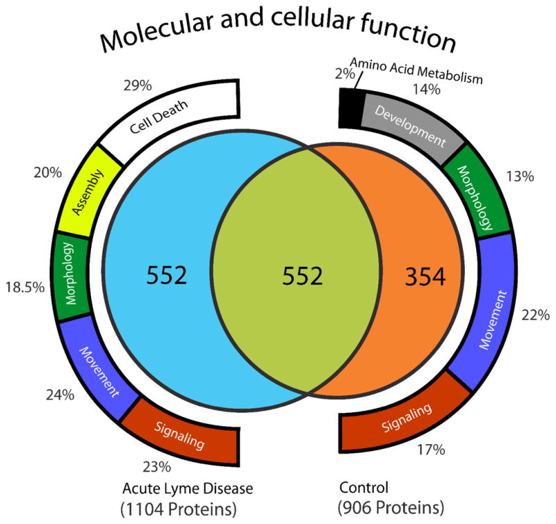
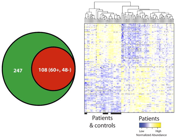
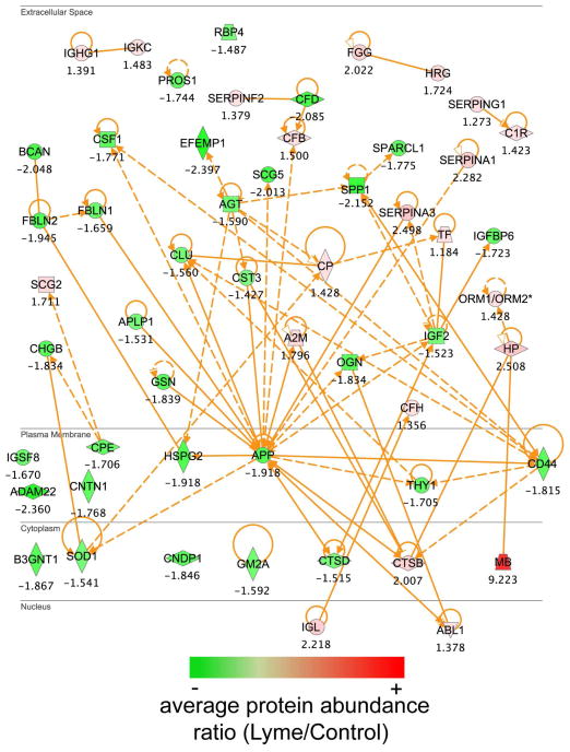
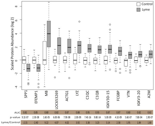
During acute Lyme disease, bacteria can disseminate to the central nervous system (CNS), leading to the development of meningitis and other neurologic symptoms. Here we have analyzed pooled cerebrospinal fluid (CSF) allowing a deep view into the proteome for patients diagnosed with early disseminated Lyme disease and CSF inflammation. Additionally, we analyzed individual patient samples and quantified differences in protein abundance employing label-free quantitative mass spectrometry-based methods. We identified 108 proteins that differ significantly in abundance in patients with acute Lyme disease from controls. Comparison between infected patients and control subjects revealed differences in proteins in the CSF associated with cell death localized to brain synapses and others that likely originate from brain parenchyma.
Conflict of interest statement
Conflict of interest statement
Thomas E. Angel- no conflict, Jon M. Jacobs- no conflict, Robert P. Smith- no conflict, Mark S. Pasternack- no conflict, Susan Elias- no conflict, Marina A. Gritsenko- no conflict, Anil Shukla- no conflict, Edward C. Gilmore- no conflict, Carol McCarthy- no conflict, David G. Camp II- no conflict, Richard D. Smith- no conflict, H. Shaw Warren- no conflict
Figures
Similar articles
-
Astroglial and neuronal proteins in cerebrospinal fluid as markers of CNS involvement in Lyme neuroborreliosis.Eur J Neurol. 1999 Mar;6(2):169-78. doi: 10.1111/j.1468-1331.1999.tb00010.x. Eur J Neurol. 1999. PMID: 10053229
-
Oral doxycycline for Lyme neuroborreliosis with symptoms of encephalitis, myelitis, vasculitis or intracranial hypertension.Eur J Neurol. 2014 Sep;21(9):1162-7. doi: 10.1111/ene.12420. Epub 2014 Mar 29. Eur J Neurol. 2014. PMID: 24684211
-
Cerebrospinal fluid cytokines in Lyme neuroborreliosis.J Neuroinflammation. 2016 Oct 18;13(1):273. doi: 10.1186/s12974-016-0745-x. J Neuroinflammation. 2016. PMID: 27756335 Free PMC article.
-
Diagnostic value of cerebrospinal fluid CXCL13 for acute Lyme neuroborreliosis. A systematic review and meta-analysis.Clin Microbiol Infect. 2018 Dec;24(12):1234-1240. doi: 10.1016/j.cmi.2018.04.007. Epub 2018 Apr 16. Clin Microbiol Infect. 2018. PMID: 29674128
-
[Neurologic syndromes in Lyme disease].Pol Merkur Lekarski. 2000 Aug;9(50):584-8. Pol Merkur Lekarski. 2000. PMID: 11081332 Review. Polish.
Cited by
-
The Lyme disease bacterium, Borrelia burgdorferi, stimulates an inflammatory response in human choroid plexus epithelial cells.PLoS One. 2020 Jul 9;15(7):e0234993. doi: 10.1371/journal.pone.0234993. eCollection 2020. PLoS One. 2020. PMID: 32645014 Free PMC article.
-
Mass spectrometry-based proteomic techniques to identify cerebrospinal fluid biomarkers for diagnosing suspected central nervous system infections. A systematic review.J Infect. 2019 Nov;79(5):407-418. doi: 10.1016/j.jinf.2019.08.005. Epub 2019 Aug 9. J Infect. 2019. PMID: 31404562 Free PMC article.
-
The Physiological Roles of Amyloid-β Peptide Hint at New Ways to Treat Alzheimer's Disease.Front Aging Neurosci. 2018 Apr 25;10:118. doi: 10.3389/fnagi.2018.00118. eCollection 2018. Front Aging Neurosci. 2018. PMID: 29922148 Free PMC article. Review.
-
Pathogenesis of Alzheimer's disease: Involvement of the choroid plexus.Alzheimers Dement. 2023 Aug;19(8):3537-3554. doi: 10.1002/alz.12970. Epub 2023 Feb 24. Alzheimers Dement. 2023. PMID: 36825691 Free PMC article.
-
Characterization of individual mouse cerebrospinal fluid proteomes.Proteomics. 2014 May;14(9):1102-6. doi: 10.1002/pmic.201300241. Epub 2014 Mar 20. Proteomics. 2014. PMID: 24677814 Free PMC article.
References
-
- Burgdorfer W, Barbour AG, Hayes SF, Benach JL, Grunwaldt E, Davis JP. Lyme disease-a tick-borne spirochetosis? Science. 1982;216(4552):1317–9. - PubMed
-
- Alghaferi MY, Anderson JM, Park J, Auwaerter PG, Aucott JN, Norris DE, Dumler JS. Borrelia burgdorferi ospC heterogeneity among human and murine isolates from a defined region of northern Maryland and southern Pennsylvania: Lack of correlation with invasive and noninvasive genotypes. Journal of Clinical Microbiology. 2005;43(4):1879–1884. - PMC - PubMed
MeSH terms
Substances
Grants and funding
LinkOut - more resources
Full Text Sources
Other Literature Sources
