Review
. 2013 Jul 1;19(1):63-71.
doi: 10.1089/ars.2012.4864. Epub 2012 Oct 2.

Disulfide bond formation in the bacterial periplasm: major achievements and challenges ahead

Affiliations
Review

Disulfide bond formation in the bacterial periplasm: major achievements and challenges ahead

Katleen Denoncin et al. Antioxid Redox Signal. .

Abstract

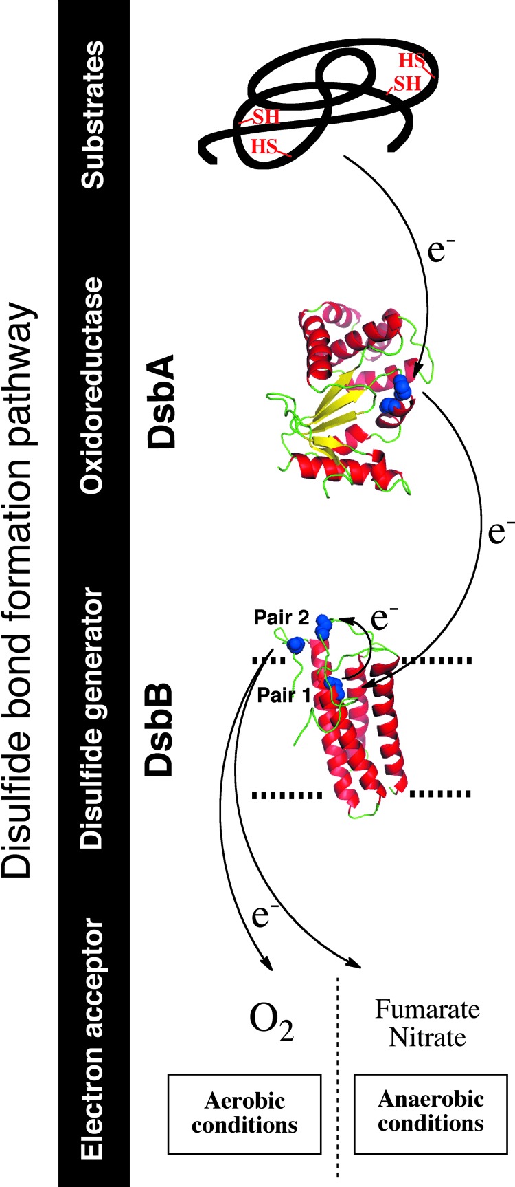
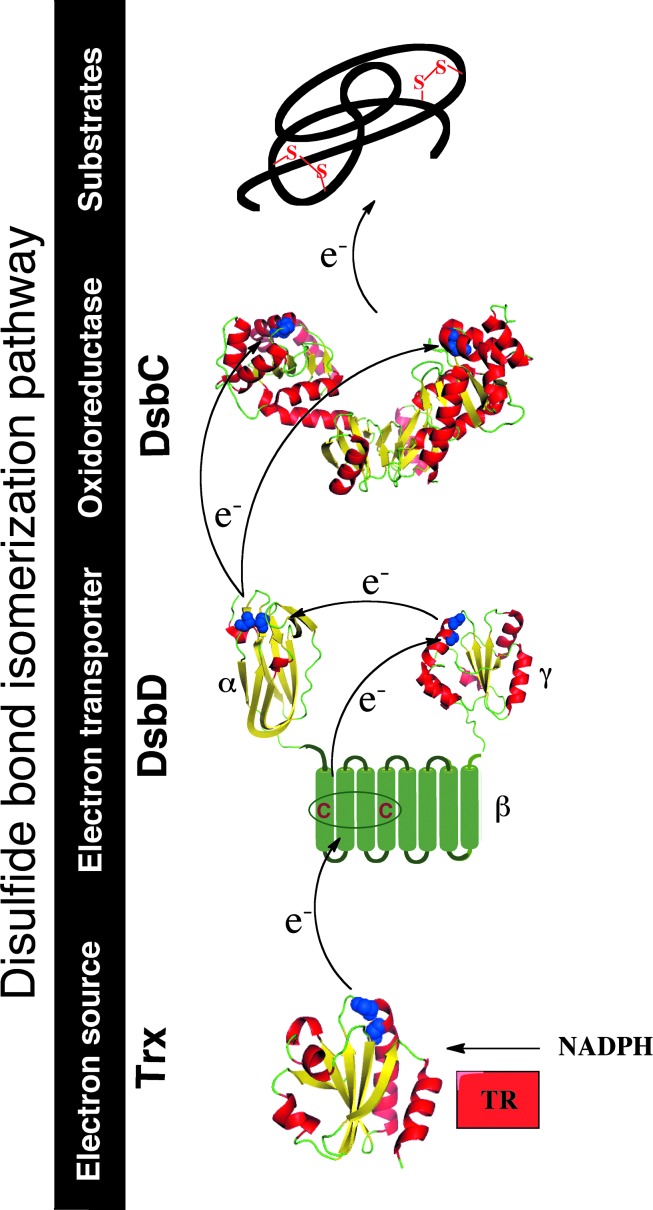
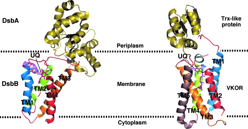
Significance: The discovery of the oxidoreductase disulfide bond protein A (DsbA) in 1991 opened the way to the unraveling of the pathways of disulfide bond formation in the periplasm of Escherichia coli and other Gram-negative bacteria. Correct oxidative protein folding in the E. coli envelope depends on both the DsbA/DsbB pathway, which catalyzes disulfide bond formation, and the DsbC/DsbD pathway, which catalyzes disulfide bond isomerization.

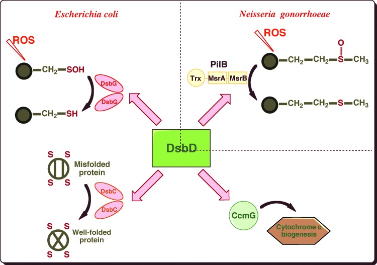
Recent advances: Recent data have revealed an unsuspected link between the oxidative protein-folding pathways and the defense mechanisms against oxidative stress. Moreover, bacterial disulfide-bond-forming systems that differ from those at play in E. coli have been discovered.

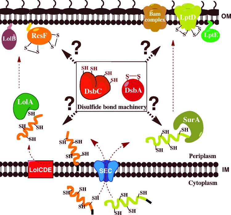
Critical issues: In this review, we discuss fundamental questions that remain unsolved, such as what is the mechanism employed by DsbD to catalyze the transfer of reducing equivalents across the membrane and how do the oxidative protein-folding catalysts DsbA and DsbC cooperate with the periplasmic chaperones in the folding of secreted proteins.

Future directions: Understanding the mechanism of DsbD will require solving the structure of the membranous domain of this protein. Another challenge of the coming years will be to put the knowledge of the disulfide formation machineries into the global cellular context to unravel the interplay between protein-folding catalysts and chaperones. Also, a thorough characterization of the disulfide bond formation machineries at work in pathogenic bacteria is necessary to design antimicrobial drugs targeting the folding pathway of virulence factors stabilized by disulfide bonds.


Figures

FIG. 1.
FIG. 1.
Disulfide bond formation in the Escherichia coli periplasm. Disulfide bonds are introduced by disulfide bond protein A (DsbA), which is then recycled by the inner membrane (IM) protein DsbB. DsbB generates disulfides de novo from quinone reduction. The electrons are then transferred to the respiratory chain. The final electron acceptor is molecular oxygen (O2) under aerobic conditions and nitrate or fumarate under anaerobic conditions. Three-dimensional structures of E. coli DsbA (protein database [PDB] entry code 1A2L) and E. coli DsbB (PDB entry code 2ZUQ) are drawn in ribbon form. Cysteine residues are drawn in space-filling form and colored in blue. Black arrows indicate the flow of electrons. The figures were generated using MacPyMol (Delano Scientific LLC 2006). To see this illustration in color, the reader is referred to the web version of this article at www.liebertpub.com/ars.
FIG. 2.
FIG. 2.
Disulfide bond isomerization in the E. coli periplasm. Non-native disulfides are repaired by the soluble homodimeric protein DsbC. DsbC is kept reduced in the periplasm by the membrane protein DsbD. DsbD receives electrons from cytoplasmic thioredoxin (Trx), which is kept reduced by thioredoxin reductase (TR), at the expense of reduced nicotine adenine dinucleotide phosphate (NADPH). The three-dimensional structures of E. coli DsbC (PDB entry code 1EEJ), DsbDα (PDB entry code 1JPE), and DsbDγ (PDB entry code 2FWF) are drawn in ribbon form. Cysteine residues are drawn in space-filling form and colored in blue. Black arrows indicate the flow of electrons. The figures were generated using MacPyMol (Delano Scientific LLC 2006). To see this illustration in color, the reader is referred to the web version of this article at www.liebertpub.com/ars.
FIG. 3.
FIG. 3.
DsbD is an electron hub that transfers reducing equivalents to several oxidoreductases present in the cell envelope. In E. coli, DsbD provides reducing equivalents to DsbC, the isomerase that corrects non-native disulfides, to CcmG, an oxidoreductase involved in the maturation of cytochrome c and to DsbG, which controls the global sulfenic acid content of the periplasm. In Neisseria gonorrhoeae, DsbD shuttles electrons to the N-terminal domain of PilB, a multidomain protein that exhibits methionine sulfoxide reductase (Msr) activity (To see this illustration in color, the reader is referred to the web version of this article at www.liebertpub.com/ars.)
FIG. 4.
FIG. 4.
Protein-folding catalysts cooperate with periplasmic chaperones in the assembly of outer membrane (OM) proteins. The β-barrel protein LptD and the lipoprotein RcsF are synthesized in the cytoplasm. They are transported unfolded across the IM by the SEC translocon. RcsF is first addressed to the IM complex LolCDE and then transferred to the chaperone LolA. LolA transports RcsF across the periplasm and delivers it to LolB, which incorporates lipoproteins into the OM. After translocation by SEC, LptD is escorted across the periplasm by the chaperone SurA. SurA delivers β-barrel proteins to the Bam machinery, which inserts them in the OM. The assembly of LptD requires the formation of the LptD/LptE complex. The oxidative folding catalysts DsbA and DsbC are involved in the formation and isomerization of the nonconsecutive disulfides of RcsF and LptD. How they cooperate with LolA and SurA is not clearly established (To see this illustration in color, the reader is referred to the web version of this article at www.liebertpub.com/ars.)
FIG. 5.
FIG. 5.
Three-dimensional structure of the RcsF protein. Two nonconsecutive disulfides are present in the three-dimensional structure of RcsF, which is drawn in ribbon form (PDB entry code 2Y1B). The first disulfide bond involves Cys74 (C1) and Cys118 (C3) and links an amino acid in the β2-strand to a residue in the β3-β4 loop. The second disulfide between Cys109 (C2) and Cys124 (C4) is a cross-strand disulfide (CSD) that connects the β3 and β4 strands. Cysteine residues are indicated on the structure. The figures were generated using MacPyMol (Delano Scientific LLC 2006). To see this illustration in color, the reader is referred to the web version of this article at www.liebertpub.com/ars.
FIG. 6.
FIG. 6.
Architecture comparison between the E. coli DsbB-DsbA complex and Synechococcus vitamin K epoxide reductase (VKOR) fused to its Trx-like partner. Three-dimensional structures of the E. coli DsbB-DsbA complex (PDB entry code 2HI7) and Synechococcus VKOR fused to its Trx-like partner (PDB entry code 3KP9) are shown by ribbon diagram. E. coli DsbB is characterized by a four-helix bundle scaffold, whereas Synechococcus VKOR has five transmembrane α-helices. The electron-accepting cofactor, ubiquinone (UQ), is represented by magenta sticks. Dotted lines indicate membrane boundaries. The figures were generated using MacPyMol (Delano Scientific LLC 2006). To see this illustration in color, the reader is referred to the web version of this article at www.liebertpub.com/ars.

References

    1. Bader M. Muse W. Ballou DP. Gassner C. Bardwell JC. Oxidative protein folding is driven by the electron transport system. Cell. 1999;98:217–227. - PubMed
    1. Bardwell JC. Lee JO. Jander G. Martin N. Belin D. Beckwith J. A pathway for disulfide bond formation in vivo. Proc Natl Acad Sci U S A. 1993;90:1038–1042. - PMC - PubMed
    1. Bardwell JC. McGovern K. Beckwith J. Identification of a protein required for disulfide bond formation in vivo. Cell. 1991;67:581–589. - PubMed
    1. Brot N. Collet JF. Johnson LC. Jonsson TJ. Weissbach H. Lowther WT. The thioredoxin domain of Neisseria gonorrhoeae PilB can use electrons from DsbD to reduce downstream methionine sulfoxide reductases. J Biol Chem. 2006;281:32668–32675. - PubMed
    1. Chimalakonda G. Ruiz N. Chng SS. Garner RA. Kahne D. Silhavy TJ. Lipoprotein LptE is required for the assembly of LptD by the beta-barrel assembly machine in the outer membrane of Escherichia coli. Proc Natl Acad Sci U S A. 2011;108:2492–2497. - PMC - PubMed

Publication types

LinkOut - more resources