Review
. 2013 Jul 20;19(3):285-98.
doi: 10.1089/ars.2012.4883. Epub 2012 Sep 24.

Mitochondria-mediated energy adaption in cancer: the H(+)-ATP synthase-geared switch of metabolism in human tumors

Affiliations
Review

Mitochondria-mediated energy adaption in cancer: the H(+)-ATP synthase-geared switch of metabolism in human tumors

María Sánchez-Aragó et al. Antioxid Redox Signal. .

Abstract

Significance: Since the signing of the National Cancer Act in 1971, cancer still remains a major cause of death despite significant progresses made in understanding the biology and treatment of the disease. After many years of ostracism, the peculiar energy metabolism of tumors has been recognized as an additional phenotypic trait of the cancer cell.

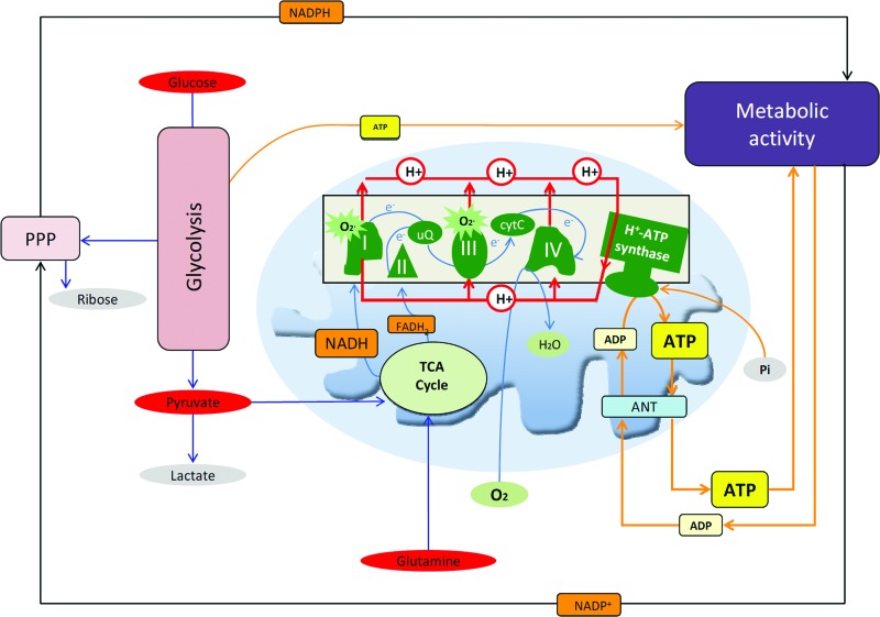
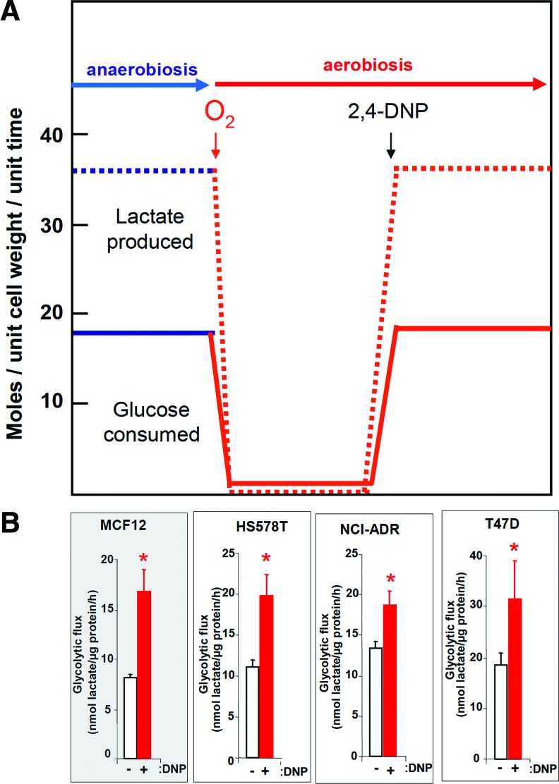
Recent advances: While the enhanced aerobic glycolysis of carcinomas has already been translated to bedside for precise tumor imaging and staging of cancer patients, accepting that an impaired bioenergetic function of mitochondria is pivotal to understand energy metabolism of tumors and in its progression is debated. However, mitochondrial bioenergetics and cell death are tightly connected.

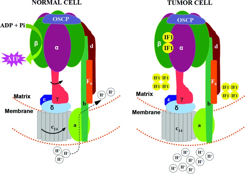
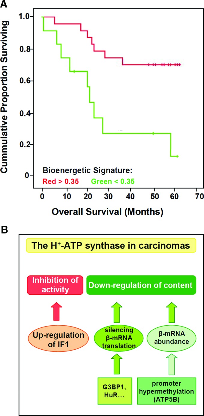
Critical issues: Recent clinical findings indicate that H(+)-ATP synthase, a core component of mitochondrial oxidative phosphorylation, is repressed at both the protein and activity levels in human carcinomas. This review summarizes the relevance that mitochondrial function has to understand energy metabolism of tumors and explores the connection between the bioenergetic function of the organelle and the activity of mitochondria as tumor suppressors.

Future directions: The reversible nature of energy metabolism in tumors highlights the relevance that the microenvironment has for tumor progression. Moreover, the stimulation of mitochondrial activity or the inhibition of glycolysis suppresses tumor growth. Future research should elucidate the mechanisms promoting the silencing of oxidative phosphorylation in carcinomas. The aim is the development of new therapeutic strategies tackling energy metabolism to eradicate tumors or at least, to maintain tumor dormancy and transform cancer into a chronic disease.


Figures

FIG. 1.
FIG. 1.
Overview of energy metabolism. The scheme shows the relevant pathways involved in the metabolism of glucose and glutamine (blue lines). Glucose is catabolized by the pentose phosphate pathway (PPP) to obtain reducing power (NADPH) and ribose for nucleic acid biosynthesis and oxidize by glycolysis to generate pyruvate and other metabolic intermediates. In the cytoplasm, the generated pyruvate can be reduced to lactate and further exported from the cell or completely oxidized in the mitochondria by the tricarboxylic acid cycle (TCA cycle). The transfer of electrons obtained in oxidations (NADH/FADH2) to molecular oxygen by respiratory complexes (green) of the inner mitochondrial membrane is depicted by light blue lines. The formation of the proton gradient generated by respiration and its utilization for the synthesis of ATP by the H+-ATP synthase in oxidative phosphorylation is highlighted by a red circuit. The entrance of ADP in exchange with ATP by the adenine nucleotide translocase (ANT) and inorganic phosphate is also shown (yellow lines). Metabolic activity utilizes carbon skeletons, NADPH, and ATP. To see this illustration in color, the reader is referred to the web version of this article at www.liebertpub.com/ars
FIG. 2.
FIG. 2.
H+-ATP synthase in normal and tumor cells. Schematic illustration of the organization of the mitochondrial H+-ATP synthase. Subunits are color-coded and labeled. F1 is the globular catalytic domain made of subunits α, β, and the three central stalk subunits, γ, δ, and ɛ. The F0 domain is comprised of the c oligomer, subunit a, and the peripheral stalk subunits b, d, F6, and OSCP. The minor subunits (e, f, g, and A6L) are not shown, but they all span the membrane and probably bind close to the a subunit. The rotor is made up of the c-ring and the central stalk. In normal cells, the re-entrance of protons through the a subunit promotes the rotation of the c-ring and of the attached central stalk, driving the conformational changes in the β-α-F1-ATPase subunits for synthesis of ATP. In tumor cells, the physiological inhibitor IF1 (yellow) is highly overexpressed, binding the catalytic α/β interface and preventing synthesis of ATP and promoting mitochondrial hyperpolarization. To see this illustration in color, the reader is referred to the web version of this article at www.liebertpub.com/ars
FIG. 3.
FIG. 3.
Mitochondrial activity defines the rate of glucose consumption. (A) In absence of oxidative phosphorylation (anaerobiosis, blue lines) the cellular rates of glucose consumption and of lactate production are very high. The availability of oxygen (aerobiosis, red lines) triggers the onset of oxidative phosphorylation and the repression of glucose consumption and lactate production rates. Uncoupling the mitochondrial proton gradient with 2,4-dinitrophenol (2,4-DNP) re-establish the original glucose consumption and lactate production rates. (B) Plots represent the glycolytic flux measured in breast normal (MCF12) and cancer (NCI-ADR, HS578T and T47D) cells in the absence (-) or presence (+) of 2,4-DNP. The glycolytic flux is highly increased when the bioenergetic activity of mitochondria is impaired. To see this illustration in color, the reader is referred to the web version of this article at www.liebertpub.com/ars
FIG. 4.
FIG. 4.
H+-ATP synthase in cancer biology. (A) The bioenergetic signature (β-F1-ATPase/GAPDH ratio) of colon carcinomas is a significant marker of the prognosis of colon cancer patients [re-drawn from (3)]. (B) Different mechanisms affect the activity and/or content of H+-ATP synthase in human carcinomas. The overexpression of the ATPase inhibitor factor 1 (IF1) in human carcinomas limits the activity of the H+-ATP synthase and promotes the metabolic switch in cancer cells. Silencing of β-F1-ATPase mRNA (β-mRNA) translation in carcinomas is mediated by the interaction of the mRNA with different mRNA binding proteins such as G3BP1. The reduction of β-mRNA availability by hypermethylation of the promoter of β-F1-ATPase gene (ATP5B) in chronic myeloid leukemia cells has also been described. To see this illustration in color, the reader is referred to the web version of this article at www.liebertpub.com/ars
FIG. 5.
FIG. 5.
Overview of the integration of energy metabolism and cell death. The scheme shows the connections between energy metabolism and the three major cell-death pathways: apoptosis (red), necrosis (blue), and autophagy (green). The availability of oxygen and glucose could trigger cell death or signal for proliferation. The cellular levels of reactive oxygen species (ROS) produced during mitochondrial respiration determine cell fate. The depicted high and low ROS levels should be considered above a basal ROS production as a result of an impaired mitochondrial function. Low levels of ROS are fundamental for transcription of genes involved in cell growth, whereas higher ROS levels results in oxidative damage that can participate in apoptosis, necrosis, or autophagy. Similarly, ATP generated by glycolysis and oxidative phosphorylation holds the balance between life and death. High ATP amounts are necessary for cell survival and the inhibition of autophagy, but also for the correct execution of apoptosis. When ATP is low, such as during PARP-1 hyper-activation, cells die by necrosis. Cyt c, AIF, and H+-ATP synthase are important proteins of oxidative phosphorylation that have emerged as regulators of apoptosis and necrosis. Bax, p53, and Bcl-xL are well known pro- and anti-apoptotic proteins that have been recently described as regulators of oxidative phosphorylation. To see this illustration in color, the reader is referred to the web version of this article at www.liebertpub.com/ars
FIG. 6.
FIG. 6.
The activity of H+-ATP synthase regulates the efficiency of cell death. Illustration of the rapid (∼3 h) staurosporine (STS)-induced cell death in liver C9 cells. Mitochondria-tagged gfp cells were treated with STS in the absence or presence of oligomycin (OL) or left untreated (CRL). STS induces the rapid fragmentation and swelling of mitochondria, resulting in significant nuclear DNA fragmentation and cell death. Treatment of the cells with oligomycin (OL) was unable to prevent the changes in mitochondrial morphology but significantly delayed nuclear DNA-fragmentation and cell death. To see this illustration in color, the reader is referred to the web version of this article at www.liebertpub.com/ars
FIG. 7.
FIG. 7.
Energy metabolism can gear the cell-death pathway of cancer cells. Cancer cells have different susceptibility to death stimulus as a result of their metabolic phenotype. A first event to death after priming the cells is the rapid dismantling and fragmentation of the mitochondrial tubular network into small mitochondria (blue). Tumor cells with relevant oxidative phosphorylation (OXPHOS) (i.e., high bioenergetic signature) (a) will respond with a burst in ROS production contributing to the oxidation of mitochondrial proteins (red dots) and their release from mitochondria to effectively swamp the cells into apoptosis (fragmented nucleus in blue). In contrast, cancer cells that have a high glycolytic metabolism (i.e., low bioenergetic signature) (b) will not respond with ROS production to a conventional chemotherapeutic agent, resulting in resistance to cell death. In this situation, it is suggested that metabolic inhibitors interfering with glycolysis (b1) (bromopyruvate, BrPy; iodoacetate, IA) to produce a metabolic catastrophe by depletion of cellular ATP levels can promote cell death by necrosis. Alternatively, treatment of highly glycolytic tumors with compounds that could activate the metabolism of pyruvate in mitochondria or the overall oxidative phosphorylation (b2) (dichloroacetate, DCA) could revert the metabolic phenotype of the cancer cell to produce ROS and the execution of apoptotic cell death. To see this illustration in color, the reader is referred to the web version of this article at www.liebertpub.com/ars

Similar articles

Cited by

References

    1. Acebo P. Giner D. Calvo P. Blanco-Rivero A. Ortega AD. Fernandez PL. Roncador G. Fernandez-Malave E. Chamorro M. Cuezva JM. Cancer abolishes the tissue type-specific differences in the phenotype of energetic metabolism. Transl Oncol. 2009;2:138–145. - PMC - PubMed
    1. Alavian KN. Li H. Collis L. Bonanni L. Zeng L. Sacchetti S. Lazrove E. Nabili P. Flaherty B. Graham M. Chen Y. Messerli SM. Mariggio MA. Rahner C. McNay E. Shore GC. Smith PJ. Hardwick JM. Jonas EA. Bcl-x(L) regulates metabolic efficiency of neurons through interaction with the mitochondrial F(1)F(O) ATP synthase. Nat Cell Biol. 2011;13:1224–1233. - PMC - PubMed
    1. Aldea M. Clofent J. Nunez de Arenas C. Chamorro M. Velasco M. Berrendero JR. Navarro C. Cuezva JM. Reverse phase protein microarrays quantify and validate the bioenergetic signature as biomarker in colorectal cancer. Cancer Lett. 2011;311:210–218. - PubMed
    1. Amuthan G. Biswas G. Zhang SY. Klein-Szanto A. Vijayasarathy C. Avadhani NG. Mitochondria-to-nucleus stress signaling induces phenotypic changes, tumor progression and cell invasion. EMBO J. 2001;20:1910–1920. - PMC - PubMed
    1. Arismendi-Morillo G. Electron microscopy morphology of the mitochondrial network in gliomas and their vascular microenvironment. Biochim Biophys Acta. 2011;1807:602–608. - PubMed

Substances