. 2012 Dec;39(4):441-50.
doi: 10.1016/j.jaut.2012.07.006. Epub 2012 Aug 15.

Autoreactive HSP60 epitope-specific T-cells in early human atherosclerotic lesions

Affiliations

Autoreactive HSP60 epitope-specific T-cells in early human atherosclerotic lesions

Giovanni Almanzar et al. J Autoimmun. 2012 Dec.

Abstract

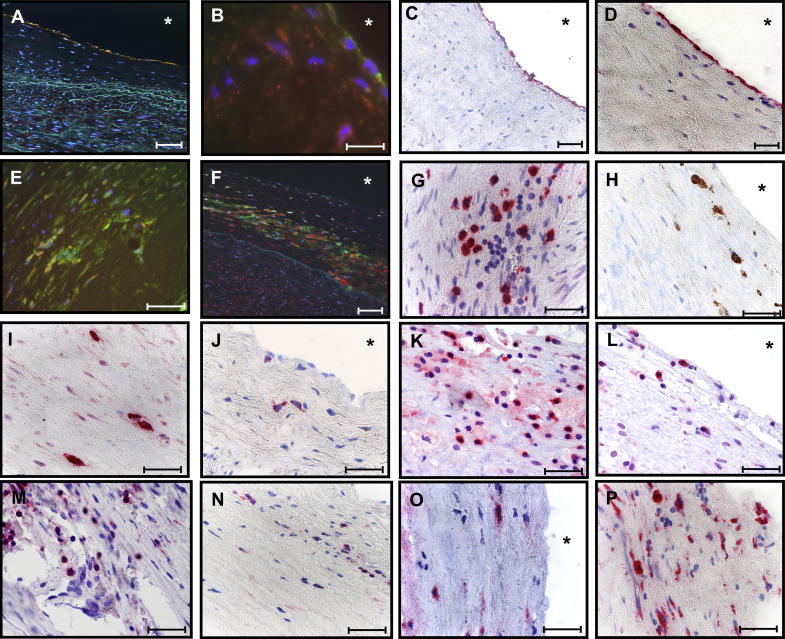
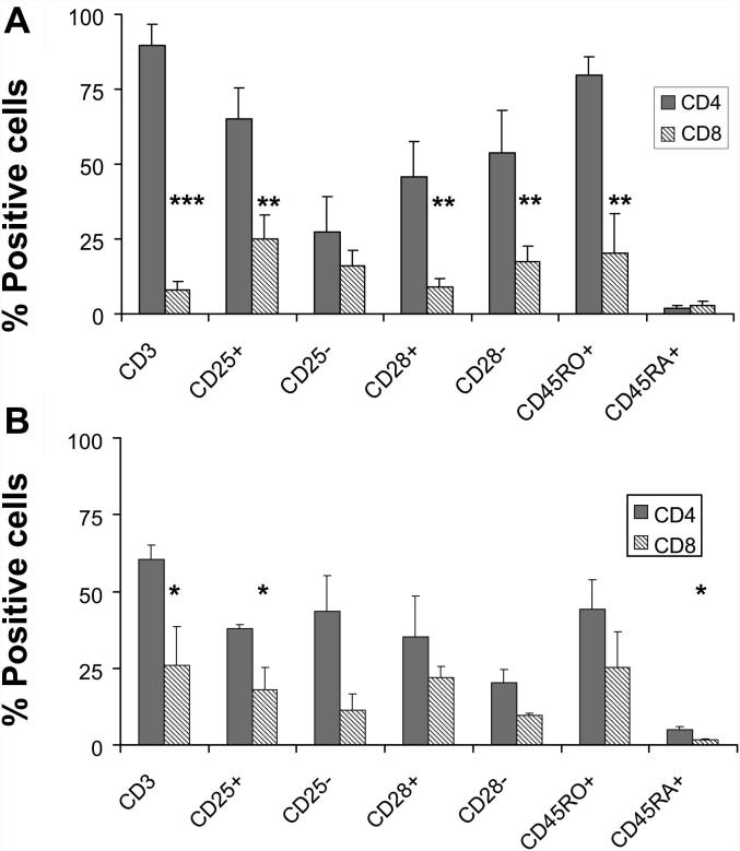
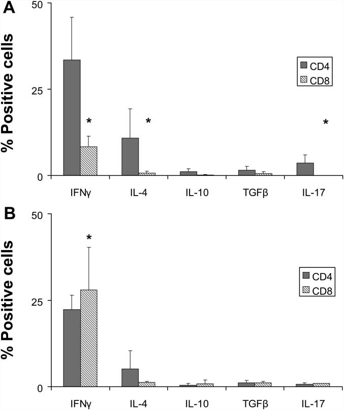
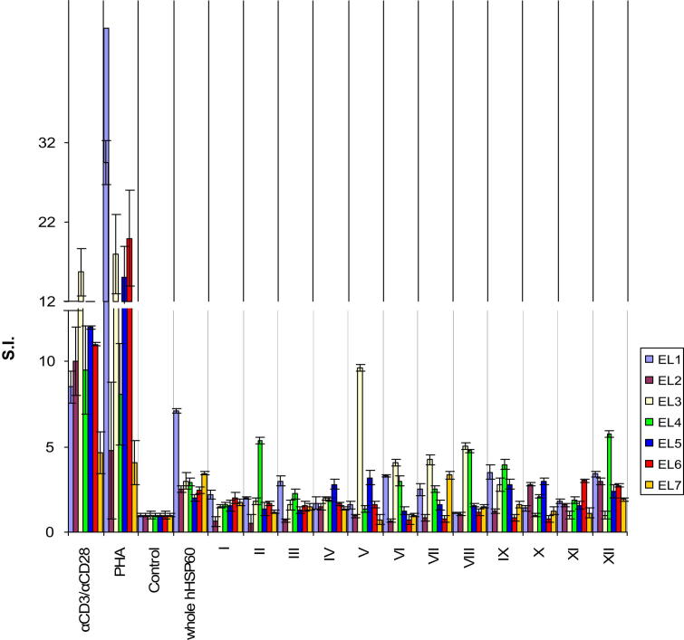
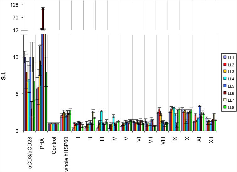
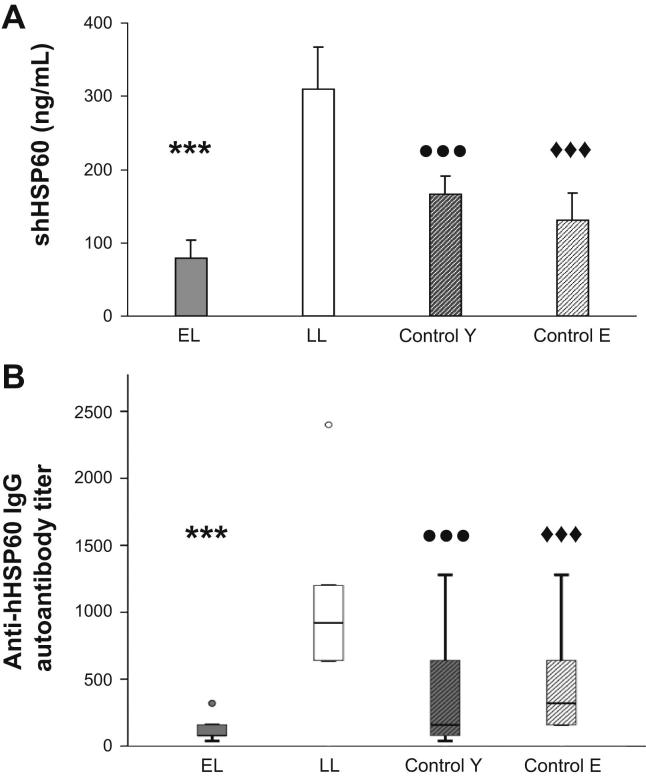
Atherosclerosis is a multifactorial chronic inflammatory disease characterized by the presence of T-cells, macrophages, and dendritic cells in the arterial intima. Classical risk factors lead to over-expression of stress proteins, especially heat shock protein 60 (HSP60). HSP60 on the surface of arterial endothelial cells (ECs) then becomes a target for pre-existing adaptive anti-HSP60 immunity resulting in infiltration of the intima by mononuclear cells. In the present study, T-cells derived from early, clinically still inapparent human atherosclerotic lesions were analyzed phenotypically and for their reactivity against HSP60 and HSP60-derived peptides. HSP60 was detected in ECs and CD40- and HLA Class II-positive cells within the intima. Effector memory CD4(+) T-cells producing high amounts of interferon-γ and low levels of interleukin-4 were the dominant subpopulation. T-cells derived from late lesions displayed a more restricted T-cell receptor repertoire to HSP60-derived peptides than those isolated from early lesions. Increased levels of soluble HSP60 and circulating anti-human HSP60 autoantibodies were found in donors with late but not early lesions. This is the first functional study of T-cells derived from early human atherosclerotic lesions that supports the previously proposed concept that HSP60-reactive T-cells initiate atherosclerosis by recognition of atherogenic HSP60 epitopes.


Figures

Fig. 1
Fig. 1
Immunohistological analysis of human atherosclerotic lesions. HSP60 expression (red) was determined in (A) ECs at arterial bifurcation areas stressed by turbulent blood flow conditions (vWF+, green, 200×, scale bar 200 μm) with simultaneous expression of adhesion molecules as (B) VCAM-1 (CD106, green, scale bar 200 μm), (C) CD106, and (D) CD62E (40×, scale bar 100 μm). Mononuclear cells in the intima expressed (E) CD40+ (green) and (F) HLA-DR+ (green, 200×, scale bar 200 μm). Subsets of (G) CD3+, (H) macrophages (CD68+, brown), (I) mast-cells (tryptase+), (J) DCs (CD1a+), (K) CD4+ T-cells, and (L) CD8+ T-cells were identified infiltrating the intima. (M) CD3+, (N) CD4+, (O) CD8+ T-cells, and (P) CD68+ were identified in advanced atherosclerotic lesions. Images were acquired using a Nikon Eclipse E800 microscope. Original magnification 600× (scale bar 100 μm). All immunohistological stainings and analysis of these were performed at least three times. A representative immunohistological analysis from 4 EL and 8 LL donors are demonstrated here.
Fig. 2
Fig. 2
Intralesional atherosclerotic T-cells are mostly memory effector T-cells. Surface expression of CD4, CD8, CD45RO, CD45RA, CD25, and CD28 were determined in intralesional CD3+ T-cells isolated from (A) EL (n = 7) and (B) LL (n = 8). CD4+ significantly predominate over CD8+ T-cells in atherosclerotic lesions (EL p < 0.005; LL p < 0.05). Significant differences in the surface expression of phenotypic markers are indicated in EL and LL T-cells (p < 0.05; ∗∗p < 0.01; ∗∗∗p < 0.005). Phenotypic characterization was performed by four-color flow cytometry. Bars indicate the percentage of positive cells as mean ± SEM. These experiments were performed once (in triplicates) for each donor.
Fig. 3
Fig. 3
T-cells isolated from atherosclerotic lesions produce the proinflammatory cytokine IFNγ. IFNγ, IL-4, IL-10, IL-17, and TGFβ production in CD4+ and CD8+ T-cells were determined in (A) EL (n = 7) and (B) LL (n = 8) donors by intracellular cytokine analysis after unspecific stimulation with PMA ionomycin. IFNγ-, IL-4-(∗∗p < 0.01), and IL-17-producing CD4+ T-cells were predominantly present as compared with the cytokine profile of CD8+ T-cells in EL (p < 0.05). No significant differences were observed in the cytokine production profile within LL T-cells. However, a significant increase in the frequency of IFNγ-producing CD8 was observed in LL T-cells as compared with EL T-cells (p < 0.05). Bars indicate the percentage (mean ± SEM) of positive cytokine-producing cells by four-color cytometry. These experiments were performed once for each donor.
Fig. 4
Fig. 4
EL T-cells proliferate in response to whole hHSP60 protein and recognize hHSP60-derived peptide pools. The proliferative cellular response of 1 × 104 intralesional T-cells isolated from 7 EL donors was evaluated upon stimulation with either whole hHSP60 or hHSP60 peptide pools (indicated by Roman numerals, all at 10 μg/mL). PHA and monoclonal anti-CD3/anti-CD28 antibodies (αCD3/αCD28) were used for positive controls. Antigen was omitted in the negative control (Control). Data are shown as stimulation index (S.I. ± SEM). All assays were carried out in triplicates.
Fig. 5
Fig. 5
LL T-cells display restricted peptide recognition. S.I. of 1 × 104 T-cells isolated from 8 LL donors upon stimulation with either whole hHSP60 protein or hHSP60 peptide pools (indicated by Roman numerals, all at 10 μg/mL). PHA and monoclonal anti-CD3/anti-CD28 antibodies (αCD3/αCD28) were used as positive control. Antigen was omitted in the negative control (Control). Data are shown as stimulation index (S.I. ± SEM). All assays were carried out in triplicates.
Fig. 6
Fig. 6
Soluble HSP60 (sHSP60) and anti-hHSP60 IgG autoantibody production may be an indicator of the atherosclerosis stage. Levels of circulating (A) sHSP60 (ng/mL) and (B) anti-hHSP60 IgG autoantibodies (titer) were determined in the plasma of EL (n = 7) and LL (n = 8) donors, young controls (control Y, n = 26), and elderly controls (control E, n = 14) by ELISA. Values are indicated as mean ± SEM. Significant differences are shown as follows: (A) sHSP60: EL vs LL (∗∗∗p < 0.005); LL vs Control Y (•••p < 0.005); LL vs control E (♦♦♦p < 0.005), (B) Anti-hHSP60 IgG autoantibodies: EL vs LL (∗∗∗p < 0.005); LL vs Control Y (•••p < 0.005); LL vs control E (♦♦♦p < 0.005). All assays were carried out in triplicates.

Similar articles

Cited by

References

    1. Collins R.G., Velji R., Guevara N.V., Hicks M.J., Chan L., Beaudet A.L. P-selectin or intercellular adhesion molecule (ICAM)-1 deficiency substantially protects against atherosclerosis in apolipoprotein E-deficient mice. J Exp Med. 2000;191:189–194. - PMC - PubMed
    1. Boring L., Gosling J., Cleary M., Charo I.F. Decreased lesion formation in CCR2−/− mice reveals a role for chemokines in the initiation of atherosclerosis. Nature. 1998;394:894–897. - PubMed
    1. Gu L., Okada Y., Clinton S.K., Gerard C., Sukhova G.K., Libby P. Absence of monocyte chemoattractant protein-1 reduces atherosclerosis in low density lipoprotein receptor-deficient mice. Mol Cell. 1998;2:275–281. - PubMed
    1. Young D.B. Heat-shock proteins: immunity and autoimmunity. Curr Opin Immunol. 1992;4:396–400. - PubMed
    1. Schett G., Xu Q., Amberger A., Van der Zee R., Recheis H., Willeit J. Autoantibodies against heat shock protein 60 mediate endothelial cytotoxicity. J Clin Invest. 1995;96:2569–2577. - PMC - PubMed

Publication types

MeSH terms