Review
. 2013 Jan;29(1):116-35.
doi: 10.1016/j.dental.2012.08.004. Epub 2012 Aug 16.

Optimizing dentin bond durability: control of collagen degradation by matrix metalloproteinases and cysteine cathepsins

Affiliations
Review

Optimizing dentin bond durability: control of collagen degradation by matrix metalloproteinases and cysteine cathepsins

Leo Tjäderhane et al. Dent Mater. 2013 Jan.

Abstract

Objectives: Contemporary adhesives lose their bond strength to dentin regardless of the bonding system used. This loss relates to the hydrolysis of collagen matrix of the hybrid layers. The preservation of the collagen matrix integrity is a key issue in the attempts to improve the dentin bonding durability.

Methods: Dentin contains collagenolytic enzymes, matrix metalloproteinases (MMPs) and cysteine cathepsins, which are responsible for the hydrolytic degradation of collagen matrix in the bonded interface.

Results: The identities, roles and function of collagenolytic enzymes in mineralized dentin has been gathered only within last 15 years, but they have already been demonstrated to have an important role in dental hard tissue pathologies, including the degradation of the hybrid layer. Identifying responsible enzymes facilitates the development of new, more efficient methods to improve the stability of dentin-adhesive bond and durability of bond strength.

Significance: Understanding the nature and role of proteolytic degradation of dentin-adhesive interfaces has improved immensely and has practically grown to a scientific field of its own within only 10 years, holding excellent promise that stable resin-dentin bonds will be routinely available in a daily clinical setting already in a near future.


Figures

Figure 1
Figure 1
Representation of the progressive hydration of the collagen Gly-Ala peptide. Top row presents the perpendicular and the bottom row parallel view to the molecular axis at the same hydration level. (a) A view of the non-hydrated collagen, with the three peptide chains shown in different colors. (b) The first shell of water molecules (blue spheres), directly hydrogen-bonded to carbonyl, hydroxyl or amide groups on the peptide surface. (c) The second shell of water molecules, hydrogen bond to the water in the first shell, demonstrating the filling of the superhelical groove. (d) The third shell of water molecules. (Reproduced from [8] with permission.)
Figure 2
Figure 2
Schematic of parallel collagen fibrils in demineralized dentin cut in cross-section to show the size of interfibrillar spaces. These spaces are not empty but contain proteoglycan hydrogels that may act as molecular sieves. Many dental adhesives are mixtures of comonomers. The large black dots on the right indicate dimethacrylates like BisGMA, while the smaller black dots indicate HEMA. BisGMA seems to only penetrate the top half of hybrid layers (reproduced from [170], with permission). The schematic on the left shows that the interior of collagen fibrils is made up of hundreds of collagen molecules separated by water-filled spaces about 1.8 nm wide. Albumin, with a molecular diameter of 6 nm cannot enter “collagen water” but would be excluded as being too large (MW 68 kDa). Glucose (MW 180 Da), BGP (bone Gla protein or osteocalcin, MW 5700 Da) can equilibrate with collagen water even though the molecular diameter of osteocalcin is the same as the width of intermolecular spaces (1.8 nm in both cases) because collagen molecules are in constant rotational vibration (modified from [15]).
Figure 3
Figure 3
A conserved cysteine residue (C) in the pro-domain coordinates with the Zn2+ ion at the functional site of the catalytic domain. The pro-domain is removed by cleavage in the pro-domain and between the pro-domain and the catalytic domain. The “propeller” is a hemopexin domain contained by most MMPs, and is attached to the catalytic domain by flexible hinge domain. The hemopexin domain e.g. mediates protein–protein interactions, contributes to substrate recognition, enzyme activation, and protease localization. Modified from [25]).
Figure 4
Figure 4. Steps involved in collagenolysis by collagenase
A) Collagenase bound to triple helical collagen via cooperative interaction of catalytic and hemopexin domain, but is unable to cleave in its native state due to the smallness of the cleft of the active site. B) Unwinding the triple-helical conformation allows single α-chain to be presented to the active site of the catalytic domain for peptide hydrolysis. C) All three α-chain are successively hydrolyzed, resulting with collagen molecule to be cut into ¼ and ⅓ fragments. (Adapted from [27]).
Figure 5
Figure 5
Gelatinolytic activity in dentinal tubules (green fluorescence) and in the hybrid layer at the top of the tubules of intact human tooth as seen with confocal fluorescence microscope.
Figure 6
Figure 6
Cathepsin B in dentinal tubules. Dentin tissue cross-section visualized by Differential Interference Contrast (DIC) (A). Intratubular Cathepsin B were immunolabeled using rabbit anti-human cathepsin B as primary antibody and mouse anti-rabbit IgG conjugated with Alexa Fluor 594® as a secondary antibody at the red channel (B). Merged Images (C). T = Tubules and scale bar = 5μm.
Figure 7
Figure 7
Schematic presentation of possible mechnisms of activation and function of cysteine cathepsins and MMPs in dentin (modified from the Online-only Appendix 2 in [37]). Green blocks with MMPs (red triangles) or cathepsins (yellow arrowheads) indicate inactivity of enzyme either as a proform or in complex with specific (TIMPs, cystatins) or non-specific (e.g. α2 macroglobulin) inhibitors. Activation of protein (removal of green blocks) may represent either elimination of inhibitor, transform from latent to active form, or both. Glycosaminoglycans (GAGs) in dentin may also affect the enzyme activity either by activation or inhibition, depending on enzyme and GAGs in question [106,127]. A) pH changes caused by etching acid or acidic monomers convert dentin-bound proMMPs and/or MMP-TIMP complexes (d-MMP) into active MMP. Respectively, dentin-bound cathepsin (d-Cat) becomes active in acidic pH. B) At least cathepsin B directly cleaves and inactivates MMP-specific tissue inhibitors TIMP-1 and TIMP-2 [36], changing the balance between MMPs and their inhibitors. Acidic pH activates cysteine cathepsins, which in turn either proteolytically activate proMMPs or degrade TIMP inhibiting MMP, or both, resulting with active MMPs and functional activity after neutralization of pH. C) Glycosaminoglycan (GAG: black dot) activation and stabilization of cathepsin (and possibly MMPs), allowing functional activity of cathepsins even in neutral pH. D) Odontoblast- or pulp-tissue derived enzymes may enter the hybrid layer.
Figure 8
Figure 8
Collagen and chondroitin 6-sulphate distribution in dentin. Dentin tissue was visualized by confocal microscopy in longitudinal- (A, B and C) and cross-section (D, E and F). Dentin morphology was shown by Differential Interference Contrast (DIC), (A and E). Molecularly well- structured collagen was visualized by its intrinsic fluorescence at the green channel (B and F). Chondroitin 6-Sulphate were immunolabeled using rabbit anti-human chondroitin 6-sulphate as primary antibody and mouse anti-rabbit IgG conjugated with Alexa Fluor 594® as a secondary antibody at the red channel (C and G). Colocalization between collagen and chondroitin 6-sulphate can be seen as the yellow color formed by the overlapping channels green and red in merged images (D) or interacting in the tubular area (H). The three-dimensional model was constructed from the upper panel’s images and shows how collagen and chondroitin 6-sulphate molecules are anatomically distributed on dentinal tissues (I). CS = Chondroitin 6-Sulphate; scale bars = 5 μm (A, B, C and D) or 2 μm (E, F, G and H).
Figure 9
Figure 9
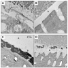
TEM analysis of hybrid layer in primary molars restored six months earlier without (controls) or with 2% CHX pretreatment (experimental group), using 2-step E&R adhesive and composite resin. A) Demineralized section of the control tooth hybrid layer stained with phosphotungstic acid and uranyl acetate. The hybrid layer (H) is almost completely degenerated and partially even missing, with empty regions occupied by epoxy resin (asterisk). C: resin composite; A: adhesive; H: hybrid layer; D: dentin. B) The section respective to A from the experimental (i.e. CHX-treatment) group. The hybrid layer (H) appears normal. C) Undemineralized, unstained, silver-impregnated section from the control tooth demonstrates a degraded hybrid layer with extensive leakage seen as black silver deposits that almost completely fill the hybrid layer. P, polyalkenoic acid copolymer. The silver filled the water-filled spaces that replaced collagen fibrils. D) An experimental tooth pretreated with CHX, with sparsely distributed silver deposits (pointer) in the hybrid layer. Arrows: polyalkenoic acid copolymer that is characteristic of 3M-ESPE adhesives. (Reproduced from [152], with permission.)
Figure 10
Figure 10
Field emission in-lens SEM (FEI-SEM) micrographs of unfixed, partially decalcified dentin after a pre-embedding immunolabeling with antibodies for MMP-2, -9 and -3. Labeling is seen as electron-dense white spots (pointers). (a,d) Low magnification view (20 000x) of the partially decalcified dentin surface showing open tubular orifices (T) surrounded by a thick collar of organic matrix and intertubular porous dentin (ITD). MMP-2 and -9 labeling mainly localized in peritubular dentin. (b,e) A higher magnification (50 000x), showing MMP-2 (b) and -9 (e) along the collagen fibrils. (c,f) High magnification (100 000x) FEI-SEM micrographs, revealing the relationship between MMP-2 and -9 and the collagen meshwork. The specimen shows a moderate labeling for MMP-9 (e), which is uniformly weaker than MMP-2 staining (c). (g–i) FEI-SEM micrographs of partially decalcified dentin after a pre-embedding immunolabelling an antibody for MMP-3. (g) Low magnification (30 000x) showing a patent dentinal tubule (DT) and the porous intertubular dentine (ITD). MMP-3 is mainly localized within the intertubular dentine. (h) Higher magnification (50 000x) showing MMP-3 located along the collagen fibrils. (i) High magnification FEI-SEM micrograph (100 000x), revealing MMP-3 distribution within the intertubular dentine. (Figures a–f reproduced from [55] and g–i from [57], with permission).
Figure 11
Figure 11
Three-dimensional model of the acquired image using in situ zymography with the cross-section of composite resin (R), hybrid layer (HL: arrow) and dentin (D). In in situ gelatin zymography, samples are overlayed with quenched florescent-conjugated gelatin gel, and any gelatinolytic activity in the sample releases fluorescence (green color). Intense gelatinolytic activity is present in dentinal tubules and especially at the bottom of the hybrid layer as a 1- to 2-μm-thick, well-defined layer. (Reproduced from [58], with permission).

References

    1. Shono Y, Terashita M, Shimada J, Kozono Y, Carvalho RM, Russell CM, Pashley DH. Durability of resin-dentin bonds. Journal of Adhesive Dentistry. 1999;1:211–8. - PubMed
    1. Hashimoto M. A review - micromorphological evidence of degradation in resin-dentin bonds and potential preventional solutions. Journal of Biomedical Materials Research Part B, Applied Biomaterials. 2010;92:268–80. - PubMed
    1. Loguercio AD, Moura SK, Pellizzaro A, Dal-Bianco K, Patzlaff RT, Grande RHM, Reis A. Durability of enamel bonding using two-step self-etch systems on ground and unground enamel. Operative Dentistry. 2008;33:79–88. - PubMed
    1. Breschi L, Mazzoni A, Ruggeri A, Cadenaro M, Di Lenarda R, De Stefano Dorigo E. Dental adhesion review: aging and stability of the bonded interface. Dental Materials. 2008;24:90–101. - PubMed
    1. Pashley DH, Tay FR, Breschi L, Tjäderhane L, Carvalho RM, Carrilho M, Tezvergil-Mutluay A. State of the art etch-and-rinse adhesives. Dental Materials. 2011;27:1–16. - PMC - PubMed

Publication types