. 2012 Sep;19(9):876-83.
doi: 10.1038/nsmb.2379. Epub 2012 Aug 14.

BIRC7-E2 ubiquitin conjugate structure reveals the mechanism of ubiquitin transfer by a RING dimer

Affiliations

BIRC7-E2 ubiquitin conjugate structure reveals the mechanism of ubiquitin transfer by a RING dimer

Hao Dou et al. Nat Struct Mol Biol. 2012 Sep.

Abstract

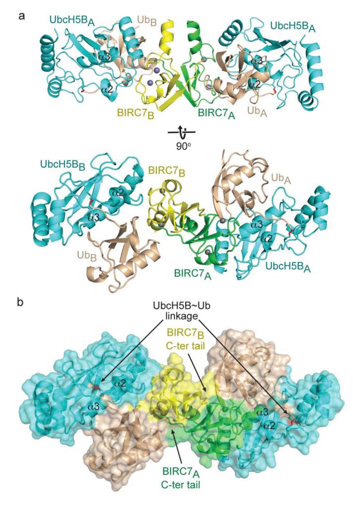
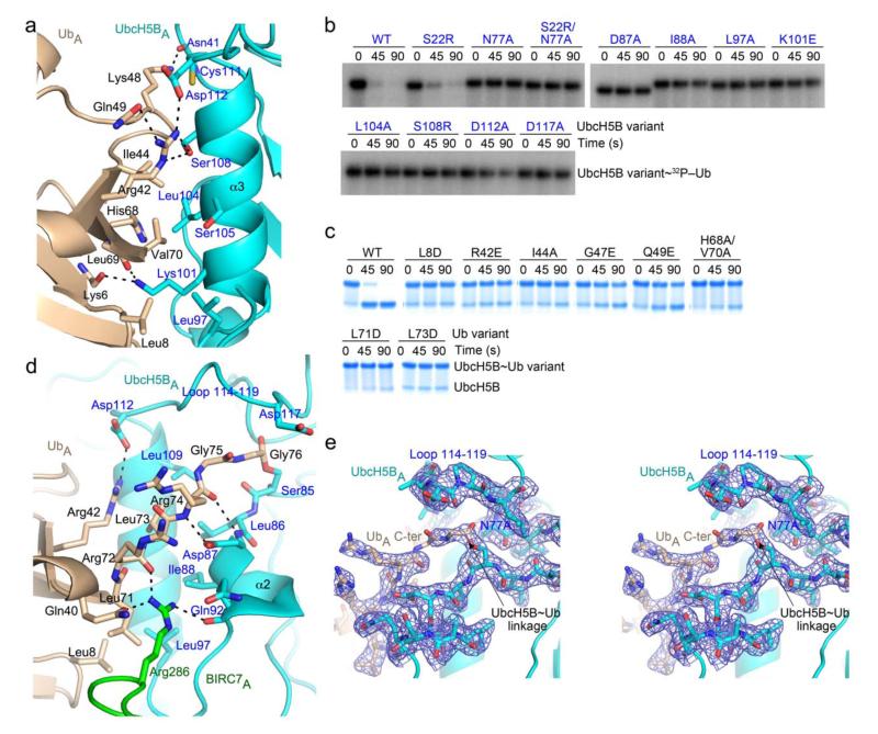
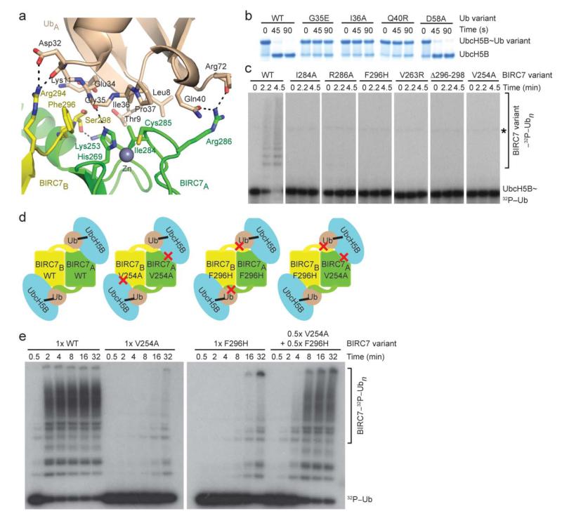
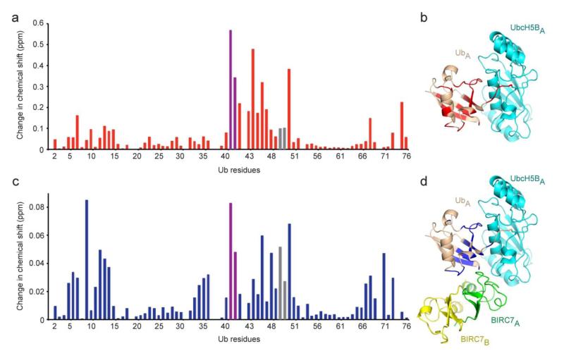
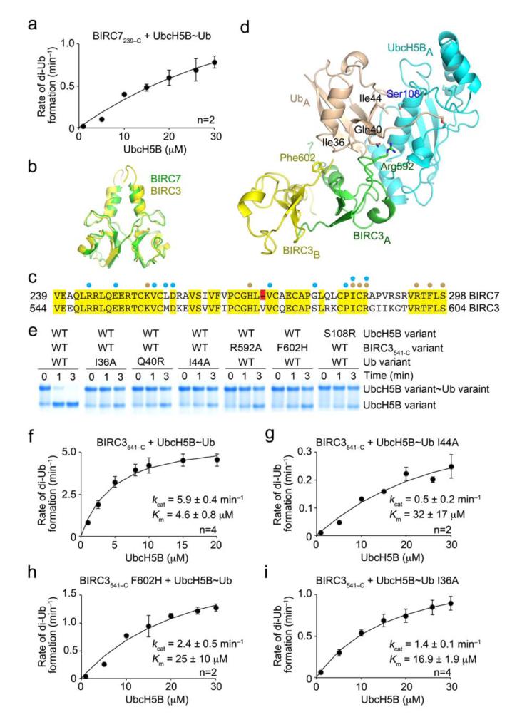
Certain RING ubiquitin ligases (E3s) dimerize to facilitate ubiquitin (Ub) transfer from ubiquitin-conjugating enzyme (E2) to substrate, but structural evidence on how this process promotes Ub transfer is lacking. Here we report the structure of the human dimeric RING domain from BIRC7 in complex with the E2 UbcH5B covalently linked to Ub (UbcH5B∼Ub). The structure reveals extensive noncovalent donor Ub interactions with UbcH5B and both subunits of the RING domain dimer that stabilize the globular body and C-terminal tail of Ub. Mutations that disrupt these noncovalent interactions or RING dimerization reduce UbcH5B∼Ub binding affinity and ubiquitination activity. Moreover, NMR analyses demonstrate that BIRC7 binding to UbcH5B∼Ub induces peak-shift perturbations in the donor Ub consistent with the crystallographically-observed Ub interactions. Our results provide structural insights into how dimeric RING E3s recruit E2∼Ub and optimize the donor Ub configuration for transfer.


Figures

Figure 1
Figure 1
Structure of BIRC7239–C–UbcH5BRAS~Ub. (a) Cartoon representation of the complex with the two crystallographic heterotrimers differentiated with A or B subscripts. Top and bottom panels are related by 90° rotation about the x-axis. Zn2+ atoms are depicted as grey spheres, Ub is colored wheat, BIRC7A green, BIRC7B yellow, and UbcH5B cyan with the covalent linkage highlighted in red. α2 and α3 from UbcH5B are labeled. (b) Surface representation of the complex, colored and oriented as in a bottom panel. UbcH5B~Ub linkages and the BIRC7 dimer C-terminal (C-ter) tails are indicated with arrows.
Figure 2
Figure 2
UbcH5B–Ub interactions within the complex. (a) Close-up view of donor UbA Ile44 patch–UbcH5BA α3 interactions. (b) Non-reduced autoradiograms of pulse-chase reactions showing the disappearance of UbcH5B~32P-Ub with full-length BIRC7, L-lysine, and UbcH5B variants over time. (c) As in b but for Ub variants and visualized by SDS-PAGE without radiolabeling. (d) Close-up view of UbcH5BA active site–UbA tail interactions. (e) Stereo view of the UbcH5BA~UbA linkage, UbcH5BA loop 114–119 and UbA tail–UbcH5BA interactions with key residues shown as sticks and labelled. 2Fo-Fc electron density (blue) contoured at 1.0 σ is shown. For a, d and e, key residues are shown as sticks and coloring is as in Fig. 1 with N atoms blue, O atoms red, and S atoms yellow. Putative hydrogen bonds are shown as dashed lines.
Figure 3
Figure 3
Donor Ub–RING interactions are required for BIRC7-catalyzed reactions. (a) Close-up view of donor UbA Ile36 patch–BIRC7 dimer interactions. (b) Non-reduced SDS-PAGE of pulse-chase reactions showing the disappearance of UbcH5B~Ub with full-length BIRC7, L-lysine, and Ub variants over time. (c) Non-reduced autoradiograms of pulse-chase reactions showing the simultaneous formation of 32P-Ub products and disappearance of UbcH5B~32P-Ub with full-length BIRC7 variants over time. An asterisk indicates the E1~Ub band. (d) Experimental design to validate the cross-dimer–donor Ub interactions. Based on the structure, we designed BIRC7 mutants expected to disrupt the BIRC7 RING–UbcH5B interaction (V254A) or the BIRC7 tail–Ub interaction (F296H), as indicated by red crosses. A heterodimer of the RING and tail mutants should be more active than a homodimer of either mutant. (e) Reduced autoradiograms showing the formation of 32P-Ub products with wild-type, mutant homodimers, and the hetero-mutant BIRC7 dimer over time.
Figure 4
Figure 4
Ub interaction surface in BIRC7239–C–UbcH5BRAS~Ub identified by NMR. (a) Changes in chemical shift per residue of 15N-Ub following covalent linkage to UbcH5BRAS determined by 1H–15N HSQC NMR. Changes were calculated according to the equation [(0.15 δN)2 + δH2]1/2. (b) Mapping of changes in a (> 1σ, 0.028 ppm) onto the Ub (colored red) in the UbcH5BA~UbA portion of BIRC7239–C–UbcH5BRAS~Ub. (c) Changes in chemical shift per residue of Ub in BIRC7239–C–UbcH5BRAS~15N-Ub compared to UbcH5BRAS~15N-Ub, calculated as in a. (d) Mapping of changes in c (> 1σ, 0.01 ppm) onto the Ub (colored blue) in the BIRC7 dimer– UbcH5BA~UbA portion of BIRC7239–C–UbcH5BRAS~Ub. For a and c, the violet and grey bars represent Gln40 and Gln49 sidechain chemical shift perturbations, respectively. For molecules, coloring is as described in Fig. 1.
Figure 5
Figure 5
Effect of donor Ub interactions on the kinetics of Ub transfer. (a) Single turnover kinetics of di-Ub formation catalyzed by BIRC7239–C. The rate of di-Ub formation was plotted against UbcH5B concentration. (b) Superposition of the structure of BIRC3541–C dimer (yellow, PDB 3EB6) onto BIRC7239–C dimer (green) from BIRC7239–C–UbcH5BRAS~Ub. BIRC3541–C dimer was generated from a symmetry-related molecule. (c) ClustalW sequence alignment of BIRC7 and BIRC3 RING domains. Identical residues are highlighted in yellow and the Val deletion in BIRC7 in red. Cyan and wheat circles indicate residues involved in contacting UbcH5B and Ub, respectively, as observed in BIRC7239–C–UbcH5BRAS~Ub and PDB 3EB6. (d) Models of UbcH5B~Ub bound to BIRC3541–C dimer generated by superposing BIRC3541–C dimer onto BIRC7239–C–UbcH5BRAS~Ub. Key residues involved in donor Ub interactions are indicated. Coloring is as described in Figs. 1 and 2. (e) Non-reduced SDS-PAGE of pulse-chase lysine discharge reactions showing the disappearance of UbcH5B~Ub with variants of BIRC3541–C, UbcH5B or Ub over time. (f–i) As in a but performed with BIRC3541–C. The rate of di-Ub formation was plotted against UbcH5B concentration for WT reaction (f), 32P-Ub I44A (g), BIRC3541–C F602H (h) and 32P-Ub I36A (i). Kinetic parameters and number of replicates, n, are indicated. Error bars indicate standard errors.

References

    1. Hershko A, Ciechanover A. The ubiquitin system. Annu Rev Biochem. 1998;67:425–79. - PubMed
    1. Pickart CM, Eddins MJ. Ubiquitin: structures, functions, mechanisms. Biochim Biophys Acta. 2004;1695:55–72. - PubMed
    1. Dye BT, Schulman BA. Structural mechanisms underlying posttranslational modification by ubiquitin-like proteins. Annu Rev Biophys Biomol Struct. 2007;36:131–50. - PubMed
    1. Wenzel DM, Lissounov A, Brzovic PS, Klevit RE. UBCH7 reactivity profile reveals parkin and HHARI to be RING/HECT hybrids. Nature. 2011;474:105–8. - PMC - PubMed
    1. Deshaies RJ, Joazeiro CA. RING domain E3 ubiquitin ligases. Annu Rev Biochem. 2009;78:399–434. - PubMed

Publication types

MeSH terms