. 2012 Aug 30;2(2):308-20.
doi: 10.1016/j.celrep.2012.07.010. Epub 2012 Aug 16.

Dispatched and scube mediate the efficient secretion of the cholesterol-modified hedgehog ligand

Affiliations

Dispatched and scube mediate the efficient secretion of the cholesterol-modified hedgehog ligand

Hanna Tukachinsky et al. Cell Rep. .

Abstract

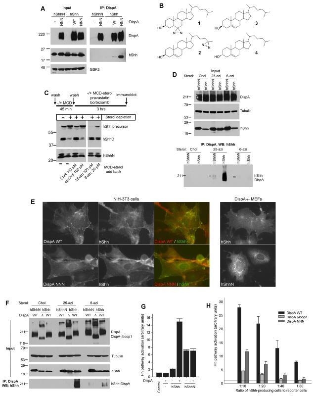
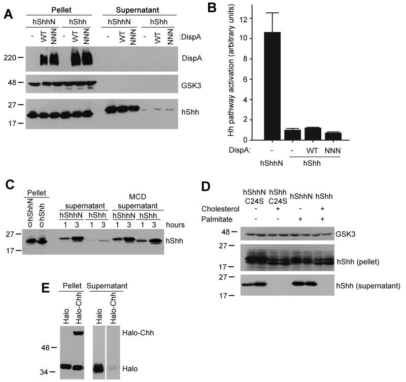
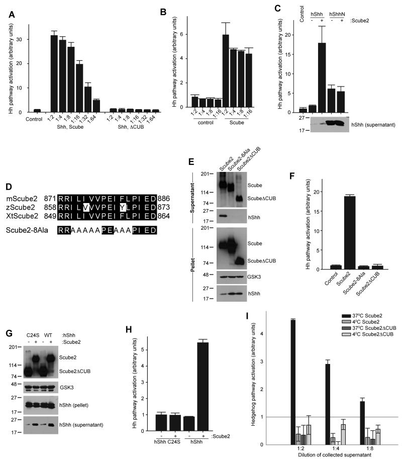
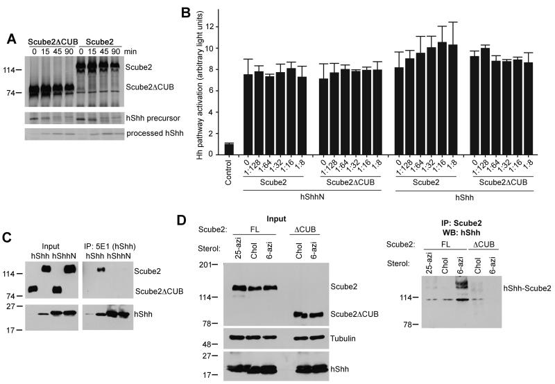
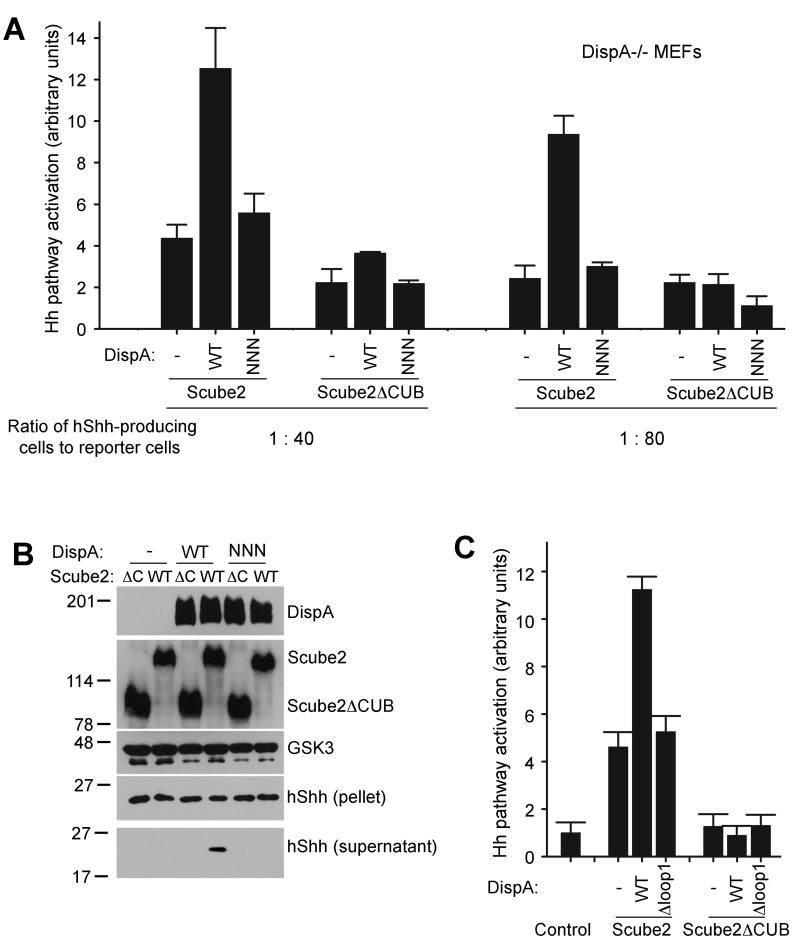
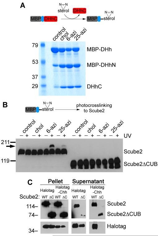
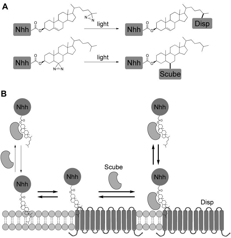
The Hedgehog (Hh) signaling pathway plays critical roles in metazoan development and in cancer. How the Hh ligand is secreted and spreads to distant cells is unclear, given its covalent modification with a hydrophobic cholesterol molecule, which makes it stick to membranes. We demonstrate that Hh ligand secretion from vertebrate cells is accomplished via two distinct and synergistic cholesterol-dependent binding events, mediated by two proteins that are essential for vertebrate Hh signaling: the membrane protein Dispatched (Disp) and a member of the Scube family of secreted proteins. Cholesterol modification is sufficient for a heterologous protein to interact with Scube and to be secreted in a Scube-dependent manner. Disp and Scube recognize different structural aspects of cholesterol similarly to how Niemann-Pick disease proteins 1 and 2 interact with cholesterol, suggesting a hand-off mechanism for transferring Hh from Disp to Scube. Thus, Disp and Scube cooperate to dramatically enhance the secretion and solubility of the cholesterol-modified Hh ligand.


Figures

Figure 1
Figure 1. Cholesterol-dependent binding of hShh to DispA is required for hShh secretion
(A) 293T cells stably co-expressing hShh or hShhN, and myc-tagged wild-type DispA (WT) or DispA-NNN (NNN) were lysed with detergent and analyzed by immunoprecipitation with anti-myc antibodies, followed by SDS-PAGE and immunoblotting with anti-hShh antibodies. (B) Structures of the photoreactive sterols, 6-azicholestanol (1) and 25-azicholesterol (2). Also shown are the structures of cholesterol (3) and the inactive diastereomer, epicholesterol (4). (C) HShh, HA-tagged at the C-terminus (hShh-HA) was stably expressed in 293T cells. The cells were sterol-depleted with methyl-β-cyclodextrin (MCD), after which cholesterol (Chol), epicholesterol (epiChol), 6-azicholestanol (6-azi) or 25-azicholesterol (25-azi) were added back as soluble MCD complexes, for 3 hours. Lysates were analyzed by SDS-PAGE and immunoblotting with HA antibodies (to detect the hShh precursor and the C-terminal fragment, hShhC) and hShh antibodies (to detect the hShh ligand). (D) 293T cells stably co-expressing myc-tagged DispA-WT and hShh or hShhN were labeled with the indicated sterols, as in (B). After incubation for 6 hours, the cells were UV-irradiated and DispA-hShh photocrosslinking was analyzed by denaturing immunoprecipitation with myc antibodies, followed by SDS-PAGE and immunoblotting for hShh. Immunoblotting for tubulin served as loading control. (E) Left panels: immunofluorescence microscopy of hShh stably co-expressed with myc-tagged DispA-WT or DispA-NNN in NIH-3T3 cells. Cells were stained with rabbit anti-hShh and mouse anti-myc antibodies, followed by goat anti-rabbit Alexa488 and goat anti-mouse Alexa594 secondary antibodies. Right panels: localization by immunofluorescence microscopy of hShh and hShhN expressed in DispA-/- MEFs. (F) As in (C) but with 293T cells stably co-expressing myc-tagged DispA or the mutant missing the first extracellular loop, DispA-Δloop1 (Δ), together with hShh or hShhN. (G) DispA-/- MEFs stably expressing hShh or hShhN, rescued by lentiviral expression of mCherry-tagged DispA or not, were co-cultured with Hh-responsive Shh Light II cells at a 1:20 ratio. Luciferase measurements were normalized to reporter cells grown alone (control). All experiments were performed in triplicate. Error bars represent the standard deviation of the mean. (H) DispA-/- MEFs stably expressing hShh, transduced with lentiviruses expressing mCherry-tagged DispA-WT, DispA-Δloop1 or DispA-NNN were analyzed as in (G), at four different ratios of hShh-producing cells to reporter cells.
Figure 2
Figure 2. DispA is not sufficient for release of cholesterol-modified hShh
(A) 293T cells stably expressing hShh and myc-tagged DispA constructs were washed of serum and were incubated with serum-free media for 6 hours. Secreted proteins were precipitated with trichloroacetic acid (TCA). Pellet and supernatant fractions were analyzed by SDS-PAGE and immunoblotting with anti-myc and anti-hShh antibodies. Blotting for GSK3 served as loading control. (B) Supernatants collected as in (A) were diluted 1:2 with serum-free media and hShh activity was assayed using Shh Light II reporter cells. Measurements were performed in triplicate and error bars represent standard deviation of the mean. (C) 293T cells expressing hShhN or hShh were incubated with serum-free media, with or without methyl-β-cyclodextrin (MCD, 100 μM), and supernatants were harvested after 1 or 3 hours. Secretion of hShh and hShhN was analyzed as in (A). (D) 293T cells transiently expressing hShh, hShhN, hShh with a palmitylation site mutation (hShhC24S), or hShhN with a palmitylation site mutation (hShhN-C24S) were incubated in serum-free media for 4 hours, and secreted proteins were TCA-precipitated. Pellet and supernatant fractions were analyzed as in (A). The samples in this panel were loaded in duplicate. (E) A secreted HA-tagged Halotag protein was fused to amino acids 190-462 of hShh (Halo-Chh); autocatalytic processing of Halo-Chh generates Halotag fused to amino acids 190-198 of hShh, modified with cholesterol. Secreted HA-Halotag fused to amino acids 190-198 of hShh (Halo) is not cholesterol-modified and serves as negative control. The Halotag constructs were expressed in 293T cells, and secreted proteins were collected into serum free media for 4 hours. Pellet and supernatant fractions were analyzed by SDS-PAGE and immunoblotting with anti-HA antibodies.
Figure 3
Figure 3. The extracellular protein Scube2 stimulates secretion of cholesterol-modified hShh
(A) Scube2 or Scube2ΔCUB was transfected into 293T cells stably expressing hShh, and 24 hours later secreted proteins were collected into serum-free media for 4 hours. HShh activity in serial dilutions of the supernatant was measured by luciferase assay in Shh Light II reporter cells. All experiments were performed in triplicate. Error bars represent the standard deviation of the mean. (B) Scube2 or Scube2ΔCUB was added in serum-free media to 293T cells stably expressing hShh, for 4 hours. Activity of secreted hShh was measured as in (A). (C) As in (A) but with 293T cells stably expressing hShh or hShhN. Secreted hShh and hShhN were collected for 6 hours, and were analyzed by reporter assay in Shh Light II cells and by immunoblotting with anti-hShh antibodies. Note the much higher amount of secreted hShhN compared to hShh. (D) Sequence of the conserved hydrophobic patch in the CUB domain of vertebrate Scube2 orthologs. Also shown is the sequence of the Scube2-8Ala mutant, in which 8 hydrophobic residues are mutated to alanines. (E) HA-tagged Scube2, Scube2-8Ala, or Scube2ΔCUB were transfected into 293T cells stably expressing hShh. Secreted proteins were collected 24 hours later, for 6 hours into serum-free media. Aliquots of the cell pellet and supernatants were analyzed by SDS-PAGE and immunoblotting for HA and hShh. (F) HShh activity in the supernatants collected in (E) was measured as in (A). (G) HShh or the palmitylation-defective mutant hShhC24S were co-expressed in 293T cells with HA-tagged Scube2 or Scube2ΔCUB. HShh and hShhC24S secretion was analyzed as in (E). (H) HShh and hShhC24S activity in the supernatants collected in (G) was measured as in (A). (I) Scube2 or Scube2ΔCUB was added in serum-free medium to 293T cells stably expressing hShh, for 1 hour at 37°C or 4°C. HShh activity in the supernatants was measured as in (A).
Figure 4
Figure 4. Cholesterol-dependent binding of Scube2 to hShh is required for hShh secretion
(A) HA-tagged Scube2 or Scube2ΔCUB was expressed in 293T cells stably expressing hShh. The cells were pulsed with 35S-methionine for 3 min and were chased with media containing unlabeled methionine for the indicated times. HShh processing was analyzed by immunoprecipitation with the 5E1 antibody, which recognizes full-length and processed hShh. Scube2 and Scube2ΔCUB were immunoprecipitated with anti-HA antibodies. The precipitated protein was analyzed by SDS-PAGE and fluorography. The same number of cells was processed for each condition. (B) The two graphs on the left show that Scube2 does not affect hShhN activity. HShhN was mixed with HA-tagged Scube2 or Scube2ΔCUB, in the indicated ratios, and its activity was measured in Shh Light II reporter cells. The two graphs on the right show Scube2 does not affect the activity of hShh pre-released with Scube2. 293T cells co-expressing hShh and HA-tagged Scube2 were used to generate serum-free hShh-Scube2 conditioned media. This media was mixed with HA-tagged Scube2 or Scube2ΔCUB, in the indicated ratios, and its activity was measured in Shh Light II reporter cells. All experiments were performed in triplicate. Error bars represent the standard deviation of the mean. (C) 293T cells co-expressing hShh or hShhN, and HA-tagged Scube2 or Scube2ΔCUB were incubated in serum-free media for 4 hours. HShh and hShhN were immunoprecipitated from the supernatant with 5E1 antibodies. A portion of the supernatant was TCA-precipitated to serve as input. All samples were analyzed by SDS-PAGE and immunoblotting with anti-HA and anti-hShh antibodies. (D) HShh or hShhN was stably co-expressed with HA-tagged Scube2 or Scube2ΔCUB in 293T cells. The cells were labeled with the indicated sterols, followed by UV irradiation. Photocrosslinked Scube2-hShh was analyzed by denaturing immunoprecipitation with HA antibodies, followed by SDS-PAGE and immunoblotting for hShh.
Figure 5
Figure 5. Scube2 synergizes with DispA to stimulate secretion of cholesterol-modified hShh
(A) DispA-/- MEFs stably expressing hShh, transduced with lentiviruses expressing mCherry-tagged wild-type DispA or DispNNN, were plated with Shh Light II reporter cells at 1:40 and 1:80 ratios. After 12 hours, Scube2 or Scube2ΔCUB was added in serum-free media. Luciferase measurements were performed 30 hours later and were normalized to untreated reporter cells. All experiments were done in triplicate. Error bars represent the standard deviation of the mean. (B) HA-tagged Scube2 or Scube2ΔCUB was transfected into 293T cells that stably express hShh, hShh and myc-tagged DispA-WT, or hShh and myc-tagged DispA-NNN. Secreted proteins were collected into serum-free media 24 hours later, for 4 hours. Aliquots of the cellular pellets and supernatants were analyzed by SDS-PAGE and immunoblotting with HA, myc and hShh antibodies. Blotting for GSK3 served as loading control. (C) As in (B), but with 293T cells expressing DispA-Δloop1 instead of DispA-NNN. Activity of secreted hShh was measured as in (A).
Figure 6
Figure 6. Cholesterol modification is sufficient for interaction with and secretion by Scube2
(A) Purified maltose-binding protein (MBP) fused to amino acids 244-471 of Drosophila Hedgehog (MBP-DHh) was used in in vitro processing reactions to generate MBP modified with either cholesterol (control), 6-azicholestanol or 25-azicholesterol. The samples were analyzed by SDS-PAGE and Coomassie staining. MBP-DHhN and DHh-C are the two fragments generated by in vitro processing. (B) The processing reactions in (A) were incubated with HA-tagged Scube2 or Scube2ΔCUB, followed by UV irradiation to induce photocrosslinking. The samples were analyzed by reducing SDS-PAGE and immunoblotting with HA antibodies. The arrow indicates the position of the photocrosslinked Scube2-MBP species. (C) Constructs that generate Halotag protein modified with cholesterol (Halotag-Chh) or not (Halotag) were co-expressed in 293T cells with myc-tagged Scube or Scube2ΔCUB. Secreted proteins were collected in serum-free media for 6 hours, followed by TCA precipitation and immunoblotting with HA and myc antibodies.
Figure 7
Figure 7. Mechanism of hShh secretion by the DispA and Scube2
(A) Summary of the results for photocrosslinking of hShh to DispA and to Scube2 in cells. DispA is crosslinked to hShh modified with 25-azicholesterol and Scube2 is crosslinked to hShh modified with 6-azicholestanol, suggesting that DispA and Scube2 recognize different aspects of the cholesterol molecule. (B) The hShh ligand associates with membranes due to its hydrophobic cholesterol anchor. HShh binds DispA in a cholesterol-dependent manner, and this interaction is required for hShh secretion, perhaps by lowering the activation energy for hShh extraction from the lipid bilayer. From DispA, hShh is transferred to the secreted protein Scube2. The fact that DispA and Scube2 recognize different features of the cholesterol molecule suggests a hand-off mechanism for this transfer, such that the cholesterol anchor of hShh never contacts directly the aqueous environment, and is thus kept soluble. This mechanism is reminiscent of the transfer of free cholesterol between the Niemann-Pick disease proteins NPC1 and NPC2 during cholesterol egress from late endosomes/lysosomes.

References

    1. Burke R, Nellen D, Bellotto M, Hafen E, Senti KA, Dickson BJ, Basler K. Dispatched, a novel sterol-sensing domain protein dedicated to the release of cholesterol-modified hedgehog from signaling cells. Cell. 1999;99:803–815. - PubMed
    1. Chamoun Z, Mann RK, Nellen D, von Kessler DP, Bellotto M, Beachy PA, Basler K. Skinny hedgehog, an acyltransferase required for palmitoylation and activity of the hedgehog signal. Science. 2001;293:2080–2084. - PubMed
    1. Chen MH, Li YJ, Kawakami T, Xu SM, Chuang PT. Palmitoylation is required for the production of a soluble multimeric Hedgehog protein complex and long-range signaling in vertebrates. Genes Dev. 2004;18:641–659. - PMC - PubMed
    1. Chen X, Tukachinsky H, Huang CH, Jao C, Chu YR, Tang HY, Mueller B, Schulman S, Rapoport TA, Salic A. Processing and turnover of the Hedgehog protein in the endoplasmic reticulum. J Cell Biol. 2011;192:825–838. - PMC - PubMed
    1. Creanga A, Glenn TD, Mann RK, Saunders AM, Talbot WS, Beachy PA. Scube/You activity mediates release of dually lipid-modified Hedgehog signal in soluble form. Genes Dev. 2012;26:1312–1325. - PMC - PubMed

Publication types

MeSH terms

Substances