. 2012 Oct 12;48(1):133-44.
doi: 10.1016/j.molcel.2012.07.012. Epub 2012 Aug 16.

Exo- and endoribonucleolytic activities of yeast cytoplasmic and nuclear RNA exosomes are dependent on the noncatalytic core and central channel

Affiliations

Exo- and endoribonucleolytic activities of yeast cytoplasmic and nuclear RNA exosomes are dependent on the noncatalytic core and central channel

Elizabeth V Wasmuth et al. Mol Cell. .

Abstract

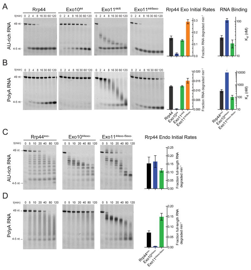
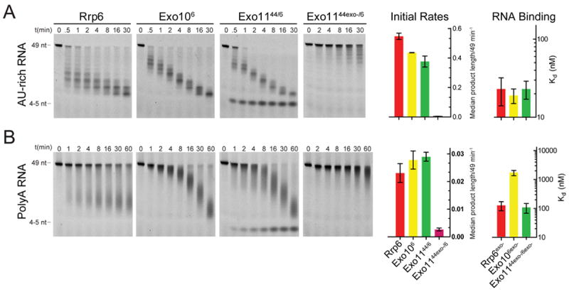
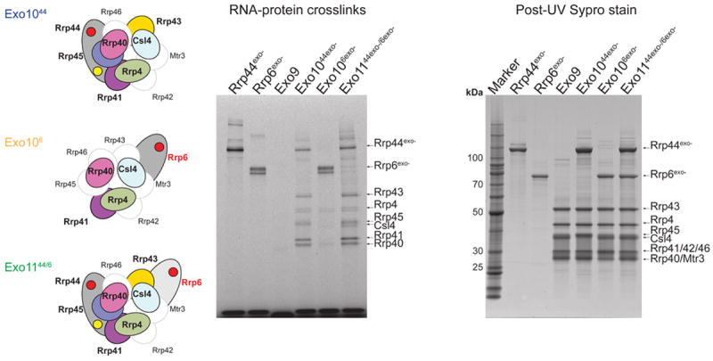
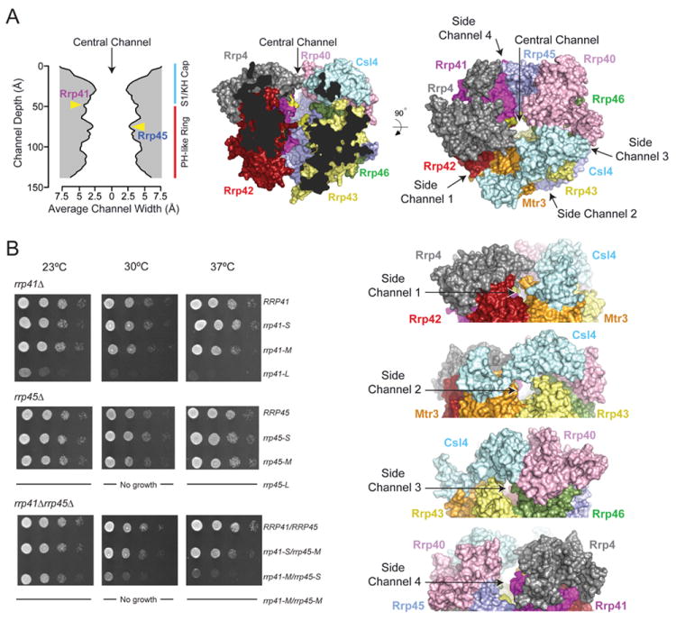
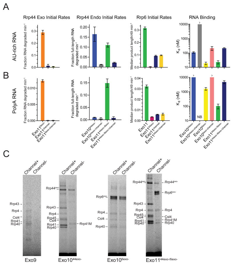
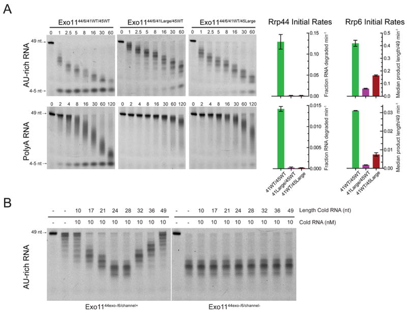
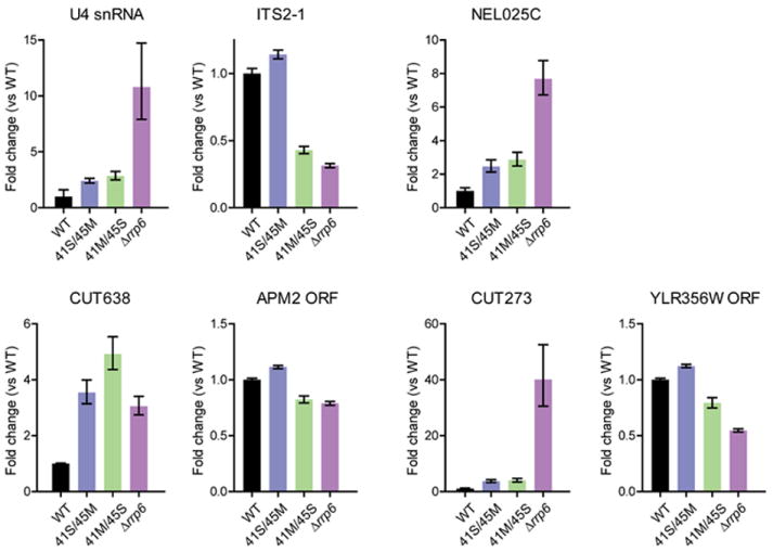
The RNA exosome is an essential multisubunit ribonuclease (RNase) that contributes to cytoplasmic and nuclear RNA decay and quality control. The 9-subunit exosome core (Exo9) features a prominent central channel formed by stacked asymmetric rings of six RNase PH-like proteins and three S1/KH domain proteins. Exo9 is catalytically inert but associates with Rrp44, an endoribonuclease and processive 3'→5' exoribonuclease, and Rrp6, a distributive 3'→5' exoribonuclease. We show that Exo9 and its central channel modulate all three yeast exosome RNase activities because channel occlusion attenuates RNA binding and RNase activities in vitro and fails to complement exosome functions in vivo. We find that Rrp6 stimulates Rrp44 RNase activities and that Rrp6 is inhibited by a mutation in the Rrp44 exoribonuclease active site in 11-subunit nuclear exosomes. These results suggest the exosome core and central channel is essential because it modulates each of the known RNase activities of the yeast RNA exosome.


Figures

Figure 1
Figure 1. The Exo9 core alters exoribonuclease, endoribonuclease and RNA binding activities of Rrp44
Association of Rrp44 with Exo9 (Exo1044) diminishes the exoribonuclease and RNA binding activities on (A) AU-rich and (B) polyA RNAs. Rrp6 stimulates Rrp44 activity in Exo1144/6 independent of Rrp6 catalytic activity. Multiple turnover RNA decay assays performed using 5’ fluorescein labeled RNAs and reaction products separated by Urea-PAGE. Representative gels and quantitation. Bar graphs depicting initial rates of Rrp44-mediated exoribonuclease activity determined from data obtained in the linear range. Bar graphs depicting dissociation constants (Kd) were derived by fluorescence polarization of catalytically dead variants of free and Exo9 core-associated Rrp44 assayed using 5’ fluorescein labeled (A) AU-rich or (B) polyA RNA. The pattern of intermediates generated by Rrp44exo- changes after association with Exo9 (Exo1044exo-) or Exo9 and Rrp6exo- (Exo1144exo-/6exo-) for (C) AU-rich and (D) polyA RNA (See Figure S3C for assays using unlabeled RNA). The distributive pattern of intermediates observed for Rrp44exo- (short and long 5’ labeled products accumulate simultaneously) is altered to patterns that appear to be generated 3’ to 5’ in Exo1044exo- and Exo1144exo-/6exo- for AU-rich RNA because longer 5’ labeled products appear before appearance of shorter products. Exo1044exo- has weaker endonuclease activity on polyA RNA but is stimulated by addition of Rrp6exo- in Exo1144exo-/6exo-. A stoichiometric ratio of enzymes and 5’-fluorescein RNA (10 nM) was incubated in reaction buffer in the presence of 3 mM MnCl2 (See Experimental Procedures). Error bars represent ± 1 standard deviation as calculated from three independent experiments. Bar graphs color coded according to Table S1.
Figure 2
Figure 2. The Exo9 core alters Rrp6 exoribonuclease activity and RNA binding
Association of Rrp6 with Exo9 (Exo106 or Exo1144/6) results in formation of a unique pattern of (A) AU-rich or (B) polyA RNA intermediates compared to Rrp6. Bar graphs representing initial rates of Rrp6-mediated exoribonuclease activity calculated by determining the median length of products generated over time and dividing the median length by substrate length (49 nt). Bar graphs depicting dissociation constants (Kd) derived by fluorescence polarization of catalytically dead variants of free and Exo9 core-associated Rrp6 using 5’ fluorescein labeled (A) AU-rich or (B) polyA RNA. Error bars represent ± 1 standard deviation as calculated from three independent experiments. Bar graphs color coded according to Table S1.
Figure 3
Figure 3. UV-RNA cross-linking reveals exosome-RNA contacts
Schematics of Exo1044exo- (top), Exo106exo- (middle) and Exo1144exo-/6exo- (bottom) shown on left. Subunits for which no cross-linking was observed are outlined, labeled and shown in white while subunits for which RNA-protein cross-links were detected are outlined, labeled and colored. Exosome subunits and exosomes were incubated with 5’ fluorescein AU-rich RNA and illuminated by UV to induce cross-linking. UV RNA-protein cross-linked products were separated by SDS-PAGE and gels were scanned to detect 5’-fluorescein RNA-protein adducts (middle), then stained with Sypro Ruby and scanned to visualize total protein (right). Subunit positions in respective gels indicated by labels and arrows. Molecular weight markers shown in left lane for the Sypro Ruby stained gel.
Figure 4
Figure 4. The exosome central channel is essential in vivo
A) The width and dimensions of the channel (left) were calculated using the dimensional analysis program, HOLE (Smart et al., 1996). Axes are labeled. The middle panel shows a view of the human Exo9 core (PDB 2NN6) in surface representation. The central channel is made apparent by removing Mtr3 and cutting away remaining surfaces of Csl4, Rrp43 and Rrp42. The positions of insertions in Rrp41 and Rrp45 are colored yellow in the structure and indicated by labels and yellow arrows in the graph depicting channel dimensions. Right panel depicts an orthogonal view of the Exo9 core in surface representation showing all nine subunits. A label and black arrow indicates the positions of the central channel and four putative side channels. Subunits colored and labeled. Below this panel are four side views of the S1/KH cap and top portion of the PH-like ring highlighting four potential side channels. B) Serial 10-fold dilutions of S. cerevisiae rrp41Δ (top), rrp45Δ (middle) or rrp41Δrrp45Δ (bottom) bearing RRP41 or RRP45 or mutant rrp41 or rrp45 alleles containing insertions ranging between 2 [S], 7 [M] and 11 [L] amino acids spotted on YPD agar grown at 23°C (left), 30°C (middle), and 37°C (right).
Figure 5
Figure 5. Channel occlusion inhibits RNA binding and RNase activities of Rrp44 and Rrp6
Bar graphs depicting initial rates calculated for Rrp6 and Rrp44 RNase on A) AU-rich and B) polyA RNA. Occluding the central channel of Exo1144/6 (Exo1144/6/channel-) with loop insertions (Rrp41-M/Rrp45-M) inhibits both Rrp44 activities while Exo1144exo-/6/channel- diminishes Rrp6 exoribonuclease activities and alleviates Rrp6 inhibition by Rrp44exo-. Rrp44 endonuclease activities are inhibited by channel occlusion in Exo1044exo- and Exo1144exo-/6exo-. Assays performed in triplicate and initial rates calculated from data obtained in the linear range (Figures S6A and S6C). Bar graphs on the right depict apparent Kd values for various exosome complexes with RNA. Error bars are ± 1 standard deviation. C) Channel occlusion in Exo9 leads to diminished cross-linking (left). Panel to the right shows that channel occlusion in Exo1044exo- leads to loss of most RNA-protein adducts to core subunits. Next panel shows that channel occlusion in Exo106exo- slightly weakens cross-linking to the S1/KH cap proteins and Rrp6. On the right, channel occlusion in Exo1144exo-/6exo- shifts the cross-linking pattern from one involving Rrp44, the PH-like and S1/KH rings to one involving Rrp6 and the S1/KH ring. Products separated by SDS-PAGE and imaged by detecting the fluorescence of the 5’ labeled AU-rich RNA. Major cross-linked species are labeled and indicated by arrows.
Figure 6
Figure 6. Rrp6 is more dependent on the upper portion of the channel to access RNA
A) RNA decay assays using AU-rich or polyA RNA for WT exosomes and exosomes reconstituted with large loop insertions at the top (41Large) or middle (45Large) of the channel. Bar graphs on right indicate initial rates of decay mediated by Rrp44 or Rrp6. Rrp44 activity is inhibited by insertions at either position while the insertion at the top of the channel (Exo1144/6 41Large/45WT) impedes Rrp6 activity more than insertion in the middle (Exo1144/6 41WT/45Large). Decay assays performed in triplicate. Error bars are ± 1 standard deviation. B) Inhibition of Rrp6 activity in Exo1144exo-/6 by 49 nt AU-rich RNA is alleviated by short competing RNAs that mimic channel occlusion in the PH-like ring. End point assay (15 minutes) of reactions containing 10 nM unlabeled AU-rich RNAs (10, 17, 21, 24, 28, 32, 36, 49 nts) that were pre-incubated with 1 nM Exo1144exo-/6 or Exo1144exo-/6/channel-. Reactions were then challenged with 10 nM 49 nt 5-fluorescein AU-rich RNA and reaction intermediates detected by denaturing PAGE. Reactions containing unlabeled 24 or 28 nt mimic channel occlusion and alleviate Rrp6 inhibition in Exo1144exo-/6 to similar levels observed in Exo1144exo-/6/channel-.
Figure 7
Figure 7. Channel occlusion results in aberrant RNA processing in vivo
Previously identified substrates of the RNA exosome were analyzed by qPCR from total RNA obtained from rrp41Δrrp45Δ strains bearing WT RRP41/RRP45 or mutant alleles containing small/medium (rrp41-S/rrp45-M), and medium/small (rrp41-M/rrp45-S) insertions and compared to levels in an rrp6Δ strain. Bar graphs show quantitation of results obtained from triplicate experiments. Error bars represent ± 1 standard deviation. Targets were normalized to Scr1 mRNA. APM2 and YLR356W ORFs serve as controls for CUT638 and CUT273, respectively.

Similar articles

Cited by

References

    1. Allmang C, Kufel J, Chanfreau G, Mitchell P, Petfalski E, Tollervey D. Functions of the exosome in rRNA, snoRNA and snRNA synthesis. EMBO J. 1999;18:5399–410. - PMC - PubMed
    1. Allmang C, Mitchell P, Petfalski E, Tollervey D. Degradation of ribosomal RNA precursors by the exosome. Nucleic Acids Res. 2000;28:1684–91. - PMC - PubMed
    1. Anderson JS, Parker RP. The 3’ to 5’ degradation of yeast mRNAs is a general mechanism for mRNA turnover that requires the SKI2 DEVH box protein and 3’ to 5’ exonucleases of the exosome complex. EMBO J. 1998;17:1497–506. - PMC - PubMed
    1. Assenholt J, Mouaikel J, Andersen KR, Brodersen DE, Libri D, Jensen TH. Exonucleolysis is required for nuclear mRNA quality control in yeast THO mutants. RNA. 2008;14:2305–13. - PMC - PubMed
    1. Basu U, Meng FL, Keim C, Grinstein V, Pefanis E, Eccleston J, Zhang T, Myers D, Wasserman CR, Wesemann DR, Januszyk K, Gregory RI, Deng H, Lima CD, Alt FW. The RNA exosome targets the AID cytidine deaminase to both strands of transcribed duplex DNA substrates. Cell. 2011;144:353–63. - PMC - PubMed

Publication types

MeSH terms

LinkOut - more resources