. 2012 Sep 28;47(6):943-53.
doi: 10.1016/j.molcel.2012.07.015. Epub 2012 Aug 16.

The seed region of a small RNA drives the controlled destruction of the target mRNA by the endoribonuclease RNase E

Affiliations

The seed region of a small RNA drives the controlled destruction of the target mRNA by the endoribonuclease RNase E

Katarzyna J Bandyra et al. Mol Cell. .

Abstract

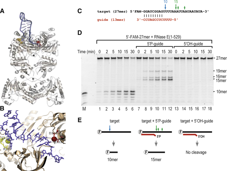
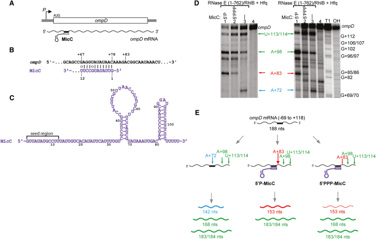
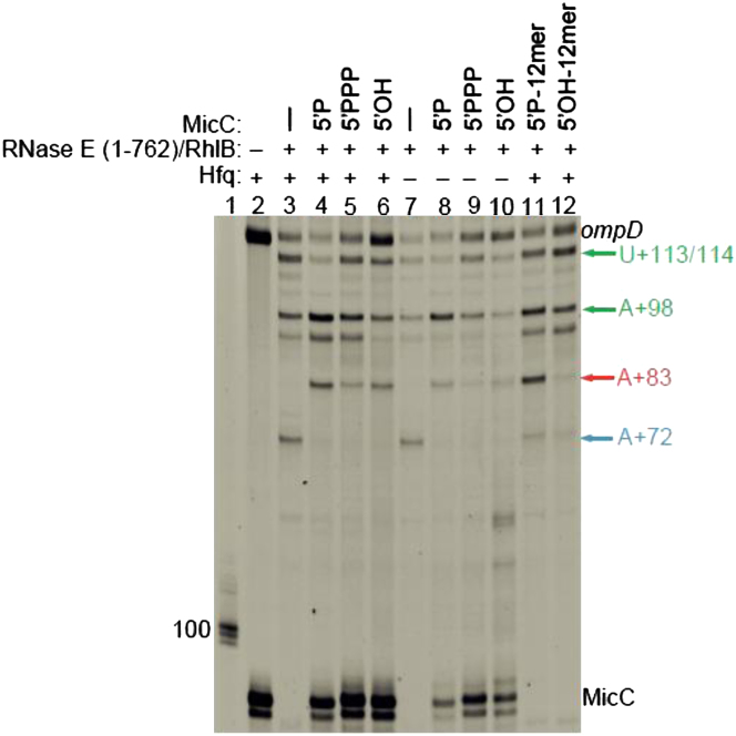
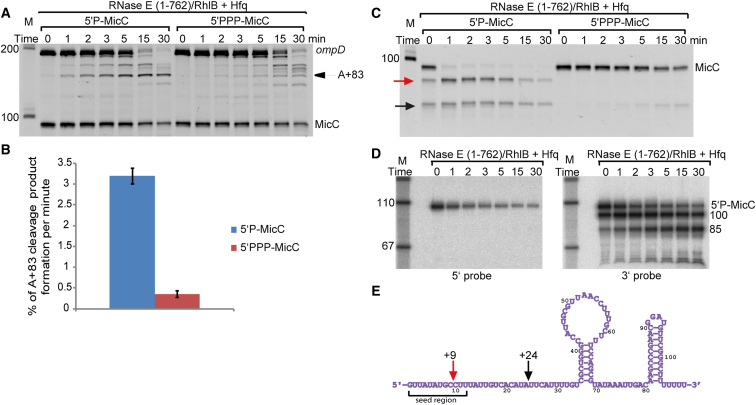
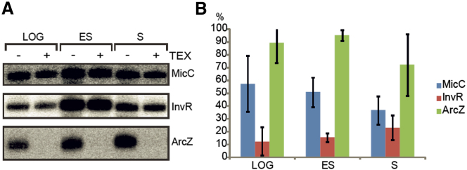
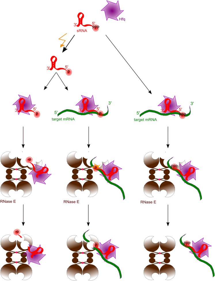
Numerous small non-coding RNAs (sRNAs) in bacteria modulate rates of translation initiation and degradation of target mRNAs, which they recognize through base-pairing facilitated by the RNA chaperone Hfq. Recent evidence indicates that the ternary complex of Hfq, sRNA and mRNA guides endoribonuclease RNase E to initiate turnover of both the RNAs. We show that a sRNA not only guides RNase E to a defined site in a target RNA, but also allosterically activates the enzyme by presenting a monophosphate group at the 5'-end of the cognate-pairing "seed." Moreover, in the absence of the target the 5'-monophosphate makes the sRNA seed region vulnerable to an attack by RNase E against which Hfq confers no protection. These results suggest that the chemical signature and pairing status of the sRNA seed region may help to both 'proofread' recognition and activate mRNA cleavage, as part of a dynamic process involving cooperation of RNA, Hfq and RNase E.


Figures

Figure 1
Figure 1
RNase E Cleavage Can Be Guided by an Artificial sRNA (A) Hypothetical model of the complex of the tetrameric catalytic domain of RNase E with a structured substrate, constructed from experimental crystal lattice interactions (Callaghan et al., 2005). (B) Expanded view that includes the 5′ sensor region and the catalytic site. The spacing from the end of the seed matching duplex region to the catalytic metal is 5 to 6 bases. The RNA substrate is colored blue; in yellow are the critical residues of the 5′ sensing pocket and 5′ phosphate atom, shown as a sphere; and in red are the active site aspartate residues and the catalytic magnesium ion (sphere). (C) The sequences of the 27-mer target RNA with a 5′ fluorescent group (FAM, black) and its partially complementary 13-mer guide (red). The guide has either a 5′ hydroxyl or a monophosphate group. (D) Denaturing gel showing the degradation products of 27-mer alone (left panel), in presence of 5′-P-13-mer guide RNA (middle panel) and in presence of 5′-OH-13-mer guide RNA (right panel). The visualization is for the FAM group so only the 5′end degradation products are visible. (E) Schematic of the cleavage patterns observed in (D). These are also indicated by arrows in (C) for the target 27-mer RNA in the presence (green) and absence (blue) of 13-mer guide RNA. (See also Figure S1.)
Figure 2
Figure 2
MicC Guides RNase E to Cleave ompD mRNA in the Coding Region (A) Schematic showing the site targeted by MicC within the ompD coding region. (B) The imperfect duplex formed by the 5′ seed region of MicC (purple) and ompD (black) showing the position of +83 nucleotide. (C) MicC secondary structure (Pfeiffer et al., 2009). The ‘seed’ recognition region is indicated. The polyU tail of sRNAs are important for mediating interactions with Hfq (Otaka et al., 2011; Sauer and Weichenrieder, 2011; Sauer et al., 2012). (D) (left panel) A denaturing gel showing main degradation products of ompD in vitro by RNase E (1-762)/RhlB and in the presence of MicC carrying 5′monophosphate (5′P) or triphosphate (5′PPP) (left panel). A sequencing gel (right panel) showing 32P-labeled products of the reactions from the left panel and mapping the RNase E (1-762)/RhlB cleavage sites in ompD. Lane 4 in both panels is a control sample loading of the ompD. The lanes labeled OH and T1 are ladders prepared by alkaline degradation and T1 nuclease digestion, respectively. (E) Schematic presentation of possible ways of RNase E cleavage of ompD mRNA in vitro. The arrows indicate the preferred cleavage positions in the presence and absence of MicC sRNA (purple). Sizes shown on the bottom panel indicate the lengths of the digestion products (U+113/114 cleavage site = 183/184 nts, A+98 = 168 nts, A+83 = 153 nts, A+72 = 142 nts products). The A+83 cleavage site is the preferred site in vivo (Pfeiffer et al., 2009). The schematic suggests cleavage preferences for the different pathways and not restricted activities. (See also Figures S2, S3, S4, and S5.)
Figure 3
Figure 3
The Imperfectly Complementary “Seed Region” of MicC Is Sufficient to Induce RNase E Mediated Cleavage of ompD. Denaturing gel showing products of ompD degradation by RNase E (1-762)/RhlB with or without Hfq (lanes 3 and 7) and in the presence of WT MicC with different state of 5′ end (lanes 4-6 in presence of Hfq and 8-10 in absence of Hfq), as well as 12 nucleotides seed region of MicC (lanes 11-12). The RNA chaperone Hfq protects the sRNA and enhances cleavage at the A+83 site. Size markers are in lane 1, control reaction without enzyme is in lane 2. (See also Figures S6, S7, and S8.) The gel was stained with SYBR gold Nucleic Acid Gel Stain, which reveals all the RNA species. The lower band of MicC is a sRNA with 1 nt shorter polyU tail due to different transcription termination by T7 polymerase on polyT stretches.
Figure 4
Figure 4
The Influence of MicC 5′ Phosphorylation State on Specific ompD Cleavage and MicC Stability (A) Time course series showing ompD degradation by RNase E (1-762)/RhlB in presence of Hfq and 5′P-MicC (left panel) or 5′PPP-MicC (right panel). (B) Graph representing efficiency of ompD A+83 cleavage by RNase E (1-762)/RhlB in presence of Hfq and 5′P or 5′PPP MicC, shown as the percent of the +83 cleavage (nM) per minute. Error bars were calculated for standard deviation for three independent experiments. (C) Time course series showing the influence of MicC 5′ phosphorylation state on its own degradation by RNase E (1-762)/RhlB in the presence of Hfq. The red arrow indicates the early cleavage product, and the black arrow indicates a second cleavage product. (D) Northern blot of the 5′P-MicC degradation as in left panel in (C), probed for 5′ end of MicC (left panel) and 3′ end of MicC (right panel). Size markers are in the left lanes (M), sizes of the cleavage products are indicated on the right. Minutes from the beginning of the reactions are indicated above the gels, 0 time point is a sample withdrawn about 5 s after enzyme addition. (See also Figure S9). (E) The cleavage sites corresponding to the red and black arrows in (C) were identified by 5′ RACE from four independent clones and are shown in the schematic of the MicC. The red arrow indicates the cleavage site within the seed region at position +9, and the black arrow indicates a second cleavage product at +24, just outside the seed region.
Figure 5
Figure 5
MicC in 5′P Form Can Be Detected In Vivo (A) Northern blot analysis of sRNAs from total RNA extracts of wild-type strain of Salmonella. The top panel shows the results for MicC at different cell growth densities (LOG, exponential phase, ES, early stationary phase; S, three hours into stationary phase). The same amounts of total RNA were either treated (+) with Terminator Exonuclease (TEX), which degrades RNA with a 5′ monophosphate, or untreated (−). Controls are shown for the sRNAs InvR, which is less TEX sensitive in logarithmic and exponential phases (middle panel), and ArcZ, which carries a 5′ monophosphate group and is sensitive to TEX treatment (bottom panel). (B) Bar chart representing the monophosphorylated fraction of sRNAs analyzed (MicC-blue, InvR-red, ArcZ-green), calculated from the percentage of integrated signal relative to the control not treated with TEX. Error bars were estimated from three independent experiments. (See also Figure S10.)
Figure 6
Figure 6
Cartoon Schematic of the Guide RNA Activation of RNase E and a Potential Proofreading Mechanism sRNA (red) is maintained in the cell in 5′PPP form. Such sRNA probably can associate with Hfq (purple) and basepair with a target mRNA (green), however 5′PPP group cannot actively stimulate RNase E (brown) (right panel). Under particular conditions (yellow lightening) the pyrophosphate from sRNA 5′ end is removed and such ‘activated’ sRNA (5′P marked with a star) efficiently guides RNase E cleavage of target mRNA (middle panel). 5′P form of sRNA would be rapidly degraded when there is no or no more target it can basepair with what would ensure ‘proofreading’ mechanism in sRNA mediated mRNA degradation.

Comment in

  • RNase E finds some sRNAs stimulating.
    De Lay N, Gottesman S. De Lay N, et al. Mol Cell. 2012 Sep 28;47(6):825-6. doi: 10.1016/j.molcel.2012.09.007. Mol Cell. 2012. PMID: 23020853 Free PMC article.

References

    1. Afonyushkin T., Vecerek B., Moll I., Bläsi U., Kaberdin V.R. Both RNase E and RNase III control the stability of sodB mRNA upon translational inhibition by the small regulatory RNA RyhB. Nucleic Acids Res. 2005;33:1678–1689. - PMC - PubMed
    1. Aiba H. Mechanism of RNA silencing by Hfq-binding small RNAs. Curr. Opin. Microbiol. 2007;10:134–139. - PubMed
    1. Argaman L., Hershberg R., Vogel J., Bejerano G., Wagner E.G., Margalit H., Altuvia S. Novel small RNA-encoding genes in the intergenic regions of Escherichia coli. Curr. Biol. 2001;11:941–950. - PubMed
    1. Balbontín R., Fiorini F., Figueroa-Bossi N., Casadesús J., Bossi L. Recognition of heptameric seed sequence underlies multi-target regulation by RybB small RNA in Salmonella enterica. Mol. Microbiol. 2010;78:380–394. - PubMed
    1. Bartel D.P. MicroRNAs: target recognition and regulatory functions. Cell. 2009;136:215–233. - PMC - PubMed

Publication types

MeSH terms

LinkOut - more resources