. 2012 Oct 12;287(42):35370-35381.
doi: 10.1074/jbc.M112.399071. Epub 2012 Aug 17.

Impact of individual proliferating cell nuclear antigen-DNA contacts on clamp loading and function on DNA

Affiliations

Impact of individual proliferating cell nuclear antigen-DNA contacts on clamp loading and function on DNA

Yayan Zhou et al. J Biol Chem. .

Abstract

Ring-shaped clamp proteins encircle DNA and affect the work of many proteins, notably processive replication by DNA polymerases. Crystal structures of clamps show several cationic residues inside the ring, and in a co-crystal of Escherichia coli β clamp-DNA, they directly contact the tilted duplex passing through (Georgescu, R. E., Kim, S. S., Yurieva, O., Kuriyan, J., Kong, X. P., and O'Donnell, M. (2008) Structure of a sliding clamp on DNA. Cell 132, 43-54). To investigate the role of these contacts in reactions involving circular clamps, we examined single arginine/lysine mutants of Saccharomyces cerevisiae proliferating cell nuclear antigen (PCNA) in replication factor C (RFC)-catalyzed loading of the clamp onto primer template DNA (ptDNA). Previous kinetic analysis has shown that ptDNA entry inside an ATP-activated RFC-PCNA complex accelerates clamp opening and ATP hydrolysis, which is followed by slow PCNA closure around DNA and product dissociation. Here we directly measured multiple steps in the reaction (PCNA opening, ptDNA binding, PCNA closure, phosphate release, and complex dissociation) to determine whether mutation of PCNA residues Arg-14, Lys-20, Arg-80, Lys-146, Arg-149, or Lys-217 to alanine affects the reaction mechanism. Contrary to earlier steady state analysis of these mutants (McNally, R., Bowman, G. D., Goedken, E. R., O'Donnell, M., and Kuriyan, J. (2010) Analysis of the role of PCNA-DNA contacts during clamp loading. BMC Struct. Biol. 10, 3), our pre-steady state data show that loss of single cationic residues can alter the rates of all DNA-linked steps in the reaction, as well as movement of PCNA on DNA. These results explain an earlier finding that individual arginines and lysines inside human PCNA are essential for polymerase δ processivity (Fukuda, K., Morioka, H., Imajou, S., Ikeda, S., Ohtsuka, E., and Tsurimoto, T. (1995) Structure-function relationship of the eukaryotic DNA replication factor, proliferating cell nuclear antigen. J. Biol. Chem. 270, 22527-22534). Mutations in the N-terminal domain have greater impact than in the C-terminal domain, indicating a positional bias in PCNA-DNA contacts that can influence its functions on DNA.


Figures

FIGURE 1.
FIGURE 1.
Clamp loading reaction, intermediate complex structures, and potential contacts between PCNA and DNA. A, minimal clamp loading pathway showing the reactants, a key RFC-PCNAopen-ptDNA intermediate formed after ATP binding, and the products, including topologically linked PCNA-ptDNA. B, MD model of S. cerevisiae RFC bound to ATPγS and open PCNA (20). C, crystal structure of bacteriophage T4 gp44/62 clamp loader bound to ADP-BeF3, an open gp45 clamp, and ptDNA (8). D, positions of the nine arginines and lysines on α helices lining the inside of a PCNA monomer (gray).
FIGURE 2.
FIGURE 2.
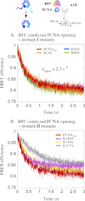
Kinetics of PCNA opening. PCNA clamp opening is measured by change in FRET between tryptophan donor (λEX = 290 nm) and AEDANS acceptor (λEM > 450 nm). Rapid mixing of RFC and PCNAWCAEDANS with ATP results in lower FRET efficiency as PCNA opens. A, data are shown for PCNAWC and domain I mutants PCNAWC-R14A, PCNAWC-K20A, and PCNAWC-R80A. An exponential fit of the PCNAWC trace yields kopen = 2.3 s−1. B, data are shown for PCNAWC and domain II mutants PCNAWC-K146A, PCNAWC-R149A, and PCNAWC-K217A. Final reactant concentrations were 0.6 μm RFC, 0.25 μm PCNAWCAEDANS, and 0.5 mm ATP.
FIGURE 3.
FIGURE 3.
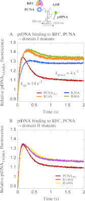
Kinetics of ptDNA binding and release. Interaction of DNA with RFC-ATP-PCNA is measured by change in fluorescence intensity of TAMRA-labeled ptDNA (λEX = 550 nm, λEM > 570 nm). Preincubation of RFC and PCNAWC with ATP (Δt = 0.02–3 s; 2-s data shown) followed by the addition of ptDNATAMRA results in rise (ptDNA binding) and then fall (complex dissociation) of the fluorescence signal. A, data are shown for PCNAWC and domain I mutants PCNAWC-R14A, PCNAWC-K20A, and PCNAWC-R80A. Double exponential fit of the PCNAWC trace yields kup = ∼14 s−1 and kdown = ∼3.9 s−1. B, data are shown for PCNAWC and domain II mutants PCNAWC-K146A and PCNAWC-R149A. Final reactant concentrations were 0.1 μm RFC, 0.4 μm PCNAWC, 0.04 μm ptDNATAMRA, and 0.5 mm ATP.
FIGURE 4.
FIGURE 4.
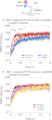
Kinetics of PCNA closure around ptDNA. Preincubation of RFC and PCNAWCAEDANS with ATP (Δt = 0.02–2 s; 2-s data shown) followed by addition of ptDNA results in FRET increase as ATP is hydrolyzed, and the clamp closes around DNA. A, data are shown for PCNAWC and domain I mutants PCNAWC-R14A, PCNAWC-K20A, and PCNAWC-R80A. Exponential fit of the PCNAWC trace yields kclose = 8 s−1. B, data are shown for PCNAWC and domain II mutants PCNAWC-K146A and PCNAWC-R149A. Final reactant concentrations were 0.6 μm RFC, 0.25 μm PCNAWCAEDANS, 0.25 μm ptDNA, and 0.5 mm ATP.
FIGURE 5.
FIGURE 5.
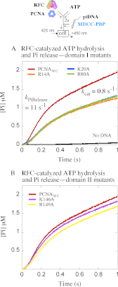
Kinetics of phosphate (Pi) release following ATP hydrolysis. RFC-catalyzed Pi release is measured by change in fluorescence intensity of MDCC-labeled PBP when it binds free PiEX = 425 nm, λEM > 450 nm). Preincubation of RFC and PCNAWC with ATP (Δt = 0.02–2 s; 2-s data shown) followed by addition of ptDNA results in a burst ATP hydrolysis and Pi release followed by a linear steady state phase. A, data are shown for PCNAWC and domain I mutants PCNAWC-R14A, PCNAWC-K20A, and PCNAWC-R80A (also, Pi release in the absence of ptDNA). An exponential + linear fit of the PCNAWC trace yields kPi Release = ∼11.4 s−1 and kcat = 0.8 s−1 (slope/3×[RFC]). B, data are shown for PCNAWC and domain II mutants PCNAWC-K146A and PCNAWC-R149A. Final reactant concentrations were 0.5 μm RFC, 1 μm PCNAWC, 2.5 μm ptDNA, 0.5 mm ATP, and 10 μm MDCC-PBP.
FIGURE 6.
FIGURE 6.
Global fitting of kinetic data to a minimal mechanism of PCNA loading. A and B, overlay of kinetic data for PCNAWC (A) and PCNAWC-R80A mutant from DNA binding/release, PCNA opening/closing, and Pi release experiments (B) provides a visual of rapid ptDNA binding to RFC-ATP-PCNA complex (formed during preincubation, Δt = 2 s), which triggers ATP hydrolysis (not shown), PCNA closure, Pi release, PCNA-ptDNA dissociation, and catalytic turnover. C–H, kinetic data measured at Δt = 0.02, 0.5, and 2 s for ptDNA binding/release (C and D), PCNA opening/closing (E and F), and Pi release for PCNAWC and PCNAWC-R80A (G and H), respectively, were all fit simultaneously to the model described in Fig. 7 and Table 2. The black lines are simulations generated by the model based on parameters listed in Table 2.
FIGURE 7.
FIGURE 7.
A clamp loading model showing the steps influenced by contact between PCNA and DNA. A, the schematic depicts each step in the mechanism used for global analysis of the kinetic data. Activated RFC is designated as *RFC; a separate step shows the equilibrium between loading-active (PCNA) and loading-inactive (**PCNA) clamp conformations; steps where the reverse rate constants are expected to be very small or are completely unknown are considered irreversible. B, a cartoon depicting the clamp loading reaction. I–III, binding of two to three ATP molecules to RFC (I) facilitates PCNA and additional ATP binding toward slow formation of active *RFC-ATP-PCNA (II) and *RFC-ATP-PCNAopen complexes (III). IV and V, ptDNA binds both intermediate complexes (IV), leading to an *RFC-ATP-PCNAopen-ptDNA complex that is ready for ATP hydrolysis (V). VI and VII, next, another slow step in the reaction designated as RFC deactivation leads to closure of PCNA around DNA (VI) and release of PCNA-ptDNA and Pi products and catalytic turnover (VII). VIII, PCNA can slip off the linear ptDNA, recycling both substrates in steady state. Slow steps in the mechanism are indicated by orange arrows. Steps IV (ptDNA binding and priming RFC for ATP hydrolysis) and VI (PCNA closure following ATP hydrolysis) are suppressed, and step VIII (PCNA-ptDNA dissociation) is enhanced by mutation of N-terminal domain I cationic residues in PCNA.

References

    1. Georgescu R. E., Kim S. S., Yurieva O., Kuriyan J., Kong X. P., O'Donnell M. (2008) Structure of a sliding clamp on DNA. Cell 132, 43–54 - PMC - PubMed
    1. McNally R., Bowman G. D., Goedken E. R., O'Donnell M., Kuriyan J. (2010) Analysis of the role of PCNA-DNA contacts during clamp loading. BMC Struct. Biol. 10, 3. - PMC - PubMed
    1. Fukuda K., Morioka H., Imajou S., Ikeda S., Ohtsuka E., Tsurimoto T. (1995) Structure-function relationship of the eukaryotic DNA replication factor, proliferating cell nuclear antigen. J. Biol. Chem. 270, 22527–22534 - PubMed
    1. Johnson A., O'Donnell M. (2005) Cellular DNA replicases. Components and dynamics at the replication fork. Annu. Rev. Biochem. 74, 283–315 - PubMed
    1. Moldovan G. L., Pfander B., Jentsch S. (2007) PCNA, the maestro of the replication fork. Cell 129, 665–679 - PubMed

Publication types

MeSH terms

LinkOut - more resources