

doi: 10.1038/ng.2382.
Epub 2012 Aug 19.
Mouse ooplasm confers context-specific reprogramming capacity
Affiliations
- PMID: 22902786
- PMCID: PMC3432711
- DOI: 10.1038/ng.2382
Item in Clipboard
Mouse ooplasm confers context-specific reprogramming capacity
Nat Genet.
2012 Sep.
Abstract
Enucleated oocytes have the distinctive ability to reprogram somatic nuclei back to totipotency. Here, we investigate genome-scale DNA methylation patterns after nuclear transfer and compare them to the dynamics at fertilization. We identify specific targets for DNA demethylation after nuclear transfer, such as germline-associated promoters, as well as unique limitations that include certain repetitive element classes.
Conflict of interest statement
The authors declare no competing financial interests.
Figures
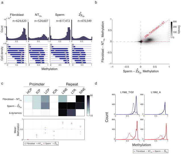
a. Histogram of methylation (top) and boxplots of methylation by CpG density (bottom) for 100 bp tiles in fibroblast, nuclear transfer reconstructed embryos (NTFib, mean of 4 replicates), sperm, and the inferred sperm value in zygote (ZySp; see Supplementary Methods). Fibroblasts and sperm show a global methylation pattern typical of somatic tissues while SCNT embryos (NTFib) exhibit a similar global shift as ZySp. Bulls-eye indicates the median, edges the 25th/75th percentile and whiskers the 2.5th/97.5th percentile. b. Scatterplot comparing global methylation dynamics between nuclear transfer (Fibroblast – NTFib) and fertilization (Sperm – ZySp). While demethylated regions appear to occur in common sites (upper right quadrant), the magnitude of demethylation is larger during fertilization as indicated by the dense cloud below the diagonal. The red triangle outlines the region where demethylation after SCNT is smaller than during fertilization. c. Heatmap (top) depicting genomic features that significantly change (dark) in Fibroblast-NTFib or Sperm-ZySp transitions, and the comparison of changes between the two transitions (Δ dynamics). Promoters are partitioned to high (H), intermediate (I) or low (L) CpG density and known imprint control regions (ICRs) are included as a control set. Color is the –log p-value. The mean methylation value for each feature set is shown in the bottom panel. Most features appear to change similarly in both processes with the exception of LINE and LTR features, which are comparably resistant to change after nuclear transfer. d. Histogram of methylation for elements in the L1Md_T and L1Md_Gf (left) and L1Md_A (right) families of the LINE-1 class. Both families are dynamic during fertilization, but only the L1Md_T/Gf families, which show larger demethylation during fertilization, show any detectable change after nuclear transfer.
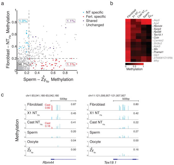
a. Scatterplot of promoter dynamics between donor fibroblasts and SCNT embryos compared to those observed at fertilization. The majority of promoters are unchanged in either process, with 1.0%, 1.1% and 1.1% of promoters dynamic in either NT specific, shared, or fertilization specific contexts. Colored dots refer to promoters that were observed to change consistently across SCNT experiments and/or during demethylation of the paternal genome after fertilization. “Other” includes promoters that either did not change or did not pass the stringent criteria for being called as dynamic. b. Dynamics specific to the Fibroblast-NT transition include several promoters that function specifically in gametes and are already hypomethylated in sperm and oocyte. c. The germ-line associated genes RNA binding motif protein 44 (Rbm44) and Testes expressed gene 19.1 (Tex19.1) are hypomethylated and expressed during gametogenesis, the early embryo, and pluripotent cell lines but de novo methylated upon gastrulation/differentiation . In fibroblasts, both gene promoters are hypermethylated and show a strong demethylation after either NT round. The level of demethylation suggests erasure in a large proportion of transplanted cells. Blue bars highlight single CpGs that are captured in all stages, red bars highlight 3 CpGs that can be associated with the Cast allele, with the mean allele-specific methylation value highlighted in red.
Comment in
-
Incomplete methylation reprogramming in SCNT embryos.Nat Genet. 2012 Sep;44(9):965-6. doi: 10.1038/ng.2393. Nat Genet. 2012. PMID: 22932499
References
-
- Reik W, Dean W, Walter J. Epigenetic reprogramming in mammalian development. Science. 2001;293:1089–1093. - PubMed
-
- Rideout WM, 3rd, Eggan K, Jaenisch R. Nuclear cloning and epigenetic reprogramming of the genome. Science. 2001;293:1093–1098. - PubMed
-
- Wakayama T, Perry AC, Zuccotti M, Johnson KR, Yanagimachi R. Full-term development of mice from enucleated oocytes injected with cumulus cell nuclei. Nature. 1998;394:369–374. - PubMed
Publication types
MeSH terms
Associated data
- Actions
Grants and funding
LinkOut - more resources
Full Text Sources
Other Literature Sources
Molecular Biology Databases
